WO2012133945A1 - Method for producing cardiomyocyte sheet from embryonic stem cells - Google Patents
Method for producing cardiomyocyte sheet from embryonic stem cells Download PDFInfo
- Publication number
- WO2012133945A1 WO2012133945A1 PCT/JP2012/059427 JP2012059427W WO2012133945A1 WO 2012133945 A1 WO2012133945 A1 WO 2012133945A1 JP 2012059427 W JP2012059427 W JP 2012059427W WO 2012133945 A1 WO2012133945 A1 WO 2012133945A1
- Authority
- WO
- WIPO (PCT)
- Prior art keywords
- cells
- sheet
- cell
- cardiomyocytes
- embryonic stem
- Prior art date
Links
Images


Classifications
-
- A—HUMAN NECESSITIES
- A61—MEDICAL OR VETERINARY SCIENCE; HYGIENE
- A61K—PREPARATIONS FOR MEDICAL, DENTAL OR TOILETRY PURPOSES
- A61K35/00—Medicinal preparations containing materials or reaction products thereof with undetermined constitution
- A61K35/12—Materials from mammals; Compositions comprising non-specified tissues or cells; Compositions comprising non-embryonic stem cells; Genetically modified cells
- A61K35/34—Muscles; Smooth muscle cells; Heart; Cardiac stem cells; Myoblasts; Myocytes; Cardiomyocytes
-
- A—HUMAN NECESSITIES
- A61—MEDICAL OR VETERINARY SCIENCE; HYGIENE
- A61K—PREPARATIONS FOR MEDICAL, DENTAL OR TOILETRY PURPOSES
- A61K35/00—Medicinal preparations containing materials or reaction products thereof with undetermined constitution
- A61K35/12—Materials from mammals; Compositions comprising non-specified tissues or cells; Compositions comprising non-embryonic stem cells; Genetically modified cells
- A61K35/44—Vessels; Vascular smooth muscle cells; Endothelial cells; Endothelial progenitor cells
-
- A—HUMAN NECESSITIES
- A61—MEDICAL OR VETERINARY SCIENCE; HYGIENE
- A61K—PREPARATIONS FOR MEDICAL, DENTAL OR TOILETRY PURPOSES
- A61K9/00—Medicinal preparations characterised by special physical form
- A61K9/70—Web, sheet or filament bases ; Films; Fibres of the matrix type containing drug
-
- A—HUMAN NECESSITIES
- A61—MEDICAL OR VETERINARY SCIENCE; HYGIENE
- A61L—METHODS OR APPARATUS FOR STERILISING MATERIALS OR OBJECTS IN GENERAL; DISINFECTION, STERILISATION OR DEODORISATION OF AIR; CHEMICAL ASPECTS OF BANDAGES, DRESSINGS, ABSORBENT PADS OR SURGICAL ARTICLES; MATERIALS FOR BANDAGES, DRESSINGS, ABSORBENT PADS OR SURGICAL ARTICLES
- A61L27/00—Materials for grafts or prostheses or for coating grafts or prostheses
- A61L27/36—Materials for grafts or prostheses or for coating grafts or prostheses containing ingredients of undetermined constitution or reaction products thereof, e.g. transplant tissue, natural bone, extracellular matrix
- A61L27/38—Materials for grafts or prostheses or for coating grafts or prostheses containing ingredients of undetermined constitution or reaction products thereof, e.g. transplant tissue, natural bone, extracellular matrix containing added animal cells
- A61L27/3804—Materials for grafts or prostheses or for coating grafts or prostheses containing ingredients of undetermined constitution or reaction products thereof, e.g. transplant tissue, natural bone, extracellular matrix containing added animal cells characterised by specific cells or progenitors thereof, e.g. fibroblasts, connective tissue cells, kidney cells
- A61L27/3834—Cells able to produce different cell types, e.g. hematopoietic stem cells, mesenchymal stem cells, marrow stromal cells, embryonic stem cells
-
- A—HUMAN NECESSITIES
- A61—MEDICAL OR VETERINARY SCIENCE; HYGIENE
- A61L—METHODS OR APPARATUS FOR STERILISING MATERIALS OR OBJECTS IN GENERAL; DISINFECTION, STERILISATION OR DEODORISATION OF AIR; CHEMICAL ASPECTS OF BANDAGES, DRESSINGS, ABSORBENT PADS OR SURGICAL ARTICLES; MATERIALS FOR BANDAGES, DRESSINGS, ABSORBENT PADS OR SURGICAL ARTICLES
- A61L27/00—Materials for grafts or prostheses or for coating grafts or prostheses
- A61L27/36—Materials for grafts or prostheses or for coating grafts or prostheses containing ingredients of undetermined constitution or reaction products thereof, e.g. transplant tissue, natural bone, extracellular matrix
- A61L27/38—Materials for grafts or prostheses or for coating grafts or prostheses containing ingredients of undetermined constitution or reaction products thereof, e.g. transplant tissue, natural bone, extracellular matrix containing added animal cells
- A61L27/3886—Materials for grafts or prostheses or for coating grafts or prostheses containing ingredients of undetermined constitution or reaction products thereof, e.g. transplant tissue, natural bone, extracellular matrix containing added animal cells comprising two or more cell types
-
- A—HUMAN NECESSITIES
- A61—MEDICAL OR VETERINARY SCIENCE; HYGIENE
- A61L—METHODS OR APPARATUS FOR STERILISING MATERIALS OR OBJECTS IN GENERAL; DISINFECTION, STERILISATION OR DEODORISATION OF AIR; CHEMICAL ASPECTS OF BANDAGES, DRESSINGS, ABSORBENT PADS OR SURGICAL ARTICLES; MATERIALS FOR BANDAGES, DRESSINGS, ABSORBENT PADS OR SURGICAL ARTICLES
- A61L27/00—Materials for grafts or prostheses or for coating grafts or prostheses
- A61L27/50—Materials characterised by their function or physical properties, e.g. injectable or lubricating compositions, shape-memory materials, surface modified materials
- A61L27/54—Biologically active materials, e.g. therapeutic substances
-
- A—HUMAN NECESSITIES
- A61—MEDICAL OR VETERINARY SCIENCE; HYGIENE
- A61P—SPECIFIC THERAPEUTIC ACTIVITY OF CHEMICAL COMPOUNDS OR MEDICINAL PREPARATIONS
- A61P43/00—Drugs for specific purposes, not provided for in groups A61P1/00-A61P41/00
-
- A—HUMAN NECESSITIES
- A61—MEDICAL OR VETERINARY SCIENCE; HYGIENE
- A61P—SPECIFIC THERAPEUTIC ACTIVITY OF CHEMICAL COMPOUNDS OR MEDICINAL PREPARATIONS
- A61P9/00—Drugs for disorders of the cardiovascular system
-
- C—CHEMISTRY; METALLURGY
- C12—BIOCHEMISTRY; BEER; SPIRITS; WINE; VINEGAR; MICROBIOLOGY; ENZYMOLOGY; MUTATION OR GENETIC ENGINEERING
- C12N—MICROORGANISMS OR ENZYMES; COMPOSITIONS THEREOF; PROPAGATING, PRESERVING, OR MAINTAINING MICROORGANISMS; MUTATION OR GENETIC ENGINEERING; CULTURE MEDIA
- C12N5/00—Undifferentiated human, animal or plant cells, e.g. cell lines; Tissues; Cultivation or maintenance thereof; Culture media therefor
- C12N5/06—Animal cells or tissues; Human cells or tissues
- C12N5/0602—Vertebrate cells
- C12N5/0652—Cells of skeletal and connective tissues; Mesenchyme
- C12N5/0657—Cardiomyocytes; Heart cells
-
- C—CHEMISTRY; METALLURGY
- C12—BIOCHEMISTRY; BEER; SPIRITS; WINE; VINEGAR; MICROBIOLOGY; ENZYMOLOGY; MUTATION OR GENETIC ENGINEERING
- C12N—MICROORGANISMS OR ENZYMES; COMPOSITIONS THEREOF; PROPAGATING, PRESERVING, OR MAINTAINING MICROORGANISMS; MUTATION OR GENETIC ENGINEERING; CULTURE MEDIA
- C12N5/00—Undifferentiated human, animal or plant cells, e.g. cell lines; Tissues; Cultivation or maintenance thereof; Culture media therefor
- C12N5/06—Animal cells or tissues; Human cells or tissues
- C12N5/0697—Artificial constructs associating cells of different lineages, e.g. tissue equivalents
-
- A—HUMAN NECESSITIES
- A61—MEDICAL OR VETERINARY SCIENCE; HYGIENE
- A61L—METHODS OR APPARATUS FOR STERILISING MATERIALS OR OBJECTS IN GENERAL; DISINFECTION, STERILISATION OR DEODORISATION OF AIR; CHEMICAL ASPECTS OF BANDAGES, DRESSINGS, ABSORBENT PADS OR SURGICAL ARTICLES; MATERIALS FOR BANDAGES, DRESSINGS, ABSORBENT PADS OR SURGICAL ARTICLES
- A61L2300/00—Biologically active materials used in bandages, wound dressings, absorbent pads or medical devices
- A61L2300/40—Biologically active materials used in bandages, wound dressings, absorbent pads or medical devices characterised by a specific therapeutic activity or mode of action
- A61L2300/412—Tissue-regenerating or healing or proliferative agents
- A61L2300/414—Growth factors
-
- A—HUMAN NECESSITIES
- A61—MEDICAL OR VETERINARY SCIENCE; HYGIENE
- A61L—METHODS OR APPARATUS FOR STERILISING MATERIALS OR OBJECTS IN GENERAL; DISINFECTION, STERILISATION OR DEODORISATION OF AIR; CHEMICAL ASPECTS OF BANDAGES, DRESSINGS, ABSORBENT PADS OR SURGICAL ARTICLES; MATERIALS FOR BANDAGES, DRESSINGS, ABSORBENT PADS OR SURGICAL ARTICLES
- A61L2430/00—Materials or treatment for tissue regeneration
- A61L2430/20—Materials or treatment for tissue regeneration for reconstruction of the heart, e.g. heart valves
-
- C—CHEMISTRY; METALLURGY
- C12—BIOCHEMISTRY; BEER; SPIRITS; WINE; VINEGAR; MICROBIOLOGY; ENZYMOLOGY; MUTATION OR GENETIC ENGINEERING
- C12N—MICROORGANISMS OR ENZYMES; COMPOSITIONS THEREOF; PROPAGATING, PRESERVING, OR MAINTAINING MICROORGANISMS; MUTATION OR GENETIC ENGINEERING; CULTURE MEDIA
- C12N2501/00—Active agents used in cell culture processes, e.g. differentation
- C12N2501/01—Modulators of cAMP or cGMP, e.g. non-hydrolysable analogs, phosphodiesterase inhibitors, cholera toxin
-
- C—CHEMISTRY; METALLURGY
- C12—BIOCHEMISTRY; BEER; SPIRITS; WINE; VINEGAR; MICROBIOLOGY; ENZYMOLOGY; MUTATION OR GENETIC ENGINEERING
- C12N—MICROORGANISMS OR ENZYMES; COMPOSITIONS THEREOF; PROPAGATING, PRESERVING, OR MAINTAINING MICROORGANISMS; MUTATION OR GENETIC ENGINEERING; CULTURE MEDIA
- C12N2501/00—Active agents used in cell culture processes, e.g. differentation
- C12N2501/04—Immunosuppressors, e.g. cyclosporin, tacrolimus
-
- C—CHEMISTRY; METALLURGY
- C12—BIOCHEMISTRY; BEER; SPIRITS; WINE; VINEGAR; MICROBIOLOGY; ENZYMOLOGY; MUTATION OR GENETIC ENGINEERING
- C12N—MICROORGANISMS OR ENZYMES; COMPOSITIONS THEREOF; PROPAGATING, PRESERVING, OR MAINTAINING MICROORGANISMS; MUTATION OR GENETIC ENGINEERING; CULTURE MEDIA
- C12N2501/00—Active agents used in cell culture processes, e.g. differentation
- C12N2501/10—Growth factors
- C12N2501/165—Vascular endothelial growth factor [VEGF]
-
- C—CHEMISTRY; METALLURGY
- C12—BIOCHEMISTRY; BEER; SPIRITS; WINE; VINEGAR; MICROBIOLOGY; ENZYMOLOGY; MUTATION OR GENETIC ENGINEERING
- C12N—MICROORGANISMS OR ENZYMES; COMPOSITIONS THEREOF; PROPAGATING, PRESERVING, OR MAINTAINING MICROORGANISMS; MUTATION OR GENETIC ENGINEERING; CULTURE MEDIA
- C12N2502/00—Coculture with; Conditioned medium produced by
- C12N2502/13—Coculture with; Conditioned medium produced by connective tissue cells; generic mesenchyme cells, e.g. so-called "embryonic fibroblasts"
- C12N2502/1329—Cardiomyocytes
-
- C—CHEMISTRY; METALLURGY
- C12—BIOCHEMISTRY; BEER; SPIRITS; WINE; VINEGAR; MICROBIOLOGY; ENZYMOLOGY; MUTATION OR GENETIC ENGINEERING
- C12N—MICROORGANISMS OR ENZYMES; COMPOSITIONS THEREOF; PROPAGATING, PRESERVING, OR MAINTAINING MICROORGANISMS; MUTATION OR GENETIC ENGINEERING; CULTURE MEDIA
- C12N2502/00—Coculture with; Conditioned medium produced by
- C12N2502/13—Coculture with; Conditioned medium produced by connective tissue cells; generic mesenchyme cells, e.g. so-called "embryonic fibroblasts"
- C12N2502/1347—Smooth muscle cells
-
- C—CHEMISTRY; METALLURGY
- C12—BIOCHEMISTRY; BEER; SPIRITS; WINE; VINEGAR; MICROBIOLOGY; ENZYMOLOGY; MUTATION OR GENETIC ENGINEERING
- C12N—MICROORGANISMS OR ENZYMES; COMPOSITIONS THEREOF; PROPAGATING, PRESERVING, OR MAINTAINING MICROORGANISMS; MUTATION OR GENETIC ENGINEERING; CULTURE MEDIA
- C12N2502/00—Coculture with; Conditioned medium produced by
- C12N2502/28—Vascular endothelial cells
-
- C—CHEMISTRY; METALLURGY
- C12—BIOCHEMISTRY; BEER; SPIRITS; WINE; VINEGAR; MICROBIOLOGY; ENZYMOLOGY; MUTATION OR GENETIC ENGINEERING
- C12N—MICROORGANISMS OR ENZYMES; COMPOSITIONS THEREOF; PROPAGATING, PRESERVING, OR MAINTAINING MICROORGANISMS; MUTATION OR GENETIC ENGINEERING; CULTURE MEDIA
- C12N2506/00—Differentiation of animal cells from one lineage to another; Differentiation of pluripotent cells
- C12N2506/02—Differentiation of animal cells from one lineage to another; Differentiation of pluripotent cells from embryonic cells
Definitions
- the present invention relates to a method for producing a myocardial sheet using embryonic stem cell-derived cardiomyocytes, endothelial cells and mural cells.
- the present invention also relates to a therapeutic agent for heart disease comprising a myocardial sheet obtained by the above method.
- Non-patent Document 1 Patent Document 2
- the expected therapeutic effect cannot be obtained because the amount of cells in the sheet is insufficient, it is considered that the sheets need to be laminated and administered (Non-patent Document 2).
- An object of the present invention relates to a method for producing a myocardial sheet using embryonic stem cell-derived cardiomyocytes, endothelial cells and mural cells, and a therapeutic agent for heart disease comprising the myocardial sheet obtained thereby. Accordingly, an object of the present invention is to provide a myocardial sheet prepared by mixing cells obtained by inducing differentiation from embryonic stem cells into cardiomyocytes, endothelial cells and mural cells.
- the inventors of the present invention manufactured embryonic stem cells from Flk / KDR-positive cells on a culture dish coated with a temperature-responsive polymer, and cardiomyocytes prepared from the Flk / KDR-positive cells.
- the present inventors succeeded in treating a myocardial infarction model with a myocardial sheet produced using cardiomyocytes derived from embryonic stem cells, endothelial cells and mural cells, and completed the present invention. It came. That is, the present invention includes the following features.
- a mixed cell comprising cardiomyocytes, endothelial cells and mural cells.
- the cell according to any one of [11] to [15], wherein the cardiomyocyte, endothelial cell and wall cell are cells produced from embryonic stem cells.
- FIG. 1 shows the results of FACS of CM and EC component before co-culture on a temperature sensitive dish and the result of FACS after sheet formation.
- APC allophycocyanin, PE; phycoerythrin, SSC; side scatter, GFP; green fluorescent protein, FITC; fluorescein isothiocyanate.
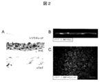
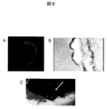
- FIG. 2 shows (A) Sirius red-stained image (upper): the formation of a layered extracellular matrix at the cell culture dish adhesion.
- HE-stained image (middle): 3-4 layers of cells constitute a sheet.
- FIG. 3 shows (A) a sheet image adhered on a MED64 probe. (B) The extracellular potential at each electrode is shown. (C) The distribution of potential measured in (B) is shown. The electrode having the highest peak negative potential (lower part of the figure) was regarded as a zero point, and color classification was performed by the time difference (seconds) from there to the peak negative potential of each electrode. The conduction from the bottom to the top is recognized.
- FIG. 4 shows the measurement results of each cytokine by ELISA on the culture supernatant.
- VEGF was shown to be extremely high compared to serum-free medium.
- SF serum free, TNFa
- tumor necrosis factor- ⁇ IGF-1
- insulin-like growth factor-1 VEGF
- basal endoral cell growth factor bFGF
- bass FGF An epidermal growth factor, HGF
- hepatocyte growth factor a hepatocyte growth factor.
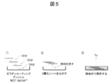
- FIG. 5 shows a schematic diagram of the cell sheet lamination method.
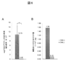
- FIG. 6 shows a graph comparing the amount of VEGF produced by sheets produced with or without cardiomyocytes (CM).
- CM cardiomyocytes
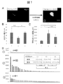
- FIG. 7 shows (A) a representative undifferentiated colony (arrow) in Group 1 (left figure: immediately after collection of the myocardial sheet) and an expanded colony of Group 2 (right figure: continuously cultured for 3 days with LIF added). . (B) The number of undifferentiated colonies in each Group is shown. (C) The average colony area in each Group is shown. (D) Show all colonies of each Group in order of size. Those of 5,000 ⁇ m 2 or more are displayed in red.
- FIG. 8 shows (A) macroscopic findings of the laminated sheet.
- B The optical microscope image of the sheet
- C Macroscopic findings after transplanting the sheet. The arrow points to the laminated sheet.
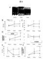
- FIG. 9 shows the results of cardiac ultrasonography.
- A The time-dependent change of the M mode figure in a myocardial sheet transplantation group is shown.
- FS left ventricular diameter shortening rate
- FAC left ventricular cross-sectional change rate
- D systolic wall thickness increase rate
- (E) shows the time course of the non-shrinkage range (AL) (infarct site range index).
- (F) shows the rate of change of diastolic left ventricular area relative to pre-treatment (PreTx) (an index of left ventricular expansion).
- PreTx before treatment, Tx2w; 2 weeks after treatment, Tx4w; 4 weeks after treatment, LV; left ventricle.
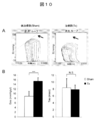
- FIG. 10 shows the results of the left ventricular volume pressure curve measurement test
- A The volume pressure curve shift accompanying the decrease in the preload in the sham group (left figure) and the treatment group (right figure).
- End systolic left ventricular elastic modulus (Ees) is indicated by the slope of a straight line indicated by an arrow.
- B Ees (left figure) and time constant (Tau) (right figure) of the Sham group (white) and the treatment group (black) are shown, respectively.
- Ees indicates left ventricular contractility (high value indicates high contractility), and Tau indicates left ventricular expandability (low value indicates high expandability).
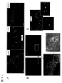
- FIG. 11 shows (A) FISH (mouse-derived cells: yellow) and cTnT immunostained image (red) on day 1 (left), day 1 (middle), and day 4 (right) after transplantation. Show. The engraftment site on the 28th day after transplantation (Tx-d28) is indicated by an arrow. (B) Connexin 43 (green) immunostained image 7 days after transplantation. The figure surrounded by the white line at the lower left shows an image of a normal myocardium.
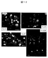
- FIG. 12 shows FISH of graft-derived engrafted cardiomyocytes on day 1 after transplantation (Tx-d1), day 3 (Tx-d3), week 1 (Tx-d7), and week 4 (Tx-d28). (Mouse-derived cells: yellow) and cTnT immunostained images (red) are shown.
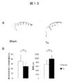
- FIG. 13 shows Sirius red stained images of (A) Sham group (left) and treatment group (Tx: right). In the treatment group, thinning of the wall is suppressed and a decrease in infarct area is observed.
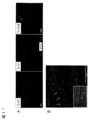
- FIG. 14 shows (A) FISH (mouse-derived cells: 1 day after transplantation (Tx-d1: left figure), 3 days (Tx-d3: middle figure), and 7 days (Tx-d7 right figure)). (Yellow), cTnT immunostained image (red), von Willebrand factor (vWF) immunostained image (green). (B) Each enlarged image of the fluorescence microscope on the third day after transplantation is shown.
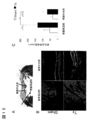
- FIG. 15 shows the measurement result of neovascular density at 4 weeks after transplantation.
- A Masson trichrome stained image of the treatment group 4 weeks after transplantation.
- FIG. 1 the infarct central part (Central-MI) and the infarct peripheral part (Peri-MI) are shown.
- B Stained images of cTnT (red) and vWF (green) in the periphery and center of the infarct in the treatment group (Tx) and the Sham group, respectively. A marked formation of a vWF-positive site with a luminal structure is observed in the treatment group in both the peripheral part and the central part.
- C Capillary density of the Central-MI and Peri-MI sites in each group shown in FIG. In the figure, **: p ⁇ 0.01, ***: p ⁇ 0.001 (unpaired t-test).
- the present invention includes (a) a step of producing Flk / KDR positive cells, cardiomyocytes, endothelial cells and mural cells from embryonic stem cells, respectively, and (b) the Flk / KDR positive cells,
- a method for producing a myocardial sheet from embryonic stem cells comprising a step of forming a myocardial sheet by mixing with endothelial cells and mural cells, and a method for producing a heart disease such as ischemic heart disease comprising a myocardial sheet obtained by this method It relates to a therapeutic agent.
- ES cells are stem cells established from the inner cell mass of early embryos (for example, blastocysts) of mammals such as humans and mice and having the pluripotency and the ability to proliferate by self-replication.
- ES cells are embryonic stem cells derived from the inner cell mass of the blastocyst, which is the embryo after the morula, at the 8-cell stage of a fertilized egg, and have the ability to differentiate into any cell that constitutes an adult, so-called differentiation differentiation. And ability to proliferate by self-replication.
- ES cells were discovered in 1981 in mice (MJ Evans and MH Kaufman (1981), Nature 292: 154-156), and then ES cell lines were also established in primates such as humans and monkeys.
- ES cells can be established by removing an inner cell mass from a blastocyst of a fertilized egg of a subject animal and culturing the inner cell mass on a fibroblast feeder.
- LIF leukemia inhibitory factor
- bFGF basic fibroblast growth factor
- DMEM / F-12 culture supplemented with 0.1 mM 2-mercaptoethanol, 0.1 mM non-essential amino acid, 2 mM L-glutamic acid, 20% KSR and 4 ng / ml ⁇ -FGF as a culture solution for ES cell production
- the solution can be used to maintain human ES cells in a humidified atmosphere at 37 ° C., 2% CO 2 /98% air (O. Fumitaka et al. (2008), Nat. Biotechnol., 26: 215- 224).
- ES cells need to be passaged every 3 to 4 days, and at this time, passage is performed by, for example, 0.25% trypsin and 0.1 mg / ml in PBS containing 1 mM CaCl 2 and 20% KSR. Can be performed using ml collagenase IV.
- ES cells can be selected by the Real-Time PCR method using the expression of gene markers such as alkaline phosphatase, Oct-3 / 4, Nanog as an index.
- gene markers such as OCT-3 / 4, NANOG, and ECAD can be used as an index (E. Kroon et al. (2008), Nat. Biotechnol., 26: 443). -452).
- Human ES cell lines are obtained from, for example, WA01 (H1) and WA09 (H9) from the WiCell Research Institute, and KhES-1, KhES-2 and KhES-3 from the Institute of Regenerative Medicine, Kyoto University (Kyoto, Japan). Is possible.
- ⁇ Differentiation medium Examples of differentiation media used for the production of Flk / KDR positive cells (also called Flk + cells), cardiomyocytes, endothelial cells and mural cells for producing myocardial sheets are described below.
- the medium can be prepared using a medium used for culturing animal cells as a basal medium.
- IMDM medium for example, IMDM medium, Medium 199 medium, Eagle's Minimum Essential Medium (EMEM) medium, ⁇ MEM medium, Dolbecco's modified Eagle's Medium (DMEM) medium, Ham's F16 medium, Ham's F16 medium, Fischer's medium, mixed media thereof, and the like are included.
- the medium may contain serum or may be serum-free.
- the medium can also contain, for example, albumin, transferrin, Knockout Serum Replacement (KSR) (serum substitute for FBS during ES cell culture), fatty acid, insulin, collagen precursor, trace element, 2-mercaptoethanol, One or more serum replacements such as 3′-thiolglycerol may be included, as well as lipids, amino acids, non-essential amino acids, vitamins, growth factors, cytokines, antibiotics, antioxidants, pyruvate, buffers, One or more substances such as inorganic salts may also be included.
- KSR Knockout Serum Replacement
- a Flk / KDR positive cell is a cell that expresses at least Flk / KDR, where Flk / KDR means Flk1 or KDR and is a receptor for vascular endothelial growth factor (VEGF). It is.
- Flk1 is exemplified by NCBI accession number NM_010612
- KDR is exemplified by NCBI accession number NM_002253.
- Flk / KDR positive cells can be prepared by inducing differentiation of embryonic stem cells by any method (Yamashita J, et al, Nature.
- cardiomyocytes mean cells expressing at least cardiac troponin (cTnT) or ⁇ MHC.
- cTnT is exemplified by NCBI accession number NM_000364 in the case of humans and NM_001130174 in the case of mice.
- ⁇ MHC is exemplified by NCBI accession number NM_002471 in the case of humans, and NM_001164171 in the case of mice.
- Embryonic stem cells can be induced to differentiate into cardiomyocytes by forming a cell mass (embryoid body) by suspension culture.
- a known method can be used as a method for inducing differentiation of cardiomyocytes from embryonic stem cells, and is not particularly specified. For example, differentiation in the presence of a substance that suppresses BMP signaling is performed.
- a method of induction (WO2005 / 033298), a method of inducing differentiation by sequentially adding Activin A and BMP (WO2007 / 002136), a method of inducing differentiation in the presence of a substance that promotes activation of the canonical Wnt signal pathway ( WO2007 / 126077) and FIk / KDR positive cells can be isolated from embryonic stem cells and cardiomyocytes can be induced to differentiate from embryonic stem cells using the method of inducing differentiation in the presence of cyclosporin A (WO2009 / 118928) .
- embryonic stem cells are adherently cultured on a culture device to produce Flk / KDR positive cells, and then differentiation is induced in the presence of cyclosporin A.
- the content of cyclosporin A in the medium is, for example, 0.1 to 30 ⁇ g / mL, and preferably 1 to 3 ⁇ g / mL, but can be any amount that allows differentiation of cardiomyocytes.
- the culture equipment is coated with a cell support material such as collagen, gelatin, poly-L-lysine, poly-D-lysine, laminin, fibronectin, Matrigel TM (Becton Dickinson). ) And the like.
- cardiomyocytes may be isolated and purified, or may be mixed with other cell types. Preferably, it is an isolated and purified cell.
- the method of isolation and purification is not particularly limited, but for example, a method of selecting using a cardiomyocyte marker such as N-cadherin as an index (Honda M, et al, Biochem Biophys Res Commun.
- endothelial cells mean cells expressing at least one of PE-CAM, VE-cadherin, and von Willebrand factor (vWF).
- the mural cell means a cell that expresses at least Smooth muscle actin (SMA).
- the PE-CAM is exemplified by NCBI accession number NM_000442 in the case of humans, and NM_001032378 in the case of mice.
- VE-cadherin is exemplified by NCBI accession number NM_001795 for humans and NM_009868 for mice.
- NCBI accession number NM_000552 is exemplified for humans
- NM_011708 is exemplified for mice.
- SMA is exemplified by NCBI accession number NM_001141945 for humans, and NM_007392 for mice.
- Embryonic stem cells can be induced to differentiate into endothelial cells or mural cells by forming a cell mass (embryoid body) by suspension culture.
- a known method can be used as a method for inducing differentiation of endothelial cells or mural cells from embryonic stem cells, and is not particularly specified.
- FLk / KDR positive cells from embryonic stem cells And cardiomyocytes can be induced to differentiate from embryonic stem cells using a method of inducing differentiation in the presence of VEGF and cAMP (Yamashita J, et al. Nature. 408: 92-6, 2000).
- the content of VEGF and cAMP in the medium is not limited as long as it can induce differentiation of endothelial cells or mural cells.
- VEGF for example, it is 25 ng / mL to 150 ng / mL, preferably 50 to 100 ng / mL.
- cAMP is used to include its derivatives (eg 8-bromo-cAMP), but its content is not limited to eg 0.1 mmol / L to 2 mmol / l, preferably 0.5 to 1 mmol / l L.
- the culture days are not particularly limited, but are 1 to 10 days, preferably 3 days after culturing Flk / KDR positive cells in the presence of VEGF and cAMP.
- the endothelial cells or mural cells may be isolated from each other and purified, or the endothelial cells or mural cells may be present in a mixture with other cell types.
- a mixed cell containing cardiomyocytes, endothelial cells and mural cells obtained using the above method can be produced.
- the content of cardiomyocytes is, for example, 10%, 20% or more, 30% or more, 40% or more, or 50% or more, 90% or less, 80% or less, 70% or less, 60% or less, or 50 % Or less.
- the preferred content is 40% or more, and the more preferred content is in the range of 40% or more and 50% or less.
- the content of endothelial cells is, for example, 1%, 2% or more, 3% or more, 4% or more, 5% or more, or 10% or more, 50% or less, 40% or less, 30% or less, 20 % Or less or 10% or less.
- a preferable content rate is 3% or more, and a more preferable content rate is the range of 3% or more and 10% or less.
- the content of mural cells is, for example, 10%, 20% or more, 30% or more, 40% or more, 50% or more, or 60% or more, 90% or less, 80% or less, 70% or less, or 60 % Or less.
- a preferable content rate is 20% or more, 30% or more, 40% or more, or 50% or more, and a more preferable content rate is a range of 50% or more and 60% or less.
- the mixed cell may be isolated as a cell culture, and is preferably a mixed cell in the form of a sheet.
- the myocardial sheet is a sheet-like cell aggregate composed of various cells forming the heart or blood vessels and connected to each other by intercellular bonding.
- examples of the various cells forming the heart or blood vessels include the above-described cardiomyocytes, endothelial cells and wall cells.
- a preferred myocardial sheet in the present invention has an electrical connection and orientation between cells, and secretes VEGF out of the cell.
- the myocardial sheet is produced by mixing and culturing cells containing at least cardiomyocytes, endothelial cells and mural cells prepared by the above-described method. At this time, the number of cells to be cultured is, for example, 1 ⁇ 10 4 to 1 ⁇ 10 6 .
- cells other than cardiomyocytes, endothelial cells, and mural cells may be included.
- the above-described embryonic stem cell-derived Flk / KDR positive cells are cultured by mixing cardiomyocytes, endothelial cells and mural cells into cells cultured for 1 to 7 days, preferably 3 days.
- VEGF may be added to the culture medium for further culturing, and the culture days at this time may be 1 to 10 days, preferably 4 days.
- the culture was coated with a temperature-responsive polymer obtained by polymerizing a (meth) acrylamide compound, an N- (or N, N-di) alkyl-substituted (meth) acrylamide derivative (Japanese Patent Laid-Open No.
- a culture device may be used, and a culture device to which poly-N-isopropylacrylamide is fixed is preferable.
- this culture equipment can also be purchased from Cellseed Co. as UpCell.
- the size of the myocardial sheet depends on the culture equipment, but preferably has a sufficient area to cover the site to be transplanted.
- the prepared myocardial sheet may be used by being laminated, and is preferably a myocardial sheet having three layers. In the lamination, the myocardial sheets are stacked in the culture solution (preferably, the myocardial sheets are shifted and stacked), and then joined by removing the culture solution.
- the myocardial sheet manufactured by the above method has, for example, the following characteristics.
- the cell composition ratio comprising at least cardiomyocytes, endothelial cells and wall cells and constituting the myocardial sheet is not limited to the following, but for example, cardiomyocytes 35-50%, endothelial cells 0.5-7%, It is 45 to 63% of wall cells.
- -Endothelial cells are scattered between cardiomyocytes.
- the myocardial sheet provided in the present invention can be used as a therapeutic agent for animal (preferably human) heart disease.
- a method for treating a heart disease is achieved by arranging a myocardial sheet so as to cover a desired portion.
- the arrangement so as to cover a desired portion can be performed using a technique well known in the art.
- the desired part if the desired part is large, it may be placed so as to surround the tissue.
- the myocardial sheet in order to obtain a desired effect, can be arranged several times in the same portion. When arranging several times, it is desirable to allow sufficient time for the myocardial sheet to engraft in the tissue and perform angiogenesis.
- Examples of the heart disease in the present invention include heart failure, ischemic heart disease, myocardial infarction, cardiomyopathy, myocarditis, hypertrophic cardiomyopathy, dilated phase hypertrophic cardiomyopathy, deficiency due to dilated cardiomyopathy, and the like. .
- Example 1 Preparation of myocardial sheet Yamashita JK, et al. FASEB J.H. 19: 1534-6, 2005.
- Flk + cells were generated by previously reported methods (Yamashita J, et al. Nature. 408: 92-6, 2000 or Yamashita JK, et al. FASEB J. 19: 1534-6, 2005).
- EMG7 or 20D17 was cultured for 4 days on gelatin-coated dishes using differentiation medium ( ⁇ MEM supplemented with 10% fetal bovine serum and 5 ⁇ 10 5 mol / L 2-mercaptoethanol), followed by Flk by FACS. It was prepared by purifying positive cells. Mixed cells of endothelial cells and mural cells were produced from the Flk + cells obtained by the above-described method using the method reported so far (Yamashita J, et al. Nature. 408: 92-6, 2000 or Yurgi- Kobayashi T, et al. Arterioscler Thromb Vasc Biol.26: 1977-84, 2006).
- Cardiomyocytes were prepared from the Flk + cells obtained by the method described above using the methods reported so far (WO 2009/118928 or Yan P, et al. Biochem Biophys Res Commun. 379: 115-20, 2009). Briefly, it was obtained by culturing on mitomycin C-treated OP9 cells for 4 days using a differentiation medium supplemented with 1-3 ⁇ g / mL Cyclosporin-A, and then separating the GFP positive fraction.
- the myocardial sheet was produced by the following method using the above-described plural types of cells. From 2.5 ⁇ 10 4 to 4.0 ⁇ 10 4 Flk + cells were seeded on a temperature-sensitive culture dish (UpCell, Cellu Seed) and cultured using a differentiation medium. On day 3 of culture, 5.0 ⁇ 10 5 mixed cells of the above endothelial cells and mural cells and 5.0 ⁇ 10 5 of the above cardiomyocytes were seeded in the same dish and differentiation medium supplemented with VEGF And cultured at 37 ° C. By returning to room temperature 4 days after the addition of cardiomyocytes (7 days after the start of culture), the cells were detached from the culture dish into a sheet to obtain a myocardial sheet.
- a temperature-sensitive culture dish UpCell, Cellu Seed
- FIG. 1 shows the results of quantification of the ratio of cardiomyocytes (CM), endothelial cells (EC), and mural cells (MC) based on the positive rates of GFP (FITC) and PE.
- CM cardiomyocytes
- EC endothelial cells
- MC mural cells
- Example 3 Histological evaluation of myocardial sheet
- a myocardial sheet prepared from EMG7 was fixed using 4% PFA (paraformaldehyde), blocked with 1% skim milk, and then antibody (primary Antibody: mouse anti-cTnT, rat anti-VE-Cadherin, secondary antibody: anti-mouse Alexa Flour 546, anti-rat Alexa Flour 488) are used to label CM and EC, and DAPI (4 ′, 6-diamino) After nuclei staining with 2-phenylindole), the images were observed with a multiphoton laser microscope (LSM510, Carl-Zeiss) or a fluorescence microscope (BZ-9000, Keyence) (FIGS.
- LSM510 multiphoton laser microscope
- Carl-Zeiss Carl-Zeiss
- BZ-9000 fluorescence microscope
- EMG7 Electrophysiological evaluation of myocardial sheet
- EMG7 was prepared on an electrode of a culture dish with an electrode (MED64 system, Alpha Med Science) coated with 0.1% gelatin. The myocardial sheet was left still. Subsequently, the medium was aspirated and incubated at 37 ° C. for 30 minutes to fix the electrode and the sheet, and the electric potential conduction on the sheet was recorded by measuring the electric potential of each electrode. The result is shown in FIG. By measuring the extracellular potential, it was confirmed that the pulsation was electrically continuously transmitted in one direction (FIG. 3C).
- Example 5 Cytokine production ability of myocardial sheet The amount of cytokine in the culture supernatant during formation (Condition 1) and after completion (Condition 2) of a myocardial sheet prepared from EMG7 was measured using ELISA (enzyme-linked immunosorbent assay). TNF ⁇ , IGF-1, VEGF, IL-6, bFGF, IFN ⁇ , EGF, Leptin and HGF were measured (HGF: mouse HGF EIA kit, IIM, otherwise: mouse angiogenesis ELISA strip, Signosis). The result is shown in FIG.
- VEGF vascular endothelial growth factor
- ⁇ Condition 2 Spread the myocardial sheet prepared by the above method on a gelatin-coated dish and leave it to stand, suck the medium, fix the culture dish and the sheet, add the medium, and add at 37 ° C for 30 minutes. Incubate. Another myocardial sheet was spread and allowed to stand on the sheet fixed to the dish, and the medium was sucked and laminated. Repeat for 3 layers in the same way. The second and third layers were laminated by shifting the position little by little from the original sheet. Subsequently, the medium was allowed to flow along the bottom of the culture dish using a pipetteman, and the laminated cell sheets were peeled from the culture dish (FIG. 5).
- VEGF production ability of myocardial sheet RNA was extracted from the myocardial sheet prepared as described above using RNeasy mini (QIAGEN), and the expression level of vegf164 was measured by quantitative RT-PCR (Step One Plus, Appliedbiosystems, vegf164 forward primer: 5′-CCAGCACATAGGAGAGATGAGCTT-3 ′ (SEQ ID NO: 1) and reverse primer: 5′-CAAGGCTCACAGTGATTTTCTGG-3 ′ (SEQ ID NO: tAC 3 '(SEQ ID NO: 3) and reverse primer: 5'-ATGGAGCCACCGATCCCAC -3 '(SEQ ID NO: 4)).
- FIG. 6A a cell sheet prepared using neonatal mouse cardiac fibroblasts (CF) was used as a control.
- CF neonatal mouse cardiac fibroblasts
- FIG. 6A the culture supernatant is aspirated before collecting (before lowering to room temperature) the sheet added with cardiomyocytes (CM (+)) and the sheet not added (CM ( ⁇ )).
- CM (+) cardiomyocytes
- CM ( ⁇ ) the sheet not added
- the supernatant after culturing for 3 hours with serum-free medium was collected, and the amount of VEGF was measured (Quantikine mouse VEGF, R & D). The result is shown in FIG.
- CM (+) sheet secretes VEGF much more than the CM ( ⁇ ) sheet.
- Example 7 Evaluation of contamination of undifferentiated cells into myocardial sheet The myocardial sheet was collected and allowed to stand on a gelatin-coated dish, and the medium was sucked and incubated at 37 ° C for 30 minutes to fix the dish and the sheet.
- Group 1 Fluorescent immunostaining is performed immediately after collection of the myocardial sheet.
- Group 2 After recovery of myocardial sheet, continuous culture for 3 days in a medium for ES cells (Yamashita J, et al. Nature. 408: 92-6, 2000) supplemented with LIF (leukemia inhibitory factor), followed by fluorescence immunization Perform staining.
- -Group 3 Fluorescent immunostaining is performed after continuous culturing for 3 days in a medium for ES cells without addition of LIF.
- Example 8 Myocardial sheet transplantation for disease model rats 10-13 weeks old, 250-330 g athymic immunodeficient rats (F344 / N Jcl-rnu / rnu) (CLEA, Japan) An infarct (MI) model was created. The rat was subjected to respiratory management with a rat ventilator and anesthetized by isoflurane aspiration. Subsequently, the heart was exposed by pericardiotomy through left intercostal thoracotomy under artificial respiration with a small amount of oxygen, and the anterior descending branch was ligated with 6-0 polypropylene thread at the periphery of the first septal branch.
- MI infarct
- the wound was closed with 4-0 polypropylene yarn.
- the presence or absence of MI was confirmed by cardiac ultrasonography (Vivid 7, GE Yokogawa Medical). Those with a left ventricular diameter shortening rate (FS) exceeding 40% were excluded as inappropriate models.
- the myocardial sheet (FIG. 8) prepared from EMG7 was transplanted 7 days after the introduction of MI. For transplantation, three myocardial sheets were laminated and used by the method described above (FIG. 5).
- an MI model rat was introduced with anesthesia with diethyl ether, and then respiratory management was performed with a rat ventilator, and anesthesia was maintained with isoflurane.
- Thoracotomy was performed with left intercostal thoracotomy and adhesion with the lungs and chest wall was carefully peeled to expose the myocardial infarction, and a laminated myocardial sheet was transplanted into the infarct. After standing for 15 minutes, the wound was closed with 4-0 polypropylene yarn.
- the Sham operation group was exposed in the same manner to myocardial infarction, and similarly closed after 15 minutes. All 12 cases observed until 4 weeks after transplantation survived.
- Example 10 Histological evaluation by transplantation of myocardial sheet Graft cell engraftment over time, localization of engraftment cells, and evaluation of changes in graft-derived cardiomyocyte morphology (maturation) The following method was carried out at 1 day, 4 weeks. After induction of anesthesia with diethyl ether, respiratory management was performed with a rat ventilator and anesthesia was maintained with isoflurane. The chest was opened at the midline, and the superior vena cava, left superior vena cava and inferior vena cava were secured and blocked. A 23G needle was inserted into the apex of the heart, and the left ventricular blood was drained by physiological saline injection.
- the right atrium was opened to prevent overexpansion and edema of the heart.
- 4% PFA was similarly injected for 45 minutes to fix the tissue (perfusion fixation method).
- the heart was removed, infiltrated with 4% PFA, and after 4 ° C over night, it was infiltrated with 15% sucrose solution (4 ° C, 2 changes, total 24 hours). Freeze embedding was performed using a solution obtained by adding dry ice to isopentane and an OCT compound.
- a 6 ⁇ m section was prepared at the center of the infarcted region, blocked with a blocking agent (Protein Block Serum-Free, DAKO), and then antibody (primary antibody: mouse anti-cTnT, rabbit anti-GFP and secondary anti-GFP).
- Primary antibody mouse anti-cTnT, rabbit anti-GFP and secondary anti-GFP.
- Antibody GFP-positive cardiomyocytes were labeled using anti-mouse Alexa Floor 546 or anti-rat Alexa Floor 488) and observed using a fluorescence microscope (BZ-9000, Keyence).
- nexin 43 is labeled on the 1-week post-transplant model (primary antibody: rabbit anti-connexin 43, secondary antibody: anti-rabbit Alexa Floor 488), and the presence or absence of the expression of gap junction at the graft site is fluorescent.
- mice cells in rat heart tissue were detected by the following method using a FISH (fluorescence in situ hybridization) probe that recognizes a species-specific repetitive sequence.
- FISH fluorescence in situ hybridization
- Rat genomic DNA FISH probe (Cy5 label) and mouse genomic DNA FISH probe (digoxigenin label) manufactured by Chromosome Science Lab were used as the above probes.
- the tissue sections were used after pretreatment by the following method. After washing with PBS, it was fixed with 4% PFA / PBS for 15 minutes, washed with PBS, then dehydrated and dried with an alcohol series.
- the hybridized chromosome specimen was subjected to stringency wash with 50% formamide / 2 ⁇ SSC at 37 ° C., and then rat and mouse nuclear signals were detected by anti-Dig-Cy3 using the signal of mouse genomic DNA FISH probe.
- a solution prepared by diluting mouse anti-cTnT with Can Get Signal Solution 1 (TOYOBO) to a specified concentration (1: 200) was added dropwise to the FISH-finished section and reacted at 37 ° C. for 1 hour.
- the plate was washed with PBST for 5 minutes ⁇ 3 times, and 1: 500 diluted secondary antibodies (anti-rabbit-Alexa 488 and anti-mouse-Alexa 594) were added with Can Get Signal Solution 2 and reacted for 30 minutes to perform immunostaining. It was. After the reaction, the plate was washed with PBST for 5 minutes ⁇ 3 times, and nuclear staining was also performed with DAPI. After staining, the genomic DNA probe signal and the fluorescently labeled antibody were microscopically observed using a Leica CW-4000 system. As a result, the number of engrafted graft cells derived from mice decreased with time, and only a small number were observed on the 28th day.
- the site engrafted after the 7th day was mainly the periphery of the infarct where more recipient myocardium remained.
- the cardiomyocyte engraftment site it was confirmed that a gap junction was being formed on the seventh day after transplantation (FIG. 11).
- cTnT cardiac troponin T
- cardiomyocyte sarcomere supermolecular aggregate
- Example 11 Evaluation of left ventricular remodeling after infarction by myocardial sheet transplantation
- 5 sections were prepared for each individual at 50 ⁇ m intervals from the center of the infarction at a thickness of 6 ⁇ m, stained with Sirius red, and fluorescent. Microscopic observation was performed. The intraventricular cavity length and infarct site length were measured, and the ratio of infarct site was calculated. The infarct site area was divided by the infarct site length to calculate the infarct site average wall thickness.
- the treatment group and the Sham group were each performed (5 individuals each) and compared.
- the number of neovascularization (capillary density) in the treatment group and the Sham group at the 4th week was measured by comparing the infarct site into the central part (Central-MI) and the peripheral part (Peri-MI) (for each three individuals). , Random 5 views each).
- Masson trichrome staining was performed for one example of the treatment group in order to illustrate the central portion and the peripheral portion.
- the distribution of neovascularization the presence or absence of neovascularization at the graft-derived cardiomyocyte engraftment portion at the stage of 1st, 3rd, 1st week was observed.
- vWF positive cells were scattered inside the graft site, and on the third day, accumulation of vWF positive cells was observed so as to surround the myocardial cell mass of the graft site from the inside. Did not show an obvious lumen structure. Since mouse signals are not shown, accumulation from the recipient (rat) side is considered. On day 7 after transplantation, the accumulation shown on day 3 was no longer observed. Furthermore, when the state on the third day was observed at a high magnification, a partially vWF-positive luminal structure was observed in the graft myocardial cell mass, and erythrocytes were observed inside. It showed that it was growing.
- FISH and vWF immunostaining were performed simultaneously at the same site, indicating that mouse-derived cells were taken up in part of the new blood vessels. It was considered that not only recipient cells but also graft-derived cells contributed to neovascularization (FIG. 14). As described above, the graft-derived cell engraftment itself is considerably reduced at 4 weeks after transplantation, but the capillary density showed that angiogenesis was significantly promoted in the treatment group. It was also shown that the angiogenesis occurred more frequently around the infarct in the treatment group (FIG. 15).
- the myocardial sheet of the present invention can be transplanted to a diseased part of a patient's heart disease to proliferate and engraft normal cardiomyocytes and promote angiogenesis with blood flow. Therefore, the present invention is applied to regenerative medicine for the treatment of heart diseases such as heart failure, ischemic heart disease, myocardial infarction, cardiomyopathy, myocarditis, hypertrophic cardiomyopathy, dilated phase hypertrophic cardiomyopathy, dilated cardiomyopathy and the like.
- the myocardial sheet can be used.
Abstract
The present invention provides a method for producing a cardiomyocyte sheet using a group of cells derived from embryonic stem cells. The method is characterized by mixing Flk/KDR-positive cells, cardiomyocytes, endothelial cells and parietal cells all derived from embryonic stem cells and culturing the mixed cells. The cardiomyocyte sheet is so adapted as to release a VEGF therefrom, and therefore can be used as a therapeutic agent for heart diseases.
Description
本発明は、胚性幹細胞由来の心筋細胞、内皮細胞および壁細胞を用いて心筋シートを製造する方法に関する。
本発明はまた、上記の方法によって得られた心筋シートを含む心疾患治療剤に関する。 The present invention relates to a method for producing a myocardial sheet using embryonic stem cell-derived cardiomyocytes, endothelial cells and mural cells.
The present invention also relates to a therapeutic agent for heart disease comprising a myocardial sheet obtained by the above method.
成人の心筋細胞は、ほとんど増殖しないため、虚血性心疾患等で欠損した心筋細胞は不可逆的な損傷となる。現在、臨床的に使用されるどの薬剤も処置も、機能性収縮組織で心筋瘢痕を置換することにおいて、効力を示していない。そこで、正常な心筋細胞を再生するための新規な治療が所望されており、別途製造された心筋細胞を投与する補充療法が提案されている。このような補充療法において、心筋細胞はレシピエントの心臓へ生着させるためシート状にして投与することが検討されている(非特許文献1、特許文献2)。さらに、シートにおける細胞量が不十分であるため期待された治療効果が得られないことから、シートを積層化して投与する必要があると考えられている(非特許文献2)。
一方、シート作製のための心筋細胞の供給元として、胎児心筋細胞、筋芽細胞、脂肪組織由来幹細胞由来の心筋芽細胞、胚性幹細胞由来の心筋細胞を用いた方法が例示されている(特許文献2、非特許文献3)。
しかし、胚性幹細胞由来細胞のみで形成されたシートの投与による直接的な効果によって心機能が改善されたとの報告はない。 Since adult cardiomyocytes hardly proliferate, cardiomyocytes deficient due to ischemic heart disease or the like are irreversibly damaged. Currently, no drugs or treatments used clinically have shown efficacy in replacing myocardial scars with functional contractile tissue. Therefore, a novel treatment for regenerating normal cardiomyocytes is desired, and replacement therapy in which separately manufactured cardiomyocytes are administered has been proposed. In such replacement therapy, it has been studied to administer cardiomyocytes in a sheet form for engraftment in the recipient's heart (Non-patentDocument 1, Patent Document 2). Furthermore, since the expected therapeutic effect cannot be obtained because the amount of cells in the sheet is insufficient, it is considered that the sheets need to be laminated and administered (Non-patent Document 2).
On the other hand, methods using fetal cardiomyocytes, myoblasts, adipose tissue-derived stem cells, and embryonic stem cells-derived cardiomyocytes are exemplified as a supplier of cardiomyocytes for sheet preparation (patent)Document 2, Non-Patent Document 3).
However, there is no report that the cardiac function was improved by the direct effect of administration of a sheet formed only from embryonic stem cell-derived cells.
一方、シート作製のための心筋細胞の供給元として、胎児心筋細胞、筋芽細胞、脂肪組織由来幹細胞由来の心筋芽細胞、胚性幹細胞由来の心筋細胞を用いた方法が例示されている(特許文献2、非特許文献3)。
しかし、胚性幹細胞由来細胞のみで形成されたシートの投与による直接的な効果によって心機能が改善されたとの報告はない。 Since adult cardiomyocytes hardly proliferate, cardiomyocytes deficient due to ischemic heart disease or the like are irreversibly damaged. Currently, no drugs or treatments used clinically have shown efficacy in replacing myocardial scars with functional contractile tissue. Therefore, a novel treatment for regenerating normal cardiomyocytes is desired, and replacement therapy in which separately manufactured cardiomyocytes are administered has been proposed. In such replacement therapy, it has been studied to administer cardiomyocytes in a sheet form for engraftment in the recipient's heart (Non-patent
On the other hand, methods using fetal cardiomyocytes, myoblasts, adipose tissue-derived stem cells, and embryonic stem cells-derived cardiomyocytes are exemplified as a supplier of cardiomyocytes for sheet preparation (patent)
However, there is no report that the cardiac function was improved by the direct effect of administration of a sheet formed only from embryonic stem cell-derived cells.
本発明の目的は、胚性幹細胞由来の心筋細胞、内皮細胞および壁細胞を用いて心筋シートを製造する方法およびそれによって得られた心筋シートを含む心疾患治療剤に関する。したがって、本発明の課題は、胚性幹細胞から心筋細胞、内皮細胞および壁細胞へ分化誘導して得られた細胞を混合して作製された心筋シートを提供することである。
本発明者らは、上記の課題を解決すべく、胚性幹細胞をFlk/KDR陽性細胞を製造し、温度応答性ポリマーを被覆した培養皿上でこのFlk/KDR陽性細胞から作製した心筋細胞ならびに内皮細胞および壁細胞を含有する細胞群を混合し培養することで、シートを得られることを見出した。さらに、このシートを移植することで生着し、心機能を改善できることを確認した。
以上の結果から、本発明者らは、胚性幹細胞由来の心筋細胞、内皮細胞および壁細胞を用いて製造された心筋シートによる心筋梗塞モデルを治療することに成功し、本発明を完成するに至った。
すなわち、本発明は以下の特徴を包含する。
[1]以下の工程(a)および(b):
(a)胚性幹細胞からFlk/KDR陽性細胞、心筋細胞、内皮細胞および壁細胞をそれぞれ製造する工程、および
(b)前記Flk/KDR陽性細胞へ前記心筋細胞、内皮細胞および壁細胞を混合することによりシートを形成させる工程
を含む、胚性幹細胞由来の心筋細胞、内皮細胞および壁細胞からシートを製造する方法。
[2]前記Flk/KDR陽性細胞が、胚性幹細胞をゼラチン被覆培養器材上で培養することにより誘導される、[1]に記載の方法。
[3]前記工程(a)において、心筋細胞が、シクロスポリンAの存在下で前記Flk/KDR陽性細胞を培養することにより製造される、[1]または[2]に記載の方法。
[4]前記工程(a)において、内皮細胞および壁細胞が、VEGFおよびcAMPの存在下で前記Flk/KDR陽性細胞を培養することにより製造される、[1]または[2]に記載の方法。
[5]前記工程(b)において、Flk/KDR陽性細胞を1~7日間、好ましくは3日間培養した後に、心筋細胞、内皮細胞および壁細胞を混合させる、[1]~[4]のいずれかに記載の方法。
[6]前記工程(b)において、温度応答性ポリマーを被覆した培養器材を用いてシートを形成させる、[1]~[5]のいずれかに記載の方法。
[7]前記工程(b)において、混合後VEGFの存在下でさらに培養することによりシートを形成させる、[1]~[6]のいずれかに記載の方法。
[8]前記シートを積層化する工程をさらに含む、[1]~[7]のいずれかに記載の方法。
[9]前記積層化が3層である、[8]に記載の方法。
[10][1]~[9]のいずれかに記載の方法で得られた心筋シートを含む心疾患治療剤。
[11]心筋細胞、内皮細胞および壁細胞から成る混合細胞。
[12]前記心筋細胞を少なくとも40%以上含む、[11]に記載の細胞。
[13]前記内皮細胞を少なくとも3%以上含む、[11]~[12]のいずれかに記載の細胞。
[14]前記壁細胞を少なくとも50%以上含む、[11]~[13]のいずれかに記載の細胞。
[15]前記混合細胞が、シートの形状である、[11]~[14]のいずれか1項に記載の細胞。
[16]前記心筋細胞、内皮細胞および壁細胞が、胚性幹細胞から製造された細胞である、[11]~[15]のいずれかに記載の細胞。
[17]前記混合細胞が、ヒト由来の細胞である、[11]~[16]のいずれかに記載の細胞。
本明細書は本願の優先権の基礎である日本国特許出願2011−076235号の明細書および/または図面に記載される内容を包含する。 An object of the present invention relates to a method for producing a myocardial sheet using embryonic stem cell-derived cardiomyocytes, endothelial cells and mural cells, and a therapeutic agent for heart disease comprising the myocardial sheet obtained thereby. Accordingly, an object of the present invention is to provide a myocardial sheet prepared by mixing cells obtained by inducing differentiation from embryonic stem cells into cardiomyocytes, endothelial cells and mural cells.
In order to solve the above problems, the inventors of the present invention manufactured embryonic stem cells from Flk / KDR-positive cells on a culture dish coated with a temperature-responsive polymer, and cardiomyocytes prepared from the Flk / KDR-positive cells. It was found that a sheet can be obtained by mixing and culturing a cell group containing endothelial cells and mural cells. Furthermore, it was confirmed that the sheet can be transplanted to improve the cardiac function.
From the above results, the present inventors succeeded in treating a myocardial infarction model with a myocardial sheet produced using cardiomyocytes derived from embryonic stem cells, endothelial cells and mural cells, and completed the present invention. It came.
That is, the present invention includes the following features.
[1] The following steps (a) and (b):
(A) producing Flk / KDR positive cells, cardiomyocytes, endothelial cells and mural cells from embryonic stem cells, respectively, and (b) mixing the cardiomyocytes, endothelial cells and mural cells into the Flk / KDR positive cells. A method for producing a sheet from embryonic stem cell-derived cardiomyocytes, endothelial cells, and mural cells, comprising a step of forming a sheet.
[2] The method according to [1], wherein the Flk / KDR positive cells are induced by culturing embryonic stem cells on a gelatin-coated culture device.
[3] The method according to [1] or [2], wherein in the step (a), cardiomyocytes are produced by culturing the Flk / KDR positive cells in the presence of cyclosporin A.
[4] The method according to [1] or [2], wherein in the step (a), endothelial cells and mural cells are produced by culturing the Flk / KDR positive cells in the presence of VEGF and cAMP. .
[5] In any one of [1] to [4], in the step (b), the Flk / KDR positive cells are cultured for 1 to 7 days, preferably 3 days, and then cardiomyocytes, endothelial cells and wall cells are mixed. The method of crab.
[6] The method according to any one of [1] to [5], wherein in the step (b), a sheet is formed using a culture device coated with a temperature-responsive polymer.
[7] The method according to any one of [1] to [6], wherein in the step (b), a sheet is formed by further culturing in the presence of VEGF after mixing.
[8] The method according to any one of [1] to [7], further including a step of laminating the sheets.
[9] The method according to [8], wherein the layering is three layers.
[10] A therapeutic agent for heart disease comprising a myocardial sheet obtained by the method according to any one of [1] to [9].
[11] A mixed cell comprising cardiomyocytes, endothelial cells and mural cells.
[12] The cell according to [11], comprising at least 40% of the cardiomyocytes.
[13] The cell according to any one of [11] to [12], which contains at least 3% of the endothelial cells.
[14] The cell according to any one of [11] to [13], which contains at least 50% of the mural cells.
[15] The cell according to any one of [11] to [14], wherein the mixed cell is in the form of a sheet.
[16] The cell according to any one of [11] to [15], wherein the cardiomyocyte, endothelial cell and wall cell are cells produced from embryonic stem cells.
[17] The cell according to any one of [11] to [16], wherein the mixed cell is a human-derived cell.
This specification includes the contents described in the specification and / or drawings of Japanese Patent Application No. 2011-076235, which is the basis of the priority of the present application.
図1は、温度感受性皿への共培養前のCM,EC componentのそれぞれのFACSの結果およびシート形成後のFACSの結果を示す。図中、APC;Allophycocyanin,PE;phycoerythrin,SSC;side scatter,GFP;green fluorescent protein,FITC;fluorescein isothiocyanateを表す。
図2は、(A)シリウスレッド染色像(上):細胞培養皿接着部に細胞外マトリクスの層状形成を認める。H−E染色像(中):3−4層の細胞がシートを構成している。cTnT染色像(下):cTnT陽性細胞はシートの主な構成成分として存在している。(B)多光子レーザー顕微鏡によるシート断面像。(C)シート表面像。CMの間にECが散在を認める。図中、H−E;ヘマトキシリン・エオジン,cTnT;cardiac troponin T,Scale bar=200μmを表す。
図3は、(A)MED64プローブ上に接着されたシート像。(B)各電極での細胞外電位を示す。(C)(B)で測定した電位の分布を示す。peak negative potentialの最も高い電極(図の下部)をゼロ点として、そこから各電極のpeak negative potentialへの時間差(秒)にて色分けを行った。下部より上部に向かう伝導を認める。
図4は、培養上清に対するELISAによる各サイトカインの測定結果を示す。シート形成中(Condition 1)およびシート作成後(Condition 2)において、VEGFが無血清培地と比較し極めて多量であることが示された。図中、SF;serum free,TNFa;tumor necrosis factor−α,IGF−1;insulin−like growth factor−1,VEGF;vascular endotherial cell growth factor,bFGF;basic fibroblast growth factor,IFNr;interferon−γ,EGF;epitherial growth factor,HGF;hepatocyte growth factorを表す。
図5は、細胞シート積層化の方法の模式図を示す。
図6は、心筋細胞(CM)添加もしくは不添加により作製したシートのVEGFの産生量を比較したグラフを示す。(A)心線維芽細胞(CF)シートにおける定量性RT−PCRの値と比較した結果を示す。(B)培養上清を用いたELISAの結果を示す。図中、**:p<0.01を表す。
図7は、(A)Group1(左図:心筋シートを回収直後)での代表的未分化コロニー(矢印)およびGroup2(右図;LIFを加えて3日継続培養したもの)の増大コロニーを示す。(B)各Groupにおける未分化コロニー数を示す。(C)各Groupにおける平均コロニー面積を示す。(D)各Groupの全コロニーをサイズを順に示す。5,000μm2以上のものを赤で表示した。5,000μm2以上をPositiveとし5,000μm2以下をNegativeとして、各Groupのコロニー数およびその割合を表に示す。有意差検定はFisherの正確確率検定を使用した。図中、Scale bar=100μm,*:p<0.05,***:p<0.001,N.S.;有意差なしを表す。
図8は、(A)積層化後シートの肉眼的所見を示す。(B)積層化後シートの光学顕微鏡像を示す。 (C)シート移植後の肉眼的所見を示す。矢印は積層化シートを指す。
図9は、心臓超音波検査の結果を示す。(A)心筋シート移植群でのMモード図の経時的変化を示す。梗塞壁(図上部)の壁運動が移植後経時的に回復していることを認める。左室内径短縮率(FS)(B)、左室断面変化率(FAC)(C)、収縮期壁厚増加率(D)の経時的変化をそれぞれ示す(いずれも左室収縮能の指標)。(D)では梗塞壁(左図)において治療群での有意な増加を示すのに対し、非梗塞壁(右図)では変化を認めない。治療後4週(Tx4w)において治療群では非梗塞壁とほぼ同様の値まで回復しているのが示されている。(E)非収縮範囲(AL)の経時的変化を示す(梗塞部位範囲の指標)。(F)治療前(PreTx)に対する拡張期左室内腔面積の変化率を示す(左室拡大の指標)。図中、PreTx;治療前,Tx2w;治療後2週目,Tx4w;治療後4週目,LV;左室。†:p<0.05,‡:p<0.01(vs PreTx,paired t−test),*:p<0.05,**:p<0.01,***:p<0.001(vs.Sham(B)(C)(D)またはTx(E)(F),unpaired t−test)を表す。
図10は、左室容量圧曲線測定検査結果を示す(A)sham群(左図)および治療群(右図)での前負荷減少に伴う容量圧曲線偏移を示す。収縮末期左室弾性率(Ees)は矢印で示す直線の傾きで示される。(B)Sham群(白)および治療群(黒)のEes(左図)および時定数(Tau)(右図)をそれぞれ示す。Eesは左室収縮能を(高値だと収縮能が高い)、Tauは左室拡張能を(低値だと拡張能が高い)それぞれ示す。図中、***:p<0.001,N.S.:有意差なしを表す。
図11は、(A)移植後1日目(左図)、1週日目(中図)、4週日目(右図)のFISH(マウス由来細胞:黄色)およびcTnT免疫染色像(赤色)を示す。移植後28日目(Tx−d28)の生着部位を矢印で示す。(B)移植後7日目のconnexin43(緑色)免疫染色像を示す。左下の白線で囲まれた図は、正常心筋層の像を示す。正常心筋層のように整った構造にはまだなっていないが、生着心筋塊の内部にconnexin43陽性の部位を認める(矢印)。図中、Cx43;connexin 43、scale bar=200μm(A),50μm(B)を表す。
図12は、移植後1日目(Tx−d1)、3日目(Tx−d3)、1週目(Tx−d7)、4週目(Tx−d28)のグラフト由来生着心筋細胞のFISH(マウス由来細胞:黄色)およびcTnT免疫染色像(赤色)を示す。移植後1日目ではcTnT陽性部位が細胞質内に粒上に点在しているが、移植後3日目、1週目と経時的な線維状構造の形成を認める。移植後4週目ではグラフト由来心筋細胞(矢印)とレシピエント心筋細胞(矢頭)は形態的にほぼ同一に観察される。図中、scale bar=25μmを表す。
図13は、(A)Sham群(左)および治療群(Tx:右)のシリウスレッド染色像を示す。治療群において壁菲薄化が抑えられ、梗塞領域の減少を認める。(B)Sham群(白)および治療群(Tx:黒)に対する梗塞領域の割合(左図)と壁厚(右図)を示す。図中、***:p<0.001を表す。
図14は、(A)移植後1日目(Tx−d1:左図)、3日目(Tx−d3:中図)、7日目(Tx−d7右図)のFISH(マウス由来細胞:黄色)、cTnT免疫染色像(赤色)、von Willebrand factor(vWF)免疫染色像(緑色)を示す。(B)移植後3日目の蛍光顕微鏡の各拡大像を示す。GFP陽性のグラフト由来心筋細胞塊の内部に管腔構造を伴ったvWF陽性部位が一部認められる。さらに明視野にて観察すると、その管腔内に赤血球が認められる。(C)移植後3日目のFISHおよびvWF免疫染色による蛍光顕微鏡の各拡大像を示す。新生血管の一部にマウスのシグナル(黄色)を認める。図中、Scale bar=200μm(A),500μm(B)右上,50μm(B)右下を表す。
図15は、移植後4週における新生血管密度(capillary density)の測定結果を示す。(A)移植後4週における治療群のマッソン・トリクローム染色像を示す。図中、梗塞中央部(Central−MI)と梗塞周辺部(Peri−MI)を示す。(B)治療群(Tx)とSham群のそれぞれ梗塞周辺部と中央部のcTnT(赤色)、vWF(緑色)の染色像を示す。周辺部および中央部ともに治療群において管腔構造を伴うvWF陽性部位の著明な形成を認める。(C)図(A)に示す各群におけるCentral−MIおよびPeri−MI部位のcapillary densityを示す。図中、**:p<0.01,***:p<0.001(unpaired t−test)を表す。 FIG. 1 shows the results of FACS of CM and EC component before co-culture on a temperature sensitive dish and the result of FACS after sheet formation. In the figure, APC; allophycocyanin, PE; phycoerythrin, SSC; side scatter, GFP; green fluorescent protein, FITC; fluorescein isothiocyanate.
FIG. 2 shows (A) Sirius red-stained image (upper): the formation of a layered extracellular matrix at the cell culture dish adhesion. HE-stained image (middle): 3-4 layers of cells constitute a sheet. cTnT-stained image (bottom): cTnT-positive cells are present as main components of the sheet. (B) Sheet cross-sectional image by multiphoton laser microscope. (C) Sheet surface image. EC is scattered between CMs. In the figure, HE represents hematoxylin-eosin, cTnT; cardiac troponin T, Scale bar = 200 μm.
FIG. 3 shows (A) a sheet image adhered on a MED64 probe. (B) The extracellular potential at each electrode is shown. (C) The distribution of potential measured in (B) is shown. The electrode having the highest peak negative potential (lower part of the figure) was regarded as a zero point, and color classification was performed by the time difference (seconds) from there to the peak negative potential of each electrode. The conduction from the bottom to the top is recognized.
FIG. 4 shows the measurement results of each cytokine by ELISA on the culture supernatant. During sheet formation (Condition 1) and after sheet formation (Condition 2), VEGF was shown to be extremely high compared to serum-free medium. In the figure, SF; serum free, TNFa; tumor necrosis factor-α, IGF-1; insulin-like growth factor-1, VEGF; basal endoral cell growth factor; bFGF; bass FGF; An epidermal growth factor, HGF; a hepatocyte growth factor.
FIG. 5 shows a schematic diagram of the cell sheet lamination method.
FIG. 6 shows a graph comparing the amount of VEGF produced by sheets produced with or without cardiomyocytes (CM). (A) The result compared with the value of quantitative RT-PCR in a cardiac fibroblast (CF) sheet | seat is shown. (B) The result of ELISA using the culture supernatant is shown. In the figure, ** represents p <0.01.
FIG. 7 shows (A) a representative undifferentiated colony (arrow) in Group 1 (left figure: immediately after collection of the myocardial sheet) and an expanded colony of Group 2 (right figure: continuously cultured for 3 days with LIF added). . (B) The number of undifferentiated colonies in each Group is shown. (C) The average colony area in each Group is shown. (D) Show all colonies of each Group in order of size. Those of 5,000 μm 2 or more are displayed in red. The number of colonies of each group and the ratio thereof are shown in the table, with 5,000 μm 2 or more as positive and 5,000 μm 2 or less as negative. The significance test used Fisher's exact test. In the figure, Scale bar = 100 μm, *: p <0.05, ***: p <0.001, N. S. Represents no significant difference.
FIG. 8 shows (A) macroscopic findings of the laminated sheet. (B) The optical microscope image of the sheet | seat after lamination | stacking is shown. (C) Macroscopic findings after transplanting the sheet. The arrow points to the laminated sheet.
FIG. 9 shows the results of cardiac ultrasonography. (A) The time-dependent change of the M mode figure in a myocardial sheet transplantation group is shown. It is observed that the wall motion of the infarct wall (upper part of the figure) has recovered over time after transplantation. Left ventricular diameter shortening rate (FS) (B), left ventricular cross-sectional change rate (FAC) (C), and systolic wall thickness increase rate (D) are shown over time (all are indicators of left ventricular contractility). . (D) shows a significant increase in the treatment group in the infarct wall (left figure), whereas no change is observed in the non-infarcted wall (right figure). It is shown that in the 4 weeks after treatment (Tx4w), the treatment group has recovered to almost the same value as the non-infarcted wall. (E) shows the time course of the non-shrinkage range (AL) (infarct site range index). (F) shows the rate of change of diastolic left ventricular area relative to pre-treatment (PreTx) (an index of left ventricular expansion). In the figure, PreTx; before treatment, Tx2w; 2 weeks after treatment, Tx4w; 4 weeks after treatment, LV; left ventricle. †: p <0.05, ‡: p <0.01 (vs PreTx, paired t-test), **: p <0.05, **: p <0.01, ***: p <0. 001 (vs. Sham (B) (C) (D) or Tx (E) (F), unpaired t-test).
FIG. 10 shows the results of the left ventricular volume pressure curve measurement test (A) The volume pressure curve shift accompanying the decrease in the preload in the sham group (left figure) and the treatment group (right figure). End systolic left ventricular elastic modulus (Ees) is indicated by the slope of a straight line indicated by an arrow. (B) Ees (left figure) and time constant (Tau) (right figure) of the Sham group (white) and the treatment group (black) are shown, respectively. Ees indicates left ventricular contractility (high value indicates high contractility), and Tau indicates left ventricular expandability (low value indicates high expandability). ***: p <0.001, N. S. : Indicates no significant difference.
FIG. 11 shows (A) FISH (mouse-derived cells: yellow) and cTnT immunostained image (red) on day 1 (left), day 1 (middle), and day 4 (right) after transplantation. Show. The engraftment site on the 28th day after transplantation (Tx-d28) is indicated by an arrow. (B) Connexin 43 (green)immunostained image 7 days after transplantation. The figure surrounded by the white line at the lower left shows an image of a normal myocardium. Although it does not yet have an organized structure like a normal myocardium, a nexin 43 positive site is observed inside the engrafted myocardial mass (arrow). In the figure, Cx43; connexin 43, scale bar = 200 μm (A), 50 μm (B).
FIG. 12 shows FISH of graft-derived engrafted cardiomyocytes onday 1 after transplantation (Tx-d1), day 3 (Tx-d3), week 1 (Tx-d7), and week 4 (Tx-d28). (Mouse-derived cells: yellow) and cTnT immunostained images (red) are shown. On the first day after transplantation, cTnT positive sites are scattered on the grains in the cytoplasm, but formation of fibrous structures over time is observed on the third day and the first week after transplantation. At 4 weeks after transplantation, the graft-derived cardiomyocytes (arrows) and recipient cardiomyocytes (arrowheads) are observed almost identically in morphology. In the figure, scale bar = 25 μm.
FIG. 13 shows Sirius red stained images of (A) Sham group (left) and treatment group (Tx: right). In the treatment group, thinning of the wall is suppressed and a decrease in infarct area is observed. (B) The ratio (left figure) and wall thickness (right figure) of the infarct region with respect to the Sham group (white) and the treatment group (Tx: black) are shown. In the figure, *** represents p <0.001.
FIG. 14 shows (A) FISH (mouse-derived cells: 1 day after transplantation (Tx-d1: left figure), 3 days (Tx-d3: middle figure), and 7 days (Tx-d7 right figure)). (Yellow), cTnT immunostained image (red), von Willebrand factor (vWF) immunostained image (green). (B) Each enlarged image of the fluorescence microscope on the third day after transplantation is shown. A part of the vWF-positive site with a luminal structure is found inside the GFP-positive graft-derived cardiomyocyte mass. When observed in a bright field, red blood cells are observed in the lumen. (C) Each magnified image of the fluorescence microscope by FISH and vWF immunostaining on the third day after transplantation is shown. A mouse signal (yellow) is observed in a part of the new blood vessel. In the figure, Scale bar = 200 μm (A), 500 μm (B) upper right, and 50 μm (B) lower right.
FIG. 15 shows the measurement result of neovascular density at 4 weeks after transplantation. (A) Masson trichrome stained image of thetreatment group 4 weeks after transplantation. In the figure, the infarct central part (Central-MI) and the infarct peripheral part (Peri-MI) are shown. (B) Stained images of cTnT (red) and vWF (green) in the periphery and center of the infarct in the treatment group (Tx) and the Sham group, respectively. A marked formation of a vWF-positive site with a luminal structure is observed in the treatment group in both the peripheral part and the central part. (C) Capillary density of the Central-MI and Peri-MI sites in each group shown in FIG. In the figure, **: p <0.01, ***: p <0.001 (unpaired t-test).
図2は、(A)シリウスレッド染色像(上):細胞培養皿接着部に細胞外マトリクスの層状形成を認める。H−E染色像(中):3−4層の細胞がシートを構成している。cTnT染色像(下):cTnT陽性細胞はシートの主な構成成分として存在している。(B)多光子レーザー顕微鏡によるシート断面像。(C)シート表面像。CMの間にECが散在を認める。図中、H−E;ヘマトキシリン・エオジン,cTnT;cardiac troponin T,Scale bar=200μmを表す。
図3は、(A)MED64プローブ上に接着されたシート像。(B)各電極での細胞外電位を示す。(C)(B)で測定した電位の分布を示す。peak negative potentialの最も高い電極(図の下部)をゼロ点として、そこから各電極のpeak negative potentialへの時間差(秒)にて色分けを行った。下部より上部に向かう伝導を認める。
図4は、培養上清に対するELISAによる各サイトカインの測定結果を示す。シート形成中(Condition 1)およびシート作成後(Condition 2)において、VEGFが無血清培地と比較し極めて多量であることが示された。図中、SF;serum free,TNFa;tumor necrosis factor−α,IGF−1;insulin−like growth factor−1,VEGF;vascular endotherial cell growth factor,bFGF;basic fibroblast growth factor,IFNr;interferon−γ,EGF;epitherial growth factor,HGF;hepatocyte growth factorを表す。
図5は、細胞シート積層化の方法の模式図を示す。
図6は、心筋細胞(CM)添加もしくは不添加により作製したシートのVEGFの産生量を比較したグラフを示す。(A)心線維芽細胞(CF)シートにおける定量性RT−PCRの値と比較した結果を示す。(B)培養上清を用いたELISAの結果を示す。図中、**:p<0.01を表す。
図7は、(A)Group1(左図:心筋シートを回収直後)での代表的未分化コロニー(矢印)およびGroup2(右図;LIFを加えて3日継続培養したもの)の増大コロニーを示す。(B)各Groupにおける未分化コロニー数を示す。(C)各Groupにおける平均コロニー面積を示す。(D)各Groupの全コロニーをサイズを順に示す。5,000μm2以上のものを赤で表示した。5,000μm2以上をPositiveとし5,000μm2以下をNegativeとして、各Groupのコロニー数およびその割合を表に示す。有意差検定はFisherの正確確率検定を使用した。図中、Scale bar=100μm,*:p<0.05,***:p<0.001,N.S.;有意差なしを表す。
図8は、(A)積層化後シートの肉眼的所見を示す。(B)積層化後シートの光学顕微鏡像を示す。 (C)シート移植後の肉眼的所見を示す。矢印は積層化シートを指す。
図9は、心臓超音波検査の結果を示す。(A)心筋シート移植群でのMモード図の経時的変化を示す。梗塞壁(図上部)の壁運動が移植後経時的に回復していることを認める。左室内径短縮率(FS)(B)、左室断面変化率(FAC)(C)、収縮期壁厚増加率(D)の経時的変化をそれぞれ示す(いずれも左室収縮能の指標)。(D)では梗塞壁(左図)において治療群での有意な増加を示すのに対し、非梗塞壁(右図)では変化を認めない。治療後4週(Tx4w)において治療群では非梗塞壁とほぼ同様の値まで回復しているのが示されている。(E)非収縮範囲(AL)の経時的変化を示す(梗塞部位範囲の指標)。(F)治療前(PreTx)に対する拡張期左室内腔面積の変化率を示す(左室拡大の指標)。図中、PreTx;治療前,Tx2w;治療後2週目,Tx4w;治療後4週目,LV;左室。†:p<0.05,‡:p<0.01(vs PreTx,paired t−test),*:p<0.05,**:p<0.01,***:p<0.001(vs.Sham(B)(C)(D)またはTx(E)(F),unpaired t−test)を表す。
図10は、左室容量圧曲線測定検査結果を示す(A)sham群(左図)および治療群(右図)での前負荷減少に伴う容量圧曲線偏移を示す。収縮末期左室弾性率(Ees)は矢印で示す直線の傾きで示される。(B)Sham群(白)および治療群(黒)のEes(左図)および時定数(Tau)(右図)をそれぞれ示す。Eesは左室収縮能を(高値だと収縮能が高い)、Tauは左室拡張能を(低値だと拡張能が高い)それぞれ示す。図中、***:p<0.001,N.S.:有意差なしを表す。
図11は、(A)移植後1日目(左図)、1週日目(中図)、4週日目(右図)のFISH(マウス由来細胞:黄色)およびcTnT免疫染色像(赤色)を示す。移植後28日目(Tx−d28)の生着部位を矢印で示す。(B)移植後7日目のconnexin43(緑色)免疫染色像を示す。左下の白線で囲まれた図は、正常心筋層の像を示す。正常心筋層のように整った構造にはまだなっていないが、生着心筋塊の内部にconnexin43陽性の部位を認める(矢印)。図中、Cx43;connexin 43、scale bar=200μm(A),50μm(B)を表す。
図12は、移植後1日目(Tx−d1)、3日目(Tx−d3)、1週目(Tx−d7)、4週目(Tx−d28)のグラフト由来生着心筋細胞のFISH(マウス由来細胞:黄色)およびcTnT免疫染色像(赤色)を示す。移植後1日目ではcTnT陽性部位が細胞質内に粒上に点在しているが、移植後3日目、1週目と経時的な線維状構造の形成を認める。移植後4週目ではグラフト由来心筋細胞(矢印)とレシピエント心筋細胞(矢頭)は形態的にほぼ同一に観察される。図中、scale bar=25μmを表す。
図13は、(A)Sham群(左)および治療群(Tx:右)のシリウスレッド染色像を示す。治療群において壁菲薄化が抑えられ、梗塞領域の減少を認める。(B)Sham群(白)および治療群(Tx:黒)に対する梗塞領域の割合(左図)と壁厚(右図)を示す。図中、***:p<0.001を表す。
図14は、(A)移植後1日目(Tx−d1:左図)、3日目(Tx−d3:中図)、7日目(Tx−d7右図)のFISH(マウス由来細胞:黄色)、cTnT免疫染色像(赤色)、von Willebrand factor(vWF)免疫染色像(緑色)を示す。(B)移植後3日目の蛍光顕微鏡の各拡大像を示す。GFP陽性のグラフト由来心筋細胞塊の内部に管腔構造を伴ったvWF陽性部位が一部認められる。さらに明視野にて観察すると、その管腔内に赤血球が認められる。(C)移植後3日目のFISHおよびvWF免疫染色による蛍光顕微鏡の各拡大像を示す。新生血管の一部にマウスのシグナル(黄色)を認める。図中、Scale bar=200μm(A),500μm(B)右上,50μm(B)右下を表す。
図15は、移植後4週における新生血管密度(capillary density)の測定結果を示す。(A)移植後4週における治療群のマッソン・トリクローム染色像を示す。図中、梗塞中央部(Central−MI)と梗塞周辺部(Peri−MI)を示す。(B)治療群(Tx)とSham群のそれぞれ梗塞周辺部と中央部のcTnT(赤色)、vWF(緑色)の染色像を示す。周辺部および中央部ともに治療群において管腔構造を伴うvWF陽性部位の著明な形成を認める。(C)図(A)に示す各群におけるCentral−MIおよびPeri−MI部位のcapillary densityを示す。図中、**:p<0.01,***:p<0.001(unpaired t−test)を表す。 FIG. 1 shows the results of FACS of CM and EC component before co-culture on a temperature sensitive dish and the result of FACS after sheet formation. In the figure, APC; allophycocyanin, PE; phycoerythrin, SSC; side scatter, GFP; green fluorescent protein, FITC; fluorescein isothiocyanate.
FIG. 2 shows (A) Sirius red-stained image (upper): the formation of a layered extracellular matrix at the cell culture dish adhesion. HE-stained image (middle): 3-4 layers of cells constitute a sheet. cTnT-stained image (bottom): cTnT-positive cells are present as main components of the sheet. (B) Sheet cross-sectional image by multiphoton laser microscope. (C) Sheet surface image. EC is scattered between CMs. In the figure, HE represents hematoxylin-eosin, cTnT; cardiac troponin T, Scale bar = 200 μm.
FIG. 3 shows (A) a sheet image adhered on a MED64 probe. (B) The extracellular potential at each electrode is shown. (C) The distribution of potential measured in (B) is shown. The electrode having the highest peak negative potential (lower part of the figure) was regarded as a zero point, and color classification was performed by the time difference (seconds) from there to the peak negative potential of each electrode. The conduction from the bottom to the top is recognized.
FIG. 4 shows the measurement results of each cytokine by ELISA on the culture supernatant. During sheet formation (Condition 1) and after sheet formation (Condition 2), VEGF was shown to be extremely high compared to serum-free medium. In the figure, SF; serum free, TNFa; tumor necrosis factor-α, IGF-1; insulin-like growth factor-1, VEGF; basal endoral cell growth factor; bFGF; bass FGF; An epidermal growth factor, HGF; a hepatocyte growth factor.
FIG. 5 shows a schematic diagram of the cell sheet lamination method.
FIG. 6 shows a graph comparing the amount of VEGF produced by sheets produced with or without cardiomyocytes (CM). (A) The result compared with the value of quantitative RT-PCR in a cardiac fibroblast (CF) sheet | seat is shown. (B) The result of ELISA using the culture supernatant is shown. In the figure, ** represents p <0.01.
FIG. 7 shows (A) a representative undifferentiated colony (arrow) in Group 1 (left figure: immediately after collection of the myocardial sheet) and an expanded colony of Group 2 (right figure: continuously cultured for 3 days with LIF added). . (B) The number of undifferentiated colonies in each Group is shown. (C) The average colony area in each Group is shown. (D) Show all colonies of each Group in order of size. Those of 5,000 μm 2 or more are displayed in red. The number of colonies of each group and the ratio thereof are shown in the table, with 5,000 μm 2 or more as positive and 5,000 μm 2 or less as negative. The significance test used Fisher's exact test. In the figure, Scale bar = 100 μm, *: p <0.05, ***: p <0.001, N. S. Represents no significant difference.
FIG. 8 shows (A) macroscopic findings of the laminated sheet. (B) The optical microscope image of the sheet | seat after lamination | stacking is shown. (C) Macroscopic findings after transplanting the sheet. The arrow points to the laminated sheet.
FIG. 9 shows the results of cardiac ultrasonography. (A) The time-dependent change of the M mode figure in a myocardial sheet transplantation group is shown. It is observed that the wall motion of the infarct wall (upper part of the figure) has recovered over time after transplantation. Left ventricular diameter shortening rate (FS) (B), left ventricular cross-sectional change rate (FAC) (C), and systolic wall thickness increase rate (D) are shown over time (all are indicators of left ventricular contractility). . (D) shows a significant increase in the treatment group in the infarct wall (left figure), whereas no change is observed in the non-infarcted wall (right figure). It is shown that in the 4 weeks after treatment (Tx4w), the treatment group has recovered to almost the same value as the non-infarcted wall. (E) shows the time course of the non-shrinkage range (AL) (infarct site range index). (F) shows the rate of change of diastolic left ventricular area relative to pre-treatment (PreTx) (an index of left ventricular expansion). In the figure, PreTx; before treatment, Tx2w; 2 weeks after treatment, Tx4w; 4 weeks after treatment, LV; left ventricle. †: p <0.05, ‡: p <0.01 (vs PreTx, paired t-test), **: p <0.05, **: p <0.01, ***: p <0. 001 (vs. Sham (B) (C) (D) or Tx (E) (F), unpaired t-test).
FIG. 10 shows the results of the left ventricular volume pressure curve measurement test (A) The volume pressure curve shift accompanying the decrease in the preload in the sham group (left figure) and the treatment group (right figure). End systolic left ventricular elastic modulus (Ees) is indicated by the slope of a straight line indicated by an arrow. (B) Ees (left figure) and time constant (Tau) (right figure) of the Sham group (white) and the treatment group (black) are shown, respectively. Ees indicates left ventricular contractility (high value indicates high contractility), and Tau indicates left ventricular expandability (low value indicates high expandability). ***: p <0.001, N. S. : Indicates no significant difference.
FIG. 11 shows (A) FISH (mouse-derived cells: yellow) and cTnT immunostained image (red) on day 1 (left), day 1 (middle), and day 4 (right) after transplantation. Show. The engraftment site on the 28th day after transplantation (Tx-d28) is indicated by an arrow. (B) Connexin 43 (green)
FIG. 12 shows FISH of graft-derived engrafted cardiomyocytes on
FIG. 13 shows Sirius red stained images of (A) Sham group (left) and treatment group (Tx: right). In the treatment group, thinning of the wall is suppressed and a decrease in infarct area is observed. (B) The ratio (left figure) and wall thickness (right figure) of the infarct region with respect to the Sham group (white) and the treatment group (Tx: black) are shown. In the figure, *** represents p <0.001.
FIG. 14 shows (A) FISH (mouse-derived cells: 1 day after transplantation (Tx-d1: left figure), 3 days (Tx-d3: middle figure), and 7 days (Tx-d7 right figure)). (Yellow), cTnT immunostained image (red), von Willebrand factor (vWF) immunostained image (green). (B) Each enlarged image of the fluorescence microscope on the third day after transplantation is shown. A part of the vWF-positive site with a luminal structure is found inside the GFP-positive graft-derived cardiomyocyte mass. When observed in a bright field, red blood cells are observed in the lumen. (C) Each magnified image of the fluorescence microscope by FISH and vWF immunostaining on the third day after transplantation is shown. A mouse signal (yellow) is observed in a part of the new blood vessel. In the figure, Scale bar = 200 μm (A), 500 μm (B) upper right, and 50 μm (B) lower right.
FIG. 15 shows the measurement result of neovascular density at 4 weeks after transplantation. (A) Masson trichrome stained image of the
本発明を以下に詳細に説明する。
本発明は、上記のとおり、(a)胚性幹細胞からFlk/KDR陽性細胞、心筋細胞、内皮細胞および壁細胞をそれぞれ製造する工程、および(b)該Flk/KDR陽性細胞を、該心筋細胞、内皮細胞および壁細胞と混合することにより心筋シートを形成させる工程を含む、胚性幹細胞から心筋シートを製造する方法およびこの方法によって得られた心筋シートを含む虚血性心疾患などの心疾患の治療剤に関する。
<胚性幹細胞>
ES細胞は、ヒトやマウスなどの哺乳動物の初期胚(例えば胚盤胞)の内部細胞塊から樹立された、多能性と自己複製による増殖能を有する幹細胞である。
ES細胞は、受精卵の8細胞期、桑実胚後の胚である胚盤胞の内部細胞塊に由来する胚由来の幹細胞であり、成体を構成するあらゆる細胞に分化する能力、いわゆる分化多能性と、自己複製による増殖能とを有している。ES細胞は、マウスで1981年に発見され(M.J.Evans and M.H.Kaufman(1981),Nature 292:154−156)、その後、ヒト、サルなどの霊長類でもES細胞株が樹立された(J.A.Thomson et al.(1998),Science 282:1145−1147;J.A.Thomson et al.(1995),Proc.Natl.Acad.Sci.USA,92:7844−7848;J.A.Thomson et al.(1996),Biol.Reprod.,55:254−259;J.A.Thomson and V.S.Marshall(1998),Curr.Top.Dev.Biol.,38:133−165)。
ES細胞は、対象動物の受精卵の胚盤胞から内部細胞塊を取出し、内部細胞塊を線維芽細胞のフィーダー上で培養することによって樹立することができる。また、継代培養による細胞の維持は、白血病抑制因子(leukemia inhibitory factor(LIF))、塩基性線維芽細胞成長因子(basic fibroblast growth factor(bFGF))などの物質を添加した培養液を用いて行うことができる。ヒトおよびサルのES細胞の樹立と維持の方法については、例えばUSP5,843,780;Thomson JA,et al,(1995),Proc Natl.Acad.Sci.U S A.92:7844−7848;Thomson JA,et al.(1998),Science.282:1145−1147;H.Suemori et al.(2006),Biochem.Biophys.Res.Commun.,345:926−932;M.Ueno et al.(2006),Proc.Natl.Acad.Sci.USA,103:9554−9559;H.Suemori et al.(2001),Dev.Dyn.,222:273−279;H.Kawasaki et al.(2002),Proc.Natl.Acad.Sci.USA,99:1580−1585;Klimanskaya I,et al.(2006),Nature.444:481−485などに記載されている。
ES細胞作製のための培養液として、例えば0.1mM 2−メルカプトエタノール、0.1mM 非必須アミノ酸、2mM L−グルタミン酸、20% KSR及び4ng/ml β‐FGFを補充したDMEM/F−12培養液を使用し、37℃、2% CO2/98%空気の湿潤雰囲気下でヒトES細胞を維持することができる(O.Fumitaka et al.(2008),Nat.Biotechnol.,26:215−224)。また、ES細胞は、3~4日おきに継代する必要があり、このとき、継代は、例えば1mM CaCl2及び20%KSRを含有するPBS中の0.25% トリプシン及び0.1mg/ml コラゲナーゼIVを用いて行うことができる。
ES細胞の選択は、一般に、アルカリホスファターゼ、Oct−3/4、Nanogなどの遺伝子マーカーの発現を指標にしてReal−Time PCR法で行うことができる。特に、ヒトES細胞の選択では、OCT−3/4、NANOG、ECADなどの遺伝子マーカーの発現を指標とすることができる(E.Kroon et al.(2008),Nat.Biotechnol.,26:443−452)。
ヒトES細胞株は、例えばWA01(H1)およびWA09(H9)は、WiCell Reserch Instituteから、KhES−1、KhES−2及びKhES−3は、京都大学再生医科学研究所(京都、日本)から入手可能である。
<分化培地>
心筋シートを製造するためのFlk/KDR陽性細胞(Flk+細胞ともいう)、心筋細胞、内皮細胞および壁細胞の製造に使用される分化培地の例を以下に記載する。
培地は、動物細胞の培養に用いられる培地を基礎培地として調製することができる。基礎培地としては、例えばIMDM培地、Medium 199培地、Eagle’s Minimum Essential Medium(EMEM)培地、αMEM培地、Doulbecco’s modified Eagle’s Medium(DMEM)培地、Ham’s F12培地、RPMI 1640培地、Fischer’s培地、およびこれらの混合培地などが包含される。培地には、血清が含有されていてもよいし、あるいは無血清でもよい。
培地はまた、必要に応じて、例えば、アルブミン、トランスフェリン、Knockout Serum Replacement(KSR)(ES細胞培養時のFBSの血清代替物)、脂肪酸、インスリン、コラーゲン前駆体、微量元素、2−メルカプトエタノール、3’−チオールグリセロールなどの1つ以上の血清代替物を含んでもよいし、ならびに、脂質、アミノ酸、非必須アミノ酸、ビタミン、増殖因子、サイトカイン、抗生物質、抗酸化剤、ピルビン酸、緩衝剤、無機塩類などの1つ以上の物質も含有しうる。
<胚性幹細胞からFlk/KDR陽性細胞を製造する方法>
本発明においてFlk/KDR陽性細胞とは、少なくともFlk/KDRを発現している細胞であり、ここでFlk/KDRとは、Flk1またはKDRを意味し、血管内皮細胞増殖因子(VEGF)の受容体である。Flk1は、NCBIのaccession番号NM_010612が例示され、KDRは、NCBIのaccession番号NM_002253が例示される。
Flk/KDR陽性細胞は、胚性幹細胞を任意の方法で分化誘導して作製することができ(Yamashita J,et al,Nature.408,6808,2000またはYamashita JK,et al,FASEB J.19:1534−6,2005)、例えば、未分化維持をさせない条件(LIFを培地へ加えないまたはフィーダー細胞を用いない)にて、コラーゲン、ゼラチン、ポリ−L−リジン、ポリ−D−リジン、ラミニン、フィブロネクチン、マトリゲルTM(ベクトン・ディキンソン)などの物質でコーティングした培養皿を用いて、4日から5日培養することで作製することができる。
<胚性幹細胞から心筋細胞を製造する方法>
本発明において心筋細胞とは、少なくとも心筋トロポニン(cTnT)またはαMHCを発現している細胞を意味する。cTnTは、ヒトの場合NCBIのaccession番号NM_000364が例示され、マウスの場合、NM_001130174が例示される。αMHCは、ヒトの場合NCBIのaccession番号NM_002471が例示され、マウスの場合、NM_001164171が例示される。
胚性幹細胞を浮遊培養により細胞塊(胚様体)を形成させて心筋細胞への分化誘導を行うことができる。この他にも胚性幹細胞から心筋細胞を分化誘導する方法としては、公知の方法を用いることができ、特に特定されるものではないが、例えば、BMPシグナル伝達を抑制する物質の存在下で分化誘導を行う方法(WO2005/033298)、Activin AとBMPを順に添加させて分化誘導を行う方法(WO2007/002136)、カノニカルWntシグナル経路の活性化を促す物質の存在下で分化誘導を行う方法(WO2007/126077)および胚性幹細胞からFlk/KDR陽性細胞を単離し、シクロスポリンAの存在下で分化誘導を行う方法(WO2009/118928)を用いて胚性幹細胞から心筋細胞を分化誘導することができる。本発明において、好ましくは、胚性幹細胞を培養器材上で接着培養し、Flk/KDR陽性細胞を製造した後、シクロスポリンAの存在下で分化誘導する方法である。培地中のシクロスポリンAの含有量は、例えば0.1から30μg/mL、好ましくは、1から3μg/mLであるが、心筋細胞の分化誘導を可能にする任意の量とすることができる。培養器材は、細胞との接着性を向上させる目的で、その表面を、細胞支持物質、例えばコラーゲン、ゼラチン、ポリ−L−リジン、ポリ−D−リジン、ラミニン、フィブロネクチン、マトリゲルTM(ベクトン・ディキンソン)などの物質でコーティングすることができる。培養日数は、特に限定されないが、Flk/KDR陽性細胞をシクロスポリンAの存在下で培養後、1日から10日であり、好ましくは4日である。
本発明において心筋細胞は、単離精製されていてもよく、他の細胞種と混在していても良い。好ましくは、単離精製された細胞である。単離精製の方法として、特に限定されないが、例えば、N−カドヘリンなどの心筋細胞マーカーを指標として選別する方法(Honda M,et al,Biochem Biophys Res Commun.29,351,877−82,2006)、心筋細胞内のミトコンドリアを指標として選択する方法(WO2006/022377)、低栄養条件で生存することができる細胞を選抜する方法(WO2007/088874)を用いることができる。
<胚性幹細胞から内皮細胞および壁細胞を製造する方法>
本発明において内皮細胞とは、少なくともPE−CAM、VE−cadherinおよびフォン−ウィルブラント因子(vWF)のいずれか一つを発現している細胞を意味する。また、壁細胞とは、少なくともSmooth muscle actin(SMA)を発現している細胞を意味する。ここで、PE−CAMは、ヒトの場合NCBIのaccession番号NM_000442が例示され、マウスの場合、NM_001032378が例示される。VE−cadherinは、ヒトの場合NCBIのaccession番号NM_001795が例示され、マウスの場合、NM_009868が例示される。vWFは、ヒトの場合NCBIのaccession番号NM_000552が例示され、マウスの場合、NM_011708が例示される。SMAは、ヒトの場合NCBIのaccession番号NM_001141945が例示され、マウスの場合、NM_007392が例示される。
胚性幹細胞を浮遊培養により細胞塊(胚様体)を形成させて内皮細胞または壁細胞へ分化誘導を行うことができる。この他にも胚性幹細胞から内皮細胞または壁細胞を分化誘導する方法としては、公知の方法を用いることができ、特に特定されるものではないが、例えば、胚性幹細胞からFlk/KDR陽性細胞を単離し、VEGFおよびcAMPの存在下で分化誘導を行う方法(Yamashita J,et al.Nature.408:92−6,2000)を用いて胚性幹細胞から心筋細胞を分化誘導することができる。培地中のVEGFおよびcAMPの含有量は、内皮細胞または壁細胞を分化誘導可能であれば限定されないが、VEGFの場合、例えば25ng/mLから150ng/mL、好ましくは50から100ng/mLである。cAMPは、その誘導体(例えば8−bromo−cAMP)を含む意味で使用されるが、その含有量は、非制限的に例えば0.1mmol/Lから2mmol/l、好ましくは0.5から1mmol/Lである。培養日数は、特に限定されないが、Flk/KDR陽性細胞をVEGFおよびcAMPの存在下で培養後、1日から10日であり、好ましくは3日である。
本発明において内皮細胞または壁細胞は、互いに単離精製されていてもよく、内皮細胞または壁細胞が他の細胞種と混在して存在しても良い。
<心筋細胞、内皮細胞および壁細胞から成る混合細胞>
本発明では、上記の方法を用いて得られた、心筋細胞、内皮細胞および壁細胞を同時に含有する、混合細胞を製造することができる。
この時、心筋細胞の含有率として、例えば、10%、20%以上、30%以上、40%以上または50%以上であり、90%以下、80%以下、70%以下、60%以下または50%以下である。好ましい含有率は、40%以上であるより好ましい含有率は、40%以上および50%以下の範囲である。
同様に、内皮細胞の含有率として、例えば、1%、2%以上、3%以上、4%以上、5%以上または10%以上であり、50%以下、40%以下、30%以下、20%以下または10%以下である。好ましい含有率は、3%以上であり、より好ましい含有率は、3%以上および10%以下の範囲である。
同様に、壁細胞の含有率として、例えば、10%、20%以上、30%以上、40%以上、50%以上または60%以上であり、90%以下、80%以下、70%以下または60%以下である。好ましい含有率は、20%以上、30%以上、40%以上または50%以上であり、より好ましい含有率は、50%以上および60%以下の範囲である。
本発明において、前記混合細胞は、細胞培養物として単離されれば良く、好ましくは、シートの形状をした混合細胞である。
<心筋シートを製造する方法>
心筋シートとは、心臓または血管を形成する各種細胞から成り、細胞間結合により細胞同士が連結されたシート状の細胞集合体である。ここで、心臓または血管を形成する各種細胞とは上述の心筋細胞、内皮細胞および壁細胞が例示される。
本発明において好ましい心筋シートは、細胞間の電気的結合および配向性を有しており、VEGFを細胞外へ分泌している。
心筋シートは、少なくとも、上述の方法で作製された心筋細胞、内皮細胞および壁細胞を含む細胞を混合して培養することによって製造される。このとき培養に供する各細胞の細胞数は、例えば1×10⁴~1×10⁶である。本発明において、心筋細胞、内皮細胞および壁細胞以外の細胞を含んでも良い。好ましくは、上述の胚性幹細胞由来のFlk/KDR陽性細胞を1日から7日、好ましくは3日培養した細胞へ心筋細胞、内皮細胞および壁細胞を混合して培養して製造される。ここで、心筋シート移植後の腫瘍の形成を防ぐ目的で多能性を保持している未分化細胞を除外して混合することが望まれる。多能性を保持している未分化細胞は、例えば、NanogまたはOct3/4により認識することが可能である。
各細胞を混合後、培養液へVEGFを添加してさらに培養してもよく、この時の培養日数は、1日から10日でよく、好ましくは4日である。
上記培養には、(メタ)アクリルアミド化合物、N−(若しくはN,N−ジ)アルキル置換(メタ)アクリルアミド誘導体(特開2010−255001)、又はビニルエーテル誘導体を重合させた温度応答性ポリマーを被覆した培養器材を用いてもよく、好ましくは、ポリ−N−イソプロピルアクリルアミドを固定した培養器材である。尚、本培養器材は、UpCellとしてセルシード社より購入することもできる。
本発明において、心筋シートの大きさは培養器材に依拠するが、好ましくは、移植対象の部位を覆うに十分な面積を有する。
作製した心筋シートは、積層化して用いても良く、好ましくは、3層重ねた心筋シートである。積層化は、心筋シートを培養液中で重ねた(好ましくは、各心筋シートをずらして重ねた)後、培養液を除去する事によって接合させることができる。複数枚重ねる場合は、一度に同作業を行っても良く、好ましくは、1層ごとに同作業を行う。
上記の方法で製造された心筋シートは、例えば、次のような特徴を有している。
・少なくとも、心筋細胞、内皮細胞および壁細胞を含み、心筋シートを構成する細胞構成比は、以下のものに限定されないが、例えば、心筋細胞35~50%、内皮細胞0.5~7%、壁細胞45~63%である。
・内皮細胞が心筋細胞と心筋細胞の間に散在している。
・VEGFを発現し分泌する。
<心疾患の治療>
本発明で提供する心筋シートは、動物(好ましくはヒト)の心疾患の治療剤として用いることができる。心疾患の治療方法は、心筋シートを、所望の部分を覆うように配置することによって達成される。ここで、所望の部分を覆うように配置することは、当該分野において周知技術を用いて行うことができる。配置に際し、所望の部分が大きい場合は、組織を取り巻くように配置してもよい。また、心筋シートの配置は、所望の効果を得るため、同部分へ数回の配置を行うこともできる。数回の配置を行う場合、心筋シートが組織へ生着し、血管新生を行うために十分な時間をおいて行うことが望ましい。
本発明における心疾患は、心不全、虚血性心疾患、心筋梗塞、心筋症、心筋炎、肥大型心筋症、拡張相肥大型心筋症、拡張型心筋症などの疾患または障害による欠損等が挙げられる。 The present invention is described in detail below.
As described above, the present invention includes (a) a step of producing Flk / KDR positive cells, cardiomyocytes, endothelial cells and mural cells from embryonic stem cells, respectively, and (b) the Flk / KDR positive cells, A method for producing a myocardial sheet from embryonic stem cells, comprising a step of forming a myocardial sheet by mixing with endothelial cells and mural cells, and a method for producing a heart disease such as ischemic heart disease comprising a myocardial sheet obtained by this method It relates to a therapeutic agent.
<Embryonic stem cells>
ES cells are stem cells established from the inner cell mass of early embryos (for example, blastocysts) of mammals such as humans and mice and having the pluripotency and the ability to proliferate by self-replication.
ES cells are embryonic stem cells derived from the inner cell mass of the blastocyst, which is the embryo after the morula, at the 8-cell stage of a fertilized egg, and have the ability to differentiate into any cell that constitutes an adult, so-called differentiation differentiation. And ability to proliferate by self-replication. ES cells were discovered in 1981 in mice (MJ Evans and MH Kaufman (1981), Nature 292: 154-156), and then ES cell lines were also established in primates such as humans and monkeys. (JA Thomson et al. (1998), Science 282: 1145-1147; JA Thomson et al. (1995), Proc. Natl. Acad. Sci. USA, 92: 7844-7848; JA Thomson et al. (1996), Biol.Reprod., 55: 254-259; JA Thomson and V.S.Marshall (1998), Curr.Top.Dev.Biol., 38: 133. -165).
ES cells can be established by removing an inner cell mass from a blastocyst of a fertilized egg of a subject animal and culturing the inner cell mass on a fibroblast feeder. In addition, maintenance of cells by subculture is performed using a culture solution to which a substance such as leukemia inhibitory factor (LIF) or basic fibroblast growth factor (bFGF) is added. It can be carried out. For methods of establishing and maintaining human and monkey ES cells, see, for example, USP 5,843,780; Thomson JA, et al, (1995), Proc Natl. Acad. Sci. USA. 92: 7844-7848; Thomson JA, et al. (1998), Science. 282: 1145-1147; Suemori et al. (2006), Biochem. Biophys. Res. Commun. 345: 926-932; Ueno et al. (2006), Proc. Natl. Acad. Sci. USA, 103: 9554-9559; Suemori et al. (2001), Dev. Dyn. 222: 273-279; Kawasaki et al. (2002), Proc. Natl. Acad. Sci. USA, 99: 1580-1585; Klimanskaya I, et al. (2006), Nature. 444: 481-485.
For example, DMEM / F-12 culture supplemented with 0.1 mM 2-mercaptoethanol, 0.1 mM non-essential amino acid, 2 mM L-glutamic acid, 20% KSR and 4 ng / ml β-FGF as a culture solution for ES cell production The solution can be used to maintain human ES cells in a humidified atmosphere at 37 ° C., 2% CO 2 /98% air (O. Fumitaka et al. (2008), Nat. Biotechnol., 26: 215- 224). In addition, ES cells need to be passaged every 3 to 4 days, and at this time, passage is performed by, for example, 0.25% trypsin and 0.1 mg / ml in PBS containing 1 mM CaCl 2 and 20% KSR. Can be performed using ml collagenase IV.
In general, ES cells can be selected by the Real-Time PCR method using the expression of gene markers such as alkaline phosphatase, Oct-3 / 4, Nanog as an index. In particular, in the selection of human ES cells, the expression of gene markers such as OCT-3 / 4, NANOG, and ECAD can be used as an index (E. Kroon et al. (2008), Nat. Biotechnol., 26: 443). -452).
Human ES cell lines are obtained from, for example, WA01 (H1) and WA09 (H9) from the WiCell Research Institute, and KhES-1, KhES-2 and KhES-3 from the Institute of Regenerative Medicine, Kyoto University (Kyoto, Japan). Is possible.
<Differentiation medium>
Examples of differentiation media used for the production of Flk / KDR positive cells (also called Flk + cells), cardiomyocytes, endothelial cells and mural cells for producing myocardial sheets are described below.
The medium can be prepared using a medium used for culturing animal cells as a basal medium. As the basal medium, for example, IMDM medium, Medium 199 medium, Eagle's Minimum Essential Medium (EMEM) medium, αMEM medium, Dolbecco's modified Eagle's Medium (DMEM) medium, Ham's F16 medium, Ham's F16 medium, Fischer's medium, mixed media thereof, and the like are included. The medium may contain serum or may be serum-free.
The medium can also contain, for example, albumin, transferrin, Knockout Serum Replacement (KSR) (serum substitute for FBS during ES cell culture), fatty acid, insulin, collagen precursor, trace element, 2-mercaptoethanol, One or more serum replacements such as 3′-thiolglycerol may be included, as well as lipids, amino acids, non-essential amino acids, vitamins, growth factors, cytokines, antibiotics, antioxidants, pyruvate, buffers, One or more substances such as inorganic salts may also be included.
<Method for producing Flk / KDR positive cells from embryonic stem cells>
In the present invention, a Flk / KDR positive cell is a cell that expresses at least Flk / KDR, where Flk / KDR means Flk1 or KDR and is a receptor for vascular endothelial growth factor (VEGF). It is. Flk1 is exemplified by NCBI accession number NM_010612, and KDR is exemplified by NCBI accession number NM_002253.
Flk / KDR positive cells can be prepared by inducing differentiation of embryonic stem cells by any method (Yamashita J, et al, Nature. 408, 6808, 2000 or Yamashita JK, et al, FASEB J. 19: 1534-6, 2005), for example, under conditions that do not maintain undifferentiation (LIF is not added to the medium or feeder cells are not used), collagen, gelatin, poly-L-lysine, poly-D-lysine, laminin, It can be prepared by culturing for 4 to 5 days using a culture dish coated with a substance such as fibronectin or Matrigel ™ (Becton Dickinson).
<Method for producing cardiomyocytes from embryonic stem cells>
In the present invention, cardiomyocytes mean cells expressing at least cardiac troponin (cTnT) or αMHC. The cTnT is exemplified by NCBI accession number NM_000364 in the case of humans and NM_001130174 in the case of mice. αMHC is exemplified by NCBI accession number NM_002471 in the case of humans, and NM_001164171 in the case of mice.
Embryonic stem cells can be induced to differentiate into cardiomyocytes by forming a cell mass (embryoid body) by suspension culture. In addition, a known method can be used as a method for inducing differentiation of cardiomyocytes from embryonic stem cells, and is not particularly specified. For example, differentiation in the presence of a substance that suppresses BMP signaling is performed. A method of induction (WO2005 / 033298), a method of inducing differentiation by sequentially adding Activin A and BMP (WO2007 / 002136), a method of inducing differentiation in the presence of a substance that promotes activation of the canonical Wnt signal pathway ( WO2007 / 126077) and FIk / KDR positive cells can be isolated from embryonic stem cells and cardiomyocytes can be induced to differentiate from embryonic stem cells using the method of inducing differentiation in the presence of cyclosporin A (WO2009 / 118928) . In the present invention, preferably, embryonic stem cells are adherently cultured on a culture device to produce Flk / KDR positive cells, and then differentiation is induced in the presence of cyclosporin A. The content of cyclosporin A in the medium is, for example, 0.1 to 30 μg / mL, and preferably 1 to 3 μg / mL, but can be any amount that allows differentiation of cardiomyocytes. For the purpose of improving the adhesion to cells, the culture equipment is coated with a cell support material such as collagen, gelatin, poly-L-lysine, poly-D-lysine, laminin, fibronectin, Matrigel ™ (Becton Dickinson). ) And the like. Although the culture days are not particularly limited, it is 1 to 10 days, preferably 4 days after culturing Flk / KDR positive cells in the presence of cyclosporin A.
In the present invention, cardiomyocytes may be isolated and purified, or may be mixed with other cell types. Preferably, it is an isolated and purified cell. The method of isolation and purification is not particularly limited, but for example, a method of selecting using a cardiomyocyte marker such as N-cadherin as an index (Honda M, et al, Biochem Biophys Res Commun. 29, 351, 877-82, 2006) A method for selecting mitochondria in cardiomyocytes as an index (WO2006 / 022377) and a method for selecting cells that can survive under low nutrient conditions (WO2007 / 088884) can be used.
<Method for producing endothelial cells and mural cells from embryonic stem cells>
In the present invention, endothelial cells mean cells expressing at least one of PE-CAM, VE-cadherin, and von Willebrand factor (vWF). The mural cell means a cell that expresses at least Smooth muscle actin (SMA). Here, the PE-CAM is exemplified by NCBI accession number NM_000442 in the case of humans, and NM_001032378 in the case of mice. VE-cadherin is exemplified by NCBI accession number NM_001795 for humans and NM_009868 for mice. As for vWF, NCBI accession number NM_000552 is exemplified for humans, and NM_011708 is exemplified for mice. SMA is exemplified by NCBI accession number NM_001141945 for humans, and NM_007392 for mice.
Embryonic stem cells can be induced to differentiate into endothelial cells or mural cells by forming a cell mass (embryoid body) by suspension culture. In addition, a known method can be used as a method for inducing differentiation of endothelial cells or mural cells from embryonic stem cells, and is not particularly specified. For example, FLk / KDR positive cells from embryonic stem cells And cardiomyocytes can be induced to differentiate from embryonic stem cells using a method of inducing differentiation in the presence of VEGF and cAMP (Yamashita J, et al. Nature. 408: 92-6, 2000). The content of VEGF and cAMP in the medium is not limited as long as it can induce differentiation of endothelial cells or mural cells. In the case of VEGF, for example, it is 25 ng / mL to 150 ng / mL, preferably 50 to 100 ng / mL. cAMP is used to include its derivatives (eg 8-bromo-cAMP), but its content is not limited to eg 0.1 mmol / L to 2 mmol / l, preferably 0.5 to 1 mmol / l L. The culture days are not particularly limited, but are 1 to 10 days, preferably 3 days after culturing Flk / KDR positive cells in the presence of VEGF and cAMP.
In the present invention, the endothelial cells or mural cells may be isolated from each other and purified, or the endothelial cells or mural cells may be present in a mixture with other cell types.
<Mixed cells consisting of cardiomyocytes, endothelial cells and mural cells>
In the present invention, a mixed cell containing cardiomyocytes, endothelial cells and mural cells obtained using the above method can be produced.
At this time, the content of cardiomyocytes is, for example, 10%, 20% or more, 30% or more, 40% or more, or 50% or more, 90% or less, 80% or less, 70% or less, 60% or less, or 50 % Or less. The preferred content is 40% or more, and the more preferred content is in the range of 40% or more and 50% or less.
Similarly, the content of endothelial cells is, for example, 1%, 2% or more, 3% or more, 4% or more, 5% or more, or 10% or more, 50% or less, 40% or less, 30% or less, 20 % Or less or 10% or less. A preferable content rate is 3% or more, and a more preferable content rate is the range of 3% or more and 10% or less.
Similarly, the content of mural cells is, for example, 10%, 20% or more, 30% or more, 40% or more, 50% or more, or 60% or more, 90% or less, 80% or less, 70% or less, or 60 % Or less. A preferable content rate is 20% or more, 30% or more, 40% or more, or 50% or more, and a more preferable content rate is a range of 50% or more and 60% or less.
In the present invention, the mixed cell may be isolated as a cell culture, and is preferably a mixed cell in the form of a sheet.
<Method for producing myocardial sheet>
The myocardial sheet is a sheet-like cell aggregate composed of various cells forming the heart or blood vessels and connected to each other by intercellular bonding. Here, examples of the various cells forming the heart or blood vessels include the above-described cardiomyocytes, endothelial cells and wall cells.
A preferred myocardial sheet in the present invention has an electrical connection and orientation between cells, and secretes VEGF out of the cell.
The myocardial sheet is produced by mixing and culturing cells containing at least cardiomyocytes, endothelial cells and mural cells prepared by the above-described method. At this time, the number of cells to be cultured is, for example, 1 × 10 4 to 1 × 10 6 . In the present invention, cells other than cardiomyocytes, endothelial cells, and mural cells may be included. Preferably, the above-described embryonic stem cell-derived Flk / KDR positive cells are cultured by mixing cardiomyocytes, endothelial cells and mural cells into cells cultured for 1 to 7 days, preferably 3 days. Here, in order to prevent the formation of a tumor after transplantation of a myocardial sheet, it is desired to exclude and mix undifferentiated cells that retain pluripotency. Undifferentiated cells that retain pluripotency can be recognized, for example, by Nanog or Oct3 / 4.
After mixing each cell, VEGF may be added to the culture medium for further culturing, and the culture days at this time may be 1 to 10 days, preferably 4 days.
The culture was coated with a temperature-responsive polymer obtained by polymerizing a (meth) acrylamide compound, an N- (or N, N-di) alkyl-substituted (meth) acrylamide derivative (Japanese Patent Laid-Open No. 2010-255001), or a vinyl ether derivative. A culture device may be used, and a culture device to which poly-N-isopropylacrylamide is fixed is preferable. In addition, this culture equipment can also be purchased from Cellseed Co. as UpCell.
In the present invention, the size of the myocardial sheet depends on the culture equipment, but preferably has a sufficient area to cover the site to be transplanted.
The prepared myocardial sheet may be used by being laminated, and is preferably a myocardial sheet having three layers. In the lamination, the myocardial sheets are stacked in the culture solution (preferably, the myocardial sheets are shifted and stacked), and then joined by removing the culture solution. When a plurality of sheets are stacked, the same operation may be performed at a time, and preferably the same operation is performed for each layer.
The myocardial sheet manufactured by the above method has, for example, the following characteristics.
-The cell composition ratio comprising at least cardiomyocytes, endothelial cells and wall cells and constituting the myocardial sheet is not limited to the following, but for example, cardiomyocytes 35-50%, endothelial cells 0.5-7%, It is 45 to 63% of wall cells.
-Endothelial cells are scattered between cardiomyocytes.
Expresses and secretes VEGF
<Treatment of heart disease>
The myocardial sheet provided in the present invention can be used as a therapeutic agent for animal (preferably human) heart disease. A method for treating a heart disease is achieved by arranging a myocardial sheet so as to cover a desired portion. Here, the arrangement so as to cover a desired portion can be performed using a technique well known in the art. In the case of placement, if the desired part is large, it may be placed so as to surround the tissue. In addition, in order to obtain a desired effect, the myocardial sheet can be arranged several times in the same portion. When arranging several times, it is desirable to allow sufficient time for the myocardial sheet to engraft in the tissue and perform angiogenesis.
Examples of the heart disease in the present invention include heart failure, ischemic heart disease, myocardial infarction, cardiomyopathy, myocarditis, hypertrophic cardiomyopathy, dilated phase hypertrophic cardiomyopathy, deficiency due to dilated cardiomyopathy, and the like. .
以下の実施例により本発明をさらに具体的に説明するが、本発明の範囲はそれらの実施例によって制限されないものとする。
[実施例1]心筋シート作製
Yamashita JK,et al.FASEB J.19:1534−6,2005に記載のaMHCプロモーターによりEGFPの発現が制御されるよう操作されたマウスES細胞株(EMG7)を用いた。
Flk+細胞は、これまでに報告された方法により作製した(Yamashita J,et al.Nature.408:92−6,2000またはYamashita JK,et al.FASEB J.19:1534−6,2005)。簡潔には、EMG7または20D17を、分化培地(10%ウシ胎児血清および5x10⁵mol/Lの2−mercaptoethanolを添加したαMEM)を用いて、ゼラチンコーティングディッシュ上で4日間培養した後、FACSによりFlk陽性細胞を純化することで作製した。
上述の方法で得られたFlk+細胞からこれまでに報告された方法を用いて内皮細胞および壁細胞の混合細胞を作製した(Yamashita J,et al.Nature.408:92−6,2000またはYurugi−Kobayashi T,et al.Arterioscler Thromb Vasc Biol.26:1977−84,2006)。簡潔には、50ng/mlのVEGFと0.5mmol/Lの8−bromo−cAMPを添加した分化培地を用いて、ゼラチンコーティングディッシュ上で3日間培養することにより得られた。
上述の方法で得られたFlk+細胞からこれまでに報告された方法を用いて心筋細胞を作製した(WO2009/118928またはYan P,et al.Biochem Biophys Res Commun.379:115−20,2009)。簡潔には、1−3μg/mLのCyclosporin−Aを添加した分化培地を用いて、mitomycin C処理をしたOP9細胞上で4日間培養した後、GFP陽性分画を分離することにより得られた。
心筋シートは、上記の複数種の細胞を用いて、次の方法により作製された。2.5×10⁴から4.0×10⁴のFlk+細胞を温度感受性培養皿(UpCell、セルーシード社)上に播種し、分化培地を用いて培養した。培養開始3日目に、5.0×10⁵の上述の内皮細胞および壁細胞の混合細胞ならびに5.0×10⁵の上述の心筋細胞を同培養皿へ播種し、VEGFを添加した分化培地を用いて37℃にて培養した。心筋細胞添加4日後(培養開始7日目)に室温に戻すことで培養皿より細胞をシート状に剥離させ、心筋シートを得た。尚、2種の細胞混合後2日目に培地の交換を行った。
[実施例2]心筋シートの細胞構成
上述のようにEMG7より作製した心筋シートを0.25%トリプシンを用いて単離し、PE(phycoerythrin)結合の抗CD31抗体にて標識し、FACSを用いてGFP(FITC)およびPEの陽性率により心筋細胞(CM),内皮細胞(EC),壁細胞(MC)の割合について定量した結果を図1に示す。その結果、CM:43.4±6.2%,EC:3.3±2.6%,MC:53.3±8.2%と計算された。
[実施例3]心筋シートの組織学的評価
組織学的評価を行うため、EMG7より作製した心筋シートを4%PFA(paraformaldehyde)を用いて固定し、1%スキムミルクにてブロッキング後、抗体(一次抗体:mouse anti−cTnT,rat anti−VE−Cadherin、二次抗体:anti−mouse Alexa Flour 546,anti−rat Alexa Flour 488)を用いてCM,ECを標識し、DAPI(4’,6−diamino−2−phenylindole)を用いて核染色した後、多光子レーザー顕微鏡(LSM510,Carl−Zeiss)または蛍光顕微鏡(BZ−9000,Keyence)により観察した(図2BおよびC)。さらに、4%PFAにて固定し、脱水・脱脂・脱アルコールののち、パラフィン浸透ののち包埋した。6μmの切片をヘマトキシリン・エオジン染色、シリウスレッド染色あるいは前述と同様にcTnTに対する免疫染色(HRP/DAB発光)した(図2A)。その結果、細胞外マトリクス(図2A上段、シリウスレッド染色)が保持されたままシートが回収されていることが示された。シートは3−4層の細胞で成り立っていることが示された。またCMは細胞シートの主な構成成分であるが、その間にECが散在している様子が観察された。
[実施例4]心筋シートの電気生理学的評価
電気生理学的評価を行うため、0.1%ゼラチンにてコーティングした電極付き培養皿(MED64 system,アルファメッド・サイエンス)の電極上にEMG7より作製した心筋シートを静置した。続いて、培地を吸引し37℃にて30分インキュベートすることにより電極とシートを固定し、それぞれの電極の電位を測定することにより、シート上での電位の伝導を記録した。その結果を図3に示す。細胞外電位測定により、拍動が電気的に連続して一方向性に伝導することを確認した(図3C)。
[実施例5]心筋シートのサイトカイン産生能
EMG7より作製した心筋シートの形成中(Condition 1)および完成後(Condition 2)の培養上清のサイトカイン量をELISA(enzyme−linked immunosorbent assay)を用いてTNFα、IGF−1、VEGF、IL−6、bFGF、IFNγ、EGF、LeptinおよびHGFを測定した(HGF:mouse HGF EIA kit,IIM,それ以外:mouse angiogenesis ELISA strip,Signosis)。その結果を図4に示す。心筋シートの形成中および完成後のいずれにおいても、angiogenesisに関わるサイトカイン(TNFα,IGF−1,VEGF,IL−6,bFGF,IFNγ,EGF,Leptin,HGF)のうち、VEGFが非常に多く産生されることが示された。
・条件(Condition)1:心筋細胞添加2日後、無血清培地(5×10⁵mol/Lの2−mercaptoethanolを添加したαMEM)を用いて2回洗浄し、無血清培地により培養2日後、培養上清を回収し、サイトカイン量の測定に用いた。
・条件(Condition)2:ゼラチンコーティングディッシュ上に上述の方法で作製した心筋シートを広げたて静置し、培地を吸引し培養皿とシートを固定後、培地を加えて37℃にて30分インキュベートする。別の心筋シートを広げてディッシュに固定したシートの上へ静置し、培地を吸引して積層させた。同様に3層分繰り返す。2,3層目は元のシートから少しずつ位置をずらして積層化させた。続いて、ピペットマンを用いて培養皿底面を沿わせるように培地を流し、積層化された細胞シートを培養皿からはがした(図5)。このように積層化した心筋シートを無血清培地を用いて2回洗浄し、無血清培地にて培養を継続し、3時間後培養上清を回収し、サイトカイン量の測定に用いた。
[実施例6]心筋シートのVEGF産生能
上述のように作製した心筋シートをRNeasy mini(QIAGEN)を用いてRNAを抽出し、vegf164の発現量を定量性RT−PCRにて測定した(Step One Plus,Appliedbiosystems、vegf164のforward primer:5’−CCAGCACATAGGAGAGATGAGCTT−3’ (配列番号1)およびreverse primer:5’−CAAGGCTCACAGTGATTTTCTGG−3’ (配列番号2)、b−actinのforward primer:5’−CATCCGTAAAGACCTCTATGCCAAC−3’ (配列番号3)およびreverse primer:5’−ATGGAGCCACCGATCCACA−3’ (配列番号4))。この時、対照として、マウス新生仔由来心線維芽細胞(CF)を用いて作成した細胞シートを用いた。その結果を図6Aに示す。EMG7より作製した心筋シートがCFシートに比べvegf164を極めて多く発現することが示された。さらに、上述の心筋シート作製過程において心筋細胞を添加したシート(CM(+))と添加しなかったシート(CM(−))をそれぞれ回収前(室温に下げる前)に培養上清を吸引し、無血清培地にて2回洗浄した後、無血清培地にて3時間培養後の上清を回収し、VEGFの量を測定した(Quantikine mouse VEGF,R&D)。その結果を図6Bに示す。CM(+)シートがCM(−)シートに比べVEGFを極めて多く分泌することが示された。
[実施例7]心筋シートへの未分化細胞混入評価
心筋シートを回収後にゼラチンコーティングディッシュ上に静置し、培地を吸引し37℃にて30分インキュベートすることによりディッュとシートを固定した。次のように3群に分け、各群3枚ずつ作成し、4%PFAによる固定後、%スキムミルクにてブロッキングし、抗体(一次:mouse anti−Oct 3/4,rabbit anti−Nanog、二次:anti−mouse Alexa Flour 488,anti−rat Alexa Flour 546)を用いてNanogまたはOct3/4陽性細胞を標識し、蛍光顕微鏡を用いて計測した。結果を図7に示す。回収直後のシート内(Group1)に未分化細胞の小コロニーが混入していることを確認した。また、LIF(+)の環境下でのシート継続培養(Group2)では混入未分化コロニーのサイズが増大するが、LIF(−)(Group3)ではその増大は抑えられることを確認した。
・Group 1:心筋シートを回収直後に蛍光免疫染色を行う。
・Group 2:心筋シートを回収後、LIF(白血病阻害因子)を添加したES細胞用培地(Yamashita J,et al.Nature.408:92−6,2000)にて3日間継続培養後、蛍光免疫染色を行う。
・Group 3:LIFを添加しないES細胞用培地にて3日間継続培養後、蛍光免疫染色を行う。
[実施例8]疾患モデルラットに対する心筋シート移植
10−13週齢、250~330gの無胸腺免疫不全ラット(F344/N Jcl−rnu/rnu)(日本クレア)より次の方法で亜急性期心筋梗塞(MI)モデル作成した。当該ラットをラット用人工呼吸器にて呼吸管理し、イソフルラン吸引により麻酔した。続いて、少量の酸素による人工呼吸下で左肋間開胸による心膜切開より心臓を露出後、前下行枝を第一中隔枝末梢で6−0ポリプロピレン糸にて結紮した。末梢灌流領域の収縮低下および色調変化を確認(それらを認めない場合再度結紮を施行)した後、4−0ポリプロピレン糸にて閉創した。6日後に心臓超音波検査(Vivid7,GE横河メディカル)にてMIの有無を確認した。左室内径短縮率(FS)が40%を超えるものは不適モデルとして除外した。
このようにMI導入後7日目にEMG7より作製した心筋シート(図8)の移植を行った。移植には前述の方法にて3枚の心筋シートを積層化して用いた(図5)。移植は、MIモデルラットに対しジエチルエーテルにて麻酔導入後、ラット用人工呼吸器にて呼吸管理し、イソフルランにて麻酔を維持した。左肋間開胸にて開胸、肺・胸壁との癒着を慎重に剥離し心筋梗塞部を露出させ、積層化心筋シートを梗塞部に移植した。15分静置ののち4−0ポリプロピレン糸にて閉創した。Sham手術群に対しては心筋梗塞部露出を同様に行い、15分後に同様に閉創した。
移植後4週まで観察した12例は全て生存した。そのうち肉眼的に心臓を観察した9例全てにおいて、奇形腫の形成を認めなかった。残り3例も心臓超音波検査上奇形腫を認めなかった。
[実施例9]心筋シート移植による心機能評価
移植後2週および4週に心臓超音波検査および移植後4週に左室容量圧曲線測定検査(心臓カテーテル検査:MIKRO−TIP catheter system,Millar instruments)により心機能を測定した。治療群とSham群についてそれぞれ評価し比較した。
上記の心臓超音波検査は次の方法で行った(各群n=9)。ジエチルエーテルにて麻酔導入後、ラット用人工呼吸器にて呼吸管理し、イソフルランにてRR間隔120~200msecになるような深度で麻酔維持した。続いて、10Sプローブ(4.0~11.0MHz)を用いて測定した。Mモードにて拡張期および収縮期の中隔径、左室内腔径、後壁径を測定し、左室内径短縮率(fractional shortening;FS)および収縮期壁厚増加率(systolic thickening)を計算した。Bモードにて拡張期および収縮期の左室内腔面積および左室周囲長を測定し、左室断面積変化率(fractional area change;FAC)および非収縮範囲(akinetic lesion;AL)を計算した。測定時には、人工呼吸を停止し、呼吸によるバイアスを除いた。その結果、左室収縮能を示すFS,FACおよびsystolic thickeningは、治療群において、治療後2週および4週で治療前に比べ改善を認め、Sham群に比べ有意に高値であることが示された。またALで示される梗塞範囲についても治療前に比べ2週および4週で縮小し、Sham群と比較して有意に限定されていた。拡張期左室内腔面積(治療前からの変化率)については、2週の段階でSham群は治療群に対して有意に左室拡大を認めた(図9F)。
また、上記の左室容量圧曲線測定検査(心臓カテーテル検査)は次の方法で行った(各群n=8)。ジエチルエーテルにて麻酔導入後、ラット用人工呼吸器にて呼吸管理し、イソフルランにて麻酔維持した。コンダクタンスカテーテル(2Fr)を右内頸動脈より挿入し後、左室に誘導し、左室容量圧曲線を測定し時定数(Tau)を計算した。カテーテルを挿入した状態で横隔膜下にて開腹し、下大静脈を圧迫、前負荷減少による容量圧曲線偏移における収縮末期左室弾性率(elastance of end systole;Ees)を計算した。測定時には人工呼吸を停止し、呼吸によるバイアスを除いた。その結果、左室収縮能を示す収縮末期左室弾性率(Ees)は治療群でより高いことが確認され、心臓超音波検査と同様の結果が得られた。一方、拡張能を示す時定数(Tau)については治療群でやや優れる傾向を示すものの、有意差には至らなかった(図10)。
[実施例10]心筋シート移植による組織学的評価
経時的なグラフト細胞の生着、生着細胞の局在の評価およびグラフト由来心筋細胞形態の変化(成熟)の評価を移植後1日、3日、1週、4週において次の方法により行った。ジエチルエーテルにて麻酔導入後、ラット用人工呼吸器にて呼吸管理し、イソフルランにて麻酔維持した。正中にて開胸し上大静脈・左上大静脈および下大静脈を確保し遮断した。心尖部に23G針を刺入し、生理的食塩水注入にて左室内血液を流出させると同時に右心房を開放し心臓の過拡張および浮腫を防止した。右心房から生理的食塩水のみ流出することを確認した後、45分間、4%PFAを同様に注入し組織を固定した(灌流固定法)。続いて、心臓を摘出し4%PFAに浸潤、4℃over nightの後、15%スクロース液に浸潤させた(4℃、2回交換、計24時間)。イソペンタンにドライアイスを加えた溶液およびOCTコンパウンドを用いて凍結包埋を行った。さらに、梗塞部位の中心部にて6μm切片を作成し、ブロッキング剤(Protein Block Serum−Free,DAKO)によりブロッキングを行った後、抗体(一次抗体:mouse anti−cTnT,rabbit anti−GFPおよび二次抗体:anti−mouse Alexa Flour 546またはanti−rat Alexa Flour 488)を用いてGFP陽性心筋細胞を標識し、蛍光顕微鏡(BZ−9000,Keyence)を用いて観察した。
また、移植後1週モデルに対し、connexin 43を標識し(一次抗体:rabbit anti−connexin 43、二次抗体:anti−rabbit Alexa Flour 488)を用いてグラフト部位のgap junctionの発現の有無を蛍光顕微鏡(BZ−9000,Keyence)を用いて観察した。
続いて、cTnTに対する免疫染色と同時に、種特異的反復配列を認識するFISH(fluorescence in situ hybridization)プローブを用いて、ラット心臓組織中のマウス細胞を次の方法により検出した。上記のプローブはクロモソームサイエンスラボ社製ラットゲノムDNA FISHプローブ(Cy5標識)およびマウスゲノムDNA FISH プローブ(digoxigenin標識)を使用した。組織切片は、次の方法で前処理後使用した。PBSで洗浄後、4%PFA/PBSにて15分固定、PBSで洗浄後、アルコールシリーズにより脱水・乾燥させた。さらに10mM Tris−EDTA(pH9.0)溶液に浸け、電子レンジを用いて10分加熱後、10秒~1分間0.02%ペプシン/0.1N HCNで処理し、さらにPBSで洗浄、アルコールシリーズにより脱水・乾燥させた。このように前処理した標本にラットおよびマウスゲノムDNA FISHプローブをアプライした後、ホットプレート上(80℃)にて切片とプローブを同時に10分間変性処理し、37℃でハイブリダイズさせた。ハイブリダイズした染色体標本を37℃の50%ホルムアミド/2×SSCでstringency washした後、anti−Dig−Cy3によりマウスゲノムDNA FISHプローブのシグナルによりラットおよびマウス核シグナル検出した。また、FISH済切片にmouse anti−cTnTをCan Get Signal Solution 1(TOYOBO)で規定濃度に希釈(1:200)した溶液を滴下し37℃で1時間反応させた。反応後PBSTで5分×3回洗浄し、Can Get Signal Solution2で1:500希釈2次抗体(anti−rabbit−Alexa488およびanti−mouse−Alexa 594)を添加し、30分反応させ免疫染色を行った。反応後PBSTで5分×3回洗浄し、DAPIにより核染色も行った。染色後、ゲノムDNAプローブシグナルおよび蛍光標識抗体をLeica CW−4000システムを用いて顕微鏡観察した。
その結果、マウス由来の生着グラフト細胞は経時的に減少し、28日目には少数を認めるのみとなっていた。7日目以降生着している部位は主に、レシピエント心筋層がより多く残っている梗塞周辺部であった。心筋細胞生着部位については、移植後7日目の段階でgap junctionを形成しつつあることが確認された(図11)。さらに、移植後時間が経過するに伴い、心筋細胞サルコメア(超分子集合体)を構成するcardiac troponin T(cTnT)の構造が形態的に成熟してゆくことがcTnTに対する免疫染色にて示され、4週目にはレシピエント心筋細胞とほぼ形態的に同一になっていることが確認された(図12)。
[実施例11]心筋シート移植による梗塞後左室リモデリングの評価
移植4週後に、梗塞中心部より50μm間隔で6μm厚にて各個体あたり5枚の切片を作成し、シリウスレッドにより染色し蛍光顕微鏡観察を行った。心室内腔長と梗塞部位長を測定し、梗塞部位の割合を計算した。また、梗塞部位面積を梗塞部位長で割り、梗塞部位平均壁厚を計算した。治療群とSham群でそれぞれ行い(各5個体ずつ)、比較した。その結果、治療群がSham群に比べ梗塞領域が限定され、梗塞部位の壁菲薄化が抑えられることが示された(図13)。これらは治療群において梗塞後左室リモデリングが抑制されているためと示唆された。
[実施例12]心筋シート移植によるグラフト部位における血管新生の評価
移植後1日、3日、1週、4週に、前述と同様の方法で作製した切片を用いてvWFに対する免疫染色(1:500)とFISHを同時に行った。4週目での治療群とSham群における新生血管数(capillary density)につき、梗塞部位を中心部(Central−MI)と周辺部(Peri−MI)に分けて計測し比較した(各3個体につき、無作為5視野ずつ)。この時、中心部と周辺部を図示するために、治療群の1例についてはマッソン・トリクローム染色を行った。治療群については1日、3日、1週の段階での新生血管の分布(グラフト由来心筋細胞生着部での血管新生の有無等)を観察した。移植後1日目にはグラフト部位の内部にvWF陽性細胞が散在しており、3日目にはグラフト部位の心筋細胞塊を内側から囲むようにvWF陽性細胞の集積が認められたが、これらは明らかな管腔構造は示していなかった。マウスのシグナルも示していないので、レシピエント(ラット)側からの集積と考えられる。移植後7日目には、3日目に示された集積はすでに認められなかった。さらに高倍率にて3日目の状態を観察すると、グラフト心筋細胞塊の中に一部vWF陽性の管腔構造を認め、またその内部に赤血球を認め、血流を伴った新生血管がグラフト内に増生していることを示した。また同部位にてFISHとvWF免疫染色を同時に行うことにより、その新生血管の一部にマウス由来細胞が取り込まれていることが示された。新生血管形成にはレシピエント細胞のみならずグラフト由来細胞も寄与していることが考えられた(図14)。移植後4週では前述のように、グラフト由来細胞の生着自体はかなり減少しているが、capillary densityの測定により、血管新生は治療群において有意に促進されていることが示された。またその血管新生は治療群において、梗塞周辺部でより多く起こっていることが示された(図15)。 The following examples further illustrate the present invention, but the scope of the present invention is not limited by these examples.
[Example 1] Preparation of myocardial sheet Yamashita JK, et al. FASEB J.H. 19: 1534-6, 2005. A mouse ES cell line (EMG7) engineered to control the expression of EGFP by the aMHC promoter was used.
Flk + cells were generated by previously reported methods (Yamashita J, et al. Nature. 408: 92-6, 2000 or Yamashita JK, et al. FASEB J. 19: 1534-6, 2005). Briefly, EMG7 or 20D17 was cultured for 4 days on gelatin-coated dishes using differentiation medium (αMEM supplemented with 10% fetal bovine serum and 5 × 10 5 mol / L 2-mercaptoethanol), followed by Flk by FACS. It was prepared by purifying positive cells.
Mixed cells of endothelial cells and mural cells were produced from the Flk + cells obtained by the above-described method using the method reported so far (Yamashita J, et al. Nature. 408: 92-6, 2000 or Yurgi- Kobayashi T, et al. Arterioscler Thromb Vasc Biol.26: 1977-84, 2006). Briefly, it was obtained by culturing on a gelatin-coated dish for 3 days using a differentiation medium supplemented with 50 ng / ml VEGF and 0.5 mmol / L 8-bromo-cAMP.
Cardiomyocytes were prepared from the Flk + cells obtained by the method described above using the methods reported so far (WO 2009/118928 or Yan P, et al. Biochem Biophys Res Commun. 379: 115-20, 2009). Briefly, it was obtained by culturing on mitomycin C-treated OP9 cells for 4 days using a differentiation medium supplemented with 1-3 μg / mL Cyclosporin-A, and then separating the GFP positive fraction.
The myocardial sheet was produced by the following method using the above-described plural types of cells. From 2.5 × 10 4 to 4.0 × 10 4 Flk + cells were seeded on a temperature-sensitive culture dish (UpCell, Cellu Seed) and cultured using a differentiation medium. Onday 3 of culture, 5.0 × 10 5 mixed cells of the above endothelial cells and mural cells and 5.0 × 10 5 of the above cardiomyocytes were seeded in the same dish and differentiation medium supplemented with VEGF And cultured at 37 ° C. By returning to room temperature 4 days after the addition of cardiomyocytes (7 days after the start of culture), the cells were detached from the culture dish into a sheet to obtain a myocardial sheet. The medium was changed on the second day after mixing the two types of cells.
[Example 2] Cell structure of myocardial sheet A myocardial sheet prepared from EMG7 as described above was isolated using 0.25% trypsin, labeled with PE (phycoerythrin) -binding anti-CD31 antibody, and then using FACS. FIG. 1 shows the results of quantification of the ratio of cardiomyocytes (CM), endothelial cells (EC), and mural cells (MC) based on the positive rates of GFP (FITC) and PE. As a result, it was calculated that CM: 43.4 ± 6.2%, EC: 3.3 ± 2.6%, MC: 53.3 ± 8.2%.
[Example 3] Histological evaluation of myocardial sheet For histological evaluation, a myocardial sheet prepared from EMG7 was fixed using 4% PFA (paraformaldehyde), blocked with 1% skim milk, and then antibody (primary Antibody: mouse anti-cTnT, rat anti-VE-Cadherin, secondary antibody: anti-mouse Alexa Flour 546, anti-rat Alexa Flour 488) are used to label CM and EC, and DAPI (4 ′, 6-diamino) After nuclei staining with 2-phenylindole), the images were observed with a multiphoton laser microscope (LSM510, Carl-Zeiss) or a fluorescence microscope (BZ-9000, Keyence) (FIGS. 2B and C). Furthermore, it was fixed with 4% PFA, and after dehydration, degreasing and dealcoholization, it was embedded after paraffin penetration. 6 μm sections were stained with hematoxylin and eosin, sirius red, or immunostained for cTnT (HRP / DAB luminescence) as described above (FIG. 2A). As a result, it was shown that the sheet was recovered while the extracellular matrix (the upper part of FIG. 2A, Sirius red staining) was retained. The sheet was shown to consist of 3-4 layers of cells. In addition, CM is the main component of the cell sheet, but it was observed that EC was scattered between them.
[Example 4] Electrophysiological evaluation of myocardial sheet For electrophysiological evaluation, EMG7 was prepared on an electrode of a culture dish with an electrode (MED64 system, Alpha Med Science) coated with 0.1% gelatin. The myocardial sheet was left still. Subsequently, the medium was aspirated and incubated at 37 ° C. for 30 minutes to fix the electrode and the sheet, and the electric potential conduction on the sheet was recorded by measuring the electric potential of each electrode. The result is shown in FIG. By measuring the extracellular potential, it was confirmed that the pulsation was electrically continuously transmitted in one direction (FIG. 3C).
[Example 5] Cytokine production ability of myocardial sheet The amount of cytokine in the culture supernatant during formation (Condition 1) and after completion (Condition 2) of a myocardial sheet prepared from EMG7 was measured using ELISA (enzyme-linked immunosorbent assay). TNFα, IGF-1, VEGF, IL-6, bFGF, IFNγ, EGF, Leptin and HGF were measured (HGF: mouse HGF EIA kit, IIM, otherwise: mouse angiogenesis ELISA strip, Signosis). The result is shown in FIG. Among the cytokines involved in angiogenesis (TNFα, IGF-1, VEGF, IL-6, bFGF, IFNγ, EGF, Leptin, HGF) both during and after formation of the myocardial sheet, VEGF is produced in an extremely large amount. Rukoto has been shown.
Condition 1: Washed twice using a serum-free medium (αMEM supplemented with 5 × 10 5 mol / L of 2-mercaptoethanol) 2 days after the addition of cardiomyocytes, and cultured 2 days after the culture with the serum-free medium The supernatant was collected and used for measurement of cytokine amount.
・ Condition 2: Spread the myocardial sheet prepared by the above method on a gelatin-coated dish and leave it to stand, suck the medium, fix the culture dish and the sheet, add the medium, and add at 37 ° C for 30 minutes. Incubate. Another myocardial sheet was spread and allowed to stand on the sheet fixed to the dish, and the medium was sucked and laminated. Repeat for 3 layers in the same way. The second and third layers were laminated by shifting the position little by little from the original sheet. Subsequently, the medium was allowed to flow along the bottom of the culture dish using a pipetteman, and the laminated cell sheets were peeled from the culture dish (FIG. 5). The myocardial sheet thus laminated was washed twice using a serum-free medium, and the culture was continued in the serum-free medium. After 3 hours, the culture supernatant was collected and used for the measurement of cytokine amount.
[Example 6] VEGF production ability of myocardial sheet RNA was extracted from the myocardial sheet prepared as described above using RNeasy mini (QIAGEN), and the expression level of vegf164 was measured by quantitative RT-PCR (Step One Plus, Appliedbiosystems, vegf164 forward primer: 5′-CCAGCACATAGGAGAGATGAGCTT-3 ′ (SEQ ID NO: 1) and reverse primer: 5′-CAAGGCTCACAGTGATTTTCTGG-3 ′ (SEQ ID NO: tAC 3 '(SEQ ID NO: 3) and reverse primer: 5'-ATGGAGCCACCGATCCCAC -3 '(SEQ ID NO: 4)). At this time, a cell sheet prepared using neonatal mouse cardiac fibroblasts (CF) was used as a control. The result is shown in FIG. 6A. It was shown that the myocardial sheet produced from EMG7 expresses vegf164 much more than the CF sheet. Further, in the above-described myocardial sheet preparation process, the culture supernatant is aspirated before collecting (before lowering to room temperature) the sheet added with cardiomyocytes (CM (+)) and the sheet not added (CM (−)). After washing twice with serum-free medium, the supernatant after culturing for 3 hours with serum-free medium was collected, and the amount of VEGF was measured (Quantikine mouse VEGF, R & D). The result is shown in FIG. 6B. It was shown that the CM (+) sheet secretes VEGF much more than the CM (−) sheet.
[Example 7] Evaluation of contamination of undifferentiated cells into myocardial sheet The myocardial sheet was collected and allowed to stand on a gelatin-coated dish, and the medium was sucked and incubated at 37 ° C for 30 minutes to fix the dish and the sheet. Divided into 3 groups as follows, 3 each group were prepared, fixed with 4% PFA, blocked with% skim milk, antibody (primary:mouse anti-Oct 3/4, rabbit anti-Nanog, secondary : Anti-mouse Alexa Floor 488, anti-rat Alexa Floor 546) were used to label Nanog or Oct3 / 4 positive cells and counted using a fluorescence microscope. The results are shown in FIG. It was confirmed that small colonies of undifferentiated cells were mixed in the sheet immediately after collection (Group 1). In addition, it was confirmed that the size of contaminating undifferentiated colonies increased in the continuous sheet culture (Group 2) in the environment of LIF (+), but the increase was suppressed in LIF (-) (Group 3).
Group 1: Fluorescent immunostaining is performed immediately after collection of the myocardial sheet.
Group 2: After recovery of myocardial sheet, continuous culture for 3 days in a medium for ES cells (Yamashita J, et al. Nature. 408: 92-6, 2000) supplemented with LIF (leukemia inhibitory factor), followed by fluorescence immunization Perform staining.
-Group 3: Fluorescent immunostaining is performed after continuous culturing for 3 days in a medium for ES cells without addition of LIF.
[Example 8] Myocardial sheet transplantation for disease model rats 10-13 weeks old, 250-330 g athymic immunodeficient rats (F344 / N Jcl-rnu / rnu) (CLEA, Japan) An infarct (MI) model was created. The rat was subjected to respiratory management with a rat ventilator and anesthetized by isoflurane aspiration. Subsequently, the heart was exposed by pericardiotomy through left intercostal thoracotomy under artificial respiration with a small amount of oxygen, and the anterior descending branch was ligated with 6-0 polypropylene thread at the periphery of the first septal branch. After confirming the contraction reduction and color change in the peripheral perfusion region (when they were not recognized, ligation was performed again), the wound was closed with 4-0 polypropylene yarn. Six days later, the presence or absence of MI was confirmed by cardiac ultrasonography (Vivid 7, GE Yokogawa Medical). Those with a left ventricular diameter shortening rate (FS) exceeding 40% were excluded as inappropriate models.
In this manner, the myocardial sheet (FIG. 8) prepared from EMG7 was transplanted 7 days after the introduction of MI. For transplantation, three myocardial sheets were laminated and used by the method described above (FIG. 5). For transplantation, an MI model rat was introduced with anesthesia with diethyl ether, and then respiratory management was performed with a rat ventilator, and anesthesia was maintained with isoflurane. Thoracotomy was performed with left intercostal thoracotomy and adhesion with the lungs and chest wall was carefully peeled to expose the myocardial infarction, and a laminated myocardial sheet was transplanted into the infarct. After standing for 15 minutes, the wound was closed with 4-0 polypropylene yarn. The Sham operation group was exposed in the same manner to myocardial infarction, and similarly closed after 15 minutes.
All 12 cases observed until 4 weeks after transplantation survived. Among them, no teratoma formation was observed in all 9 cases where the heart was observed macroscopically. None of the remaining 3 cases showed teratoma on echocardiography.
[Example 9] Evaluation of cardiac function by myocardial sheet transplantation Cardiac ultrasonography at 2 and 4 weeks after transplantation and left ventricular volumetric pressure measurement test at 4 weeks after transplantation (cardiac catheter test: MIKRO-TIP catheter system, Millar instruments) ) To measure cardiac function. The treatment group and the Sham group were each evaluated and compared.
The above cardiac ultrasonography was performed by the following method (each group n = 9). After induction of anesthesia with diethyl ether, respiratory management was performed with a rat ventilator, and anesthesia was maintained with isoflurane at a depth such that the RR interval was 120 to 200 msec. Subsequently, measurement was performed using a 10S probe (4.0 to 11.0 MHz). Diastolic and systolic septal diameter, left ventricular lumen diameter, and posterior wall diameter are measured in M mode, and left ventricular diameter shortening rate (FS) and systolic wall thickness increasing rate (systolic thickening) are calculated. did. In B mode, the left ventricular cavity area and left ventricular perimeter during diastole and systole were measured, and the left ventricular cross-sectional area change rate (FAC) and the non-systolic range (AL) were calculated. At the time of measurement, artificial respiration was stopped and the bias due to respiration was removed. As a result, FS, FAC and systemic thickening showing the left ventricular contractility were improved in the treatment group at 2 weeks and 4 weeks after the treatment, and were significantly higher than those in the Sham group. It was. Also, the infarct range indicated by AL was reduced at 2 weeks and 4 weeks compared to before treatment, and was significantly limited as compared with the Sham group. Regarding the diastolic left ventricular cavity area (rate of change from before the treatment), the Sham group significantly expanded the left ventricle relative to the treatment group at the stage of 2 weeks (FIG. 9F).
The left ventricular volume pressure curve measurement test (cardiac catheter test) was performed by the following method (each group n = 8). After induction of anesthesia with diethyl ether, respiratory management was performed with a rat ventilator and anesthesia was maintained with isoflurane. A conductance catheter (2Fr) was inserted from the right internal carotid artery and then guided to the left ventricle, and the left ventricular capacitance curve was measured to calculate the time constant (Tau). The abdomen was opened under the diaphragm with the catheter inserted, the inferior vena cava was compressed, and the end-systolic left ventricular elastic modulus (Ees) in the displacement of the volume pressure curve due to the decrease in the preload was calculated. At the time of measurement, artificial respiration was stopped and the bias due to respiration was removed. As a result, it was confirmed that the end-systolic left ventricular elastic modulus (Ees) indicating the left ventricular contractility was higher in the treatment group, and the same result as the cardiac ultrasonography was obtained. On the other hand, the time constant (Tau) indicating the dilatability was slightly superior in the treatment group, but did not reach a significant difference (FIG. 10).
[Example 10] Histological evaluation by transplantation of myocardial sheet Graft cell engraftment over time, localization of engraftment cells, and evaluation of changes in graft-derived cardiomyocyte morphology (maturation) The following method was carried out at 1 day, 4 weeks. After induction of anesthesia with diethyl ether, respiratory management was performed with a rat ventilator and anesthesia was maintained with isoflurane. The chest was opened at the midline, and the superior vena cava, left superior vena cava and inferior vena cava were secured and blocked. A 23G needle was inserted into the apex of the heart, and the left ventricular blood was drained by physiological saline injection. At the same time, the right atrium was opened to prevent overexpansion and edema of the heart. After confirming that only physiological saline flowed out of the right atrium, 4% PFA was similarly injected for 45 minutes to fix the tissue (perfusion fixation method). Subsequently, the heart was removed, infiltrated with 4% PFA, and after 4 ° C over night, it was infiltrated with 15% sucrose solution (4 ° C, 2 changes, total 24 hours). Freeze embedding was performed using a solution obtained by adding dry ice to isopentane and an OCT compound. Further, a 6 μm section was prepared at the center of the infarcted region, blocked with a blocking agent (Protein Block Serum-Free, DAKO), and then antibody (primary antibody: mouse anti-cTnT, rabbit anti-GFP and secondary anti-GFP). Antibody: GFP-positive cardiomyocytes were labeled using anti-mouse Alexa Floor 546 or anti-rat Alexa Floor 488) and observed using a fluorescence microscope (BZ-9000, Keyence).
In addition, nexin 43 is labeled on the 1-week post-transplant model (primary antibody: rabbit anti-connexin 43, secondary antibody: anti-rabbit Alexa Floor 488), and the presence or absence of the expression of gap junction at the graft site is fluorescent. Observation was performed using a microscope (BZ-9000, Keyence).
Subsequently, simultaneously with immunostaining for cTnT, mouse cells in rat heart tissue were detected by the following method using a FISH (fluorescence in situ hybridization) probe that recognizes a species-specific repetitive sequence. Rat genomic DNA FISH probe (Cy5 label) and mouse genomic DNA FISH probe (digoxigenin label) manufactured by Chromosome Science Lab were used as the above probes. The tissue sections were used after pretreatment by the following method. After washing with PBS, it was fixed with 4% PFA / PBS for 15 minutes, washed with PBS, then dehydrated and dried with an alcohol series. Further, immerse in a 10 mM Tris-EDTA (pH 9.0) solution, heat for 10 minutes using a microwave oven, treat for 10 seconds to 1 minute with 0.02% pepsin / 0.1N HCN, and further wash with PBS. Alcohol series Was dehydrated and dried. After the rat and mouse genomic DNA FISH probe was applied to the pretreated specimen in this way, the section and the probe were simultaneously denatured for 10 minutes on a hot plate (80 ° C.) and hybridized at 37 ° C. The hybridized chromosome specimen was subjected to stringency wash with 50% formamide / 2 × SSC at 37 ° C., and then rat and mouse nuclear signals were detected by anti-Dig-Cy3 using the signal of mouse genomic DNA FISH probe. In addition, a solution prepared by diluting mouse anti-cTnT with Can Get Signal Solution 1 (TOYOBO) to a specified concentration (1: 200) was added dropwise to the FISH-finished section and reacted at 37 ° C. for 1 hour. After the reaction, the plate was washed with PBST for 5 minutes × 3 times, and 1: 500 diluted secondary antibodies (anti-rabbit-Alexa 488 and anti-mouse-Alexa 594) were added with Can GetSignal Solution 2 and reacted for 30 minutes to perform immunostaining. It was. After the reaction, the plate was washed with PBST for 5 minutes × 3 times, and nuclear staining was also performed with DAPI. After staining, the genomic DNA probe signal and the fluorescently labeled antibody were microscopically observed using a Leica CW-4000 system.
As a result, the number of engrafted graft cells derived from mice decreased with time, and only a small number were observed on the 28th day. The site engrafted after the 7th day was mainly the periphery of the infarct where more recipient myocardium remained. Regarding the cardiomyocyte engraftment site, it was confirmed that a gap junction was being formed on the seventh day after transplantation (FIG. 11). Furthermore, it is shown by immunostaining for cTnT that the structure of cardiac troponin T (cTnT) constituting cardiomyocyte sarcomere (supermolecular aggregate) is morphologically matured as time passes after transplantation, At 4 weeks, it was confirmed to be almost morphologically identical to the recipient cardiomyocytes (FIG. 12).
[Example 11] Evaluation of left ventricular remodeling after infarction by myocardial sheet transplantation Four weeks after transplantation, 5 sections were prepared for each individual at 50 μm intervals from the center of the infarction at a thickness of 6 μm, stained with Sirius red, and fluorescent. Microscopic observation was performed. The intraventricular cavity length and infarct site length were measured, and the ratio of infarct site was calculated. The infarct site area was divided by the infarct site length to calculate the infarct site average wall thickness. The treatment group and the Sham group were each performed (5 individuals each) and compared. As a result, it was shown that the infarct region was limited in the treatment group compared to the Sham group, and wall thinning at the infarct site was suppressed (FIG. 13). These results suggested that left ventricular remodeling after infarction was suppressed in the treatment group.
[Example 12] Evaluation of angiogenesis at graft site bymyocardial sheet transplantation 1 day, 3 days, 1 week and 4 weeks after transplantation, immunostaining for vWF using sections prepared in the same manner as described above (1: 500) and FISH were performed simultaneously. The number of neovascularization (capillary density) in the treatment group and the Sham group at the 4th week was measured by comparing the infarct site into the central part (Central-MI) and the peripheral part (Peri-MI) (for each three individuals). , Random 5 views each). At this time, Masson trichrome staining was performed for one example of the treatment group in order to illustrate the central portion and the peripheral portion. Regarding the treatment group, the distribution of neovascularization (the presence or absence of neovascularization at the graft-derived cardiomyocyte engraftment portion) at the stage of 1st, 3rd, 1st week was observed. On the first day after transplantation, vWF positive cells were scattered inside the graft site, and on the third day, accumulation of vWF positive cells was observed so as to surround the myocardial cell mass of the graft site from the inside. Did not show an obvious lumen structure. Since mouse signals are not shown, accumulation from the recipient (rat) side is considered. On day 7 after transplantation, the accumulation shown on day 3 was no longer observed. Furthermore, when the state on the third day was observed at a high magnification, a partially vWF-positive luminal structure was observed in the graft myocardial cell mass, and erythrocytes were observed inside. It showed that it was growing. In addition, FISH and vWF immunostaining were performed simultaneously at the same site, indicating that mouse-derived cells were taken up in part of the new blood vessels. It was considered that not only recipient cells but also graft-derived cells contributed to neovascularization (FIG. 14). As described above, the graft-derived cell engraftment itself is considerably reduced at 4 weeks after transplantation, but the capillary density showed that angiogenesis was significantly promoted in the treatment group. It was also shown that the angiogenesis occurred more frequently around the infarct in the treatment group (FIG. 15).
[実施例1]心筋シート作製
Yamashita JK,et al.FASEB J.19:1534−6,2005に記載のaMHCプロモーターによりEGFPの発現が制御されるよう操作されたマウスES細胞株(EMG7)を用いた。
Flk+細胞は、これまでに報告された方法により作製した(Yamashita J,et al.Nature.408:92−6,2000またはYamashita JK,et al.FASEB J.19:1534−6,2005)。簡潔には、EMG7または20D17を、分化培地(10%ウシ胎児血清および5x10⁵mol/Lの2−mercaptoethanolを添加したαMEM)を用いて、ゼラチンコーティングディッシュ上で4日間培養した後、FACSによりFlk陽性細胞を純化することで作製した。
上述の方法で得られたFlk+細胞からこれまでに報告された方法を用いて内皮細胞および壁細胞の混合細胞を作製した(Yamashita J,et al.Nature.408:92−6,2000またはYurugi−Kobayashi T,et al.Arterioscler Thromb Vasc Biol.26:1977−84,2006)。簡潔には、50ng/mlのVEGFと0.5mmol/Lの8−bromo−cAMPを添加した分化培地を用いて、ゼラチンコーティングディッシュ上で3日間培養することにより得られた。
上述の方法で得られたFlk+細胞からこれまでに報告された方法を用いて心筋細胞を作製した(WO2009/118928またはYan P,et al.Biochem Biophys Res Commun.379:115−20,2009)。簡潔には、1−3μg/mLのCyclosporin−Aを添加した分化培地を用いて、mitomycin C処理をしたOP9細胞上で4日間培養した後、GFP陽性分画を分離することにより得られた。
心筋シートは、上記の複数種の細胞を用いて、次の方法により作製された。2.5×10⁴から4.0×10⁴のFlk+細胞を温度感受性培養皿(UpCell、セルーシード社)上に播種し、分化培地を用いて培養した。培養開始3日目に、5.0×10⁵の上述の内皮細胞および壁細胞の混合細胞ならびに5.0×10⁵の上述の心筋細胞を同培養皿へ播種し、VEGFを添加した分化培地を用いて37℃にて培養した。心筋細胞添加4日後(培養開始7日目)に室温に戻すことで培養皿より細胞をシート状に剥離させ、心筋シートを得た。尚、2種の細胞混合後2日目に培地の交換を行った。
[実施例2]心筋シートの細胞構成
上述のようにEMG7より作製した心筋シートを0.25%トリプシンを用いて単離し、PE(phycoerythrin)結合の抗CD31抗体にて標識し、FACSを用いてGFP(FITC)およびPEの陽性率により心筋細胞(CM),内皮細胞(EC),壁細胞(MC)の割合について定量した結果を図1に示す。その結果、CM:43.4±6.2%,EC:3.3±2.6%,MC:53.3±8.2%と計算された。
[実施例3]心筋シートの組織学的評価
組織学的評価を行うため、EMG7より作製した心筋シートを4%PFA(paraformaldehyde)を用いて固定し、1%スキムミルクにてブロッキング後、抗体(一次抗体:mouse anti−cTnT,rat anti−VE−Cadherin、二次抗体:anti−mouse Alexa Flour 546,anti−rat Alexa Flour 488)を用いてCM,ECを標識し、DAPI(4’,6−diamino−2−phenylindole)を用いて核染色した後、多光子レーザー顕微鏡(LSM510,Carl−Zeiss)または蛍光顕微鏡(BZ−9000,Keyence)により観察した(図2BおよびC)。さらに、4%PFAにて固定し、脱水・脱脂・脱アルコールののち、パラフィン浸透ののち包埋した。6μmの切片をヘマトキシリン・エオジン染色、シリウスレッド染色あるいは前述と同様にcTnTに対する免疫染色(HRP/DAB発光)した(図2A)。その結果、細胞外マトリクス(図2A上段、シリウスレッド染色)が保持されたままシートが回収されていることが示された。シートは3−4層の細胞で成り立っていることが示された。またCMは細胞シートの主な構成成分であるが、その間にECが散在している様子が観察された。
[実施例4]心筋シートの電気生理学的評価
電気生理学的評価を行うため、0.1%ゼラチンにてコーティングした電極付き培養皿(MED64 system,アルファメッド・サイエンス)の電極上にEMG7より作製した心筋シートを静置した。続いて、培地を吸引し37℃にて30分インキュベートすることにより電極とシートを固定し、それぞれの電極の電位を測定することにより、シート上での電位の伝導を記録した。その結果を図3に示す。細胞外電位測定により、拍動が電気的に連続して一方向性に伝導することを確認した(図3C)。
[実施例5]心筋シートのサイトカイン産生能
EMG7より作製した心筋シートの形成中(Condition 1)および完成後(Condition 2)の培養上清のサイトカイン量をELISA(enzyme−linked immunosorbent assay)を用いてTNFα、IGF−1、VEGF、IL−6、bFGF、IFNγ、EGF、LeptinおよびHGFを測定した(HGF:mouse HGF EIA kit,IIM,それ以外:mouse angiogenesis ELISA strip,Signosis)。その結果を図4に示す。心筋シートの形成中および完成後のいずれにおいても、angiogenesisに関わるサイトカイン(TNFα,IGF−1,VEGF,IL−6,bFGF,IFNγ,EGF,Leptin,HGF)のうち、VEGFが非常に多く産生されることが示された。
・条件(Condition)1:心筋細胞添加2日後、無血清培地(5×10⁵mol/Lの2−mercaptoethanolを添加したαMEM)を用いて2回洗浄し、無血清培地により培養2日後、培養上清を回収し、サイトカイン量の測定に用いた。
・条件(Condition)2:ゼラチンコーティングディッシュ上に上述の方法で作製した心筋シートを広げたて静置し、培地を吸引し培養皿とシートを固定後、培地を加えて37℃にて30分インキュベートする。別の心筋シートを広げてディッシュに固定したシートの上へ静置し、培地を吸引して積層させた。同様に3層分繰り返す。2,3層目は元のシートから少しずつ位置をずらして積層化させた。続いて、ピペットマンを用いて培養皿底面を沿わせるように培地を流し、積層化された細胞シートを培養皿からはがした(図5)。このように積層化した心筋シートを無血清培地を用いて2回洗浄し、無血清培地にて培養を継続し、3時間後培養上清を回収し、サイトカイン量の測定に用いた。
[実施例6]心筋シートのVEGF産生能
上述のように作製した心筋シートをRNeasy mini(QIAGEN)を用いてRNAを抽出し、vegf164の発現量を定量性RT−PCRにて測定した(Step One Plus,Appliedbiosystems、vegf164のforward primer:5’−CCAGCACATAGGAGAGATGAGCTT−3’ (配列番号1)およびreverse primer:5’−CAAGGCTCACAGTGATTTTCTGG−3’ (配列番号2)、b−actinのforward primer:5’−CATCCGTAAAGACCTCTATGCCAAC−3’ (配列番号3)およびreverse primer:5’−ATGGAGCCACCGATCCACA−3’ (配列番号4))。この時、対照として、マウス新生仔由来心線維芽細胞(CF)を用いて作成した細胞シートを用いた。その結果を図6Aに示す。EMG7より作製した心筋シートがCFシートに比べvegf164を極めて多く発現することが示された。さらに、上述の心筋シート作製過程において心筋細胞を添加したシート(CM(+))と添加しなかったシート(CM(−))をそれぞれ回収前(室温に下げる前)に培養上清を吸引し、無血清培地にて2回洗浄した後、無血清培地にて3時間培養後の上清を回収し、VEGFの量を測定した(Quantikine mouse VEGF,R&D)。その結果を図6Bに示す。CM(+)シートがCM(−)シートに比べVEGFを極めて多く分泌することが示された。
[実施例7]心筋シートへの未分化細胞混入評価
心筋シートを回収後にゼラチンコーティングディッシュ上に静置し、培地を吸引し37℃にて30分インキュベートすることによりディッュとシートを固定した。次のように3群に分け、各群3枚ずつ作成し、4%PFAによる固定後、%スキムミルクにてブロッキングし、抗体(一次:mouse anti−Oct 3/4,rabbit anti−Nanog、二次:anti−mouse Alexa Flour 488,anti−rat Alexa Flour 546)を用いてNanogまたはOct3/4陽性細胞を標識し、蛍光顕微鏡を用いて計測した。結果を図7に示す。回収直後のシート内(Group1)に未分化細胞の小コロニーが混入していることを確認した。また、LIF(+)の環境下でのシート継続培養(Group2)では混入未分化コロニーのサイズが増大するが、LIF(−)(Group3)ではその増大は抑えられることを確認した。
・Group 1:心筋シートを回収直後に蛍光免疫染色を行う。
・Group 2:心筋シートを回収後、LIF(白血病阻害因子)を添加したES細胞用培地(Yamashita J,et al.Nature.408:92−6,2000)にて3日間継続培養後、蛍光免疫染色を行う。
・Group 3:LIFを添加しないES細胞用培地にて3日間継続培養後、蛍光免疫染色を行う。
[実施例8]疾患モデルラットに対する心筋シート移植
10−13週齢、250~330gの無胸腺免疫不全ラット(F344/N Jcl−rnu/rnu)(日本クレア)より次の方法で亜急性期心筋梗塞(MI)モデル作成した。当該ラットをラット用人工呼吸器にて呼吸管理し、イソフルラン吸引により麻酔した。続いて、少量の酸素による人工呼吸下で左肋間開胸による心膜切開より心臓を露出後、前下行枝を第一中隔枝末梢で6−0ポリプロピレン糸にて結紮した。末梢灌流領域の収縮低下および色調変化を確認(それらを認めない場合再度結紮を施行)した後、4−0ポリプロピレン糸にて閉創した。6日後に心臓超音波検査(Vivid7,GE横河メディカル)にてMIの有無を確認した。左室内径短縮率(FS)が40%を超えるものは不適モデルとして除外した。
このようにMI導入後7日目にEMG7より作製した心筋シート(図8)の移植を行った。移植には前述の方法にて3枚の心筋シートを積層化して用いた(図5)。移植は、MIモデルラットに対しジエチルエーテルにて麻酔導入後、ラット用人工呼吸器にて呼吸管理し、イソフルランにて麻酔を維持した。左肋間開胸にて開胸、肺・胸壁との癒着を慎重に剥離し心筋梗塞部を露出させ、積層化心筋シートを梗塞部に移植した。15分静置ののち4−0ポリプロピレン糸にて閉創した。Sham手術群に対しては心筋梗塞部露出を同様に行い、15分後に同様に閉創した。
移植後4週まで観察した12例は全て生存した。そのうち肉眼的に心臓を観察した9例全てにおいて、奇形腫の形成を認めなかった。残り3例も心臓超音波検査上奇形腫を認めなかった。
[実施例9]心筋シート移植による心機能評価
移植後2週および4週に心臓超音波検査および移植後4週に左室容量圧曲線測定検査(心臓カテーテル検査:MIKRO−TIP catheter system,Millar instruments)により心機能を測定した。治療群とSham群についてそれぞれ評価し比較した。
上記の心臓超音波検査は次の方法で行った(各群n=9)。ジエチルエーテルにて麻酔導入後、ラット用人工呼吸器にて呼吸管理し、イソフルランにてRR間隔120~200msecになるような深度で麻酔維持した。続いて、10Sプローブ(4.0~11.0MHz)を用いて測定した。Mモードにて拡張期および収縮期の中隔径、左室内腔径、後壁径を測定し、左室内径短縮率(fractional shortening;FS)および収縮期壁厚増加率(systolic thickening)を計算した。Bモードにて拡張期および収縮期の左室内腔面積および左室周囲長を測定し、左室断面積変化率(fractional area change;FAC)および非収縮範囲(akinetic lesion;AL)を計算した。測定時には、人工呼吸を停止し、呼吸によるバイアスを除いた。その結果、左室収縮能を示すFS,FACおよびsystolic thickeningは、治療群において、治療後2週および4週で治療前に比べ改善を認め、Sham群に比べ有意に高値であることが示された。またALで示される梗塞範囲についても治療前に比べ2週および4週で縮小し、Sham群と比較して有意に限定されていた。拡張期左室内腔面積(治療前からの変化率)については、2週の段階でSham群は治療群に対して有意に左室拡大を認めた(図9F)。
また、上記の左室容量圧曲線測定検査(心臓カテーテル検査)は次の方法で行った(各群n=8)。ジエチルエーテルにて麻酔導入後、ラット用人工呼吸器にて呼吸管理し、イソフルランにて麻酔維持した。コンダクタンスカテーテル(2Fr)を右内頸動脈より挿入し後、左室に誘導し、左室容量圧曲線を測定し時定数(Tau)を計算した。カテーテルを挿入した状態で横隔膜下にて開腹し、下大静脈を圧迫、前負荷減少による容量圧曲線偏移における収縮末期左室弾性率(elastance of end systole;Ees)を計算した。測定時には人工呼吸を停止し、呼吸によるバイアスを除いた。その結果、左室収縮能を示す収縮末期左室弾性率(Ees)は治療群でより高いことが確認され、心臓超音波検査と同様の結果が得られた。一方、拡張能を示す時定数(Tau)については治療群でやや優れる傾向を示すものの、有意差には至らなかった(図10)。
[実施例10]心筋シート移植による組織学的評価
経時的なグラフト細胞の生着、生着細胞の局在の評価およびグラフト由来心筋細胞形態の変化(成熟)の評価を移植後1日、3日、1週、4週において次の方法により行った。ジエチルエーテルにて麻酔導入後、ラット用人工呼吸器にて呼吸管理し、イソフルランにて麻酔維持した。正中にて開胸し上大静脈・左上大静脈および下大静脈を確保し遮断した。心尖部に23G針を刺入し、生理的食塩水注入にて左室内血液を流出させると同時に右心房を開放し心臓の過拡張および浮腫を防止した。右心房から生理的食塩水のみ流出することを確認した後、45分間、4%PFAを同様に注入し組織を固定した(灌流固定法)。続いて、心臓を摘出し4%PFAに浸潤、4℃over nightの後、15%スクロース液に浸潤させた(4℃、2回交換、計24時間)。イソペンタンにドライアイスを加えた溶液およびOCTコンパウンドを用いて凍結包埋を行った。さらに、梗塞部位の中心部にて6μm切片を作成し、ブロッキング剤(Protein Block Serum−Free,DAKO)によりブロッキングを行った後、抗体(一次抗体:mouse anti−cTnT,rabbit anti−GFPおよび二次抗体:anti−mouse Alexa Flour 546またはanti−rat Alexa Flour 488)を用いてGFP陽性心筋細胞を標識し、蛍光顕微鏡(BZ−9000,Keyence)を用いて観察した。
また、移植後1週モデルに対し、connexin 43を標識し(一次抗体:rabbit anti−connexin 43、二次抗体:anti−rabbit Alexa Flour 488)を用いてグラフト部位のgap junctionの発現の有無を蛍光顕微鏡(BZ−9000,Keyence)を用いて観察した。
続いて、cTnTに対する免疫染色と同時に、種特異的反復配列を認識するFISH(fluorescence in situ hybridization)プローブを用いて、ラット心臓組織中のマウス細胞を次の方法により検出した。上記のプローブはクロモソームサイエンスラボ社製ラットゲノムDNA FISHプローブ(Cy5標識)およびマウスゲノムDNA FISH プローブ(digoxigenin標識)を使用した。組織切片は、次の方法で前処理後使用した。PBSで洗浄後、4%PFA/PBSにて15分固定、PBSで洗浄後、アルコールシリーズにより脱水・乾燥させた。さらに10mM Tris−EDTA(pH9.0)溶液に浸け、電子レンジを用いて10分加熱後、10秒~1分間0.02%ペプシン/0.1N HCNで処理し、さらにPBSで洗浄、アルコールシリーズにより脱水・乾燥させた。このように前処理した標本にラットおよびマウスゲノムDNA FISHプローブをアプライした後、ホットプレート上(80℃)にて切片とプローブを同時に10分間変性処理し、37℃でハイブリダイズさせた。ハイブリダイズした染色体標本を37℃の50%ホルムアミド/2×SSCでstringency washした後、anti−Dig−Cy3によりマウスゲノムDNA FISHプローブのシグナルによりラットおよびマウス核シグナル検出した。また、FISH済切片にmouse anti−cTnTをCan Get Signal Solution 1(TOYOBO)で規定濃度に希釈(1:200)した溶液を滴下し37℃で1時間反応させた。反応後PBSTで5分×3回洗浄し、Can Get Signal Solution2で1:500希釈2次抗体(anti−rabbit−Alexa488およびanti−mouse−Alexa 594)を添加し、30分反応させ免疫染色を行った。反応後PBSTで5分×3回洗浄し、DAPIにより核染色も行った。染色後、ゲノムDNAプローブシグナルおよび蛍光標識抗体をLeica CW−4000システムを用いて顕微鏡観察した。
その結果、マウス由来の生着グラフト細胞は経時的に減少し、28日目には少数を認めるのみとなっていた。7日目以降生着している部位は主に、レシピエント心筋層がより多く残っている梗塞周辺部であった。心筋細胞生着部位については、移植後7日目の段階でgap junctionを形成しつつあることが確認された(図11)。さらに、移植後時間が経過するに伴い、心筋細胞サルコメア(超分子集合体)を構成するcardiac troponin T(cTnT)の構造が形態的に成熟してゆくことがcTnTに対する免疫染色にて示され、4週目にはレシピエント心筋細胞とほぼ形態的に同一になっていることが確認された(図12)。
[実施例11]心筋シート移植による梗塞後左室リモデリングの評価
移植4週後に、梗塞中心部より50μm間隔で6μm厚にて各個体あたり5枚の切片を作成し、シリウスレッドにより染色し蛍光顕微鏡観察を行った。心室内腔長と梗塞部位長を測定し、梗塞部位の割合を計算した。また、梗塞部位面積を梗塞部位長で割り、梗塞部位平均壁厚を計算した。治療群とSham群でそれぞれ行い(各5個体ずつ)、比較した。その結果、治療群がSham群に比べ梗塞領域が限定され、梗塞部位の壁菲薄化が抑えられることが示された(図13)。これらは治療群において梗塞後左室リモデリングが抑制されているためと示唆された。
[実施例12]心筋シート移植によるグラフト部位における血管新生の評価
移植後1日、3日、1週、4週に、前述と同様の方法で作製した切片を用いてvWFに対する免疫染色(1:500)とFISHを同時に行った。4週目での治療群とSham群における新生血管数(capillary density)につき、梗塞部位を中心部(Central−MI)と周辺部(Peri−MI)に分けて計測し比較した(各3個体につき、無作為5視野ずつ)。この時、中心部と周辺部を図示するために、治療群の1例についてはマッソン・トリクローム染色を行った。治療群については1日、3日、1週の段階での新生血管の分布(グラフト由来心筋細胞生着部での血管新生の有無等)を観察した。移植後1日目にはグラフト部位の内部にvWF陽性細胞が散在しており、3日目にはグラフト部位の心筋細胞塊を内側から囲むようにvWF陽性細胞の集積が認められたが、これらは明らかな管腔構造は示していなかった。マウスのシグナルも示していないので、レシピエント(ラット)側からの集積と考えられる。移植後7日目には、3日目に示された集積はすでに認められなかった。さらに高倍率にて3日目の状態を観察すると、グラフト心筋細胞塊の中に一部vWF陽性の管腔構造を認め、またその内部に赤血球を認め、血流を伴った新生血管がグラフト内に増生していることを示した。また同部位にてFISHとvWF免疫染色を同時に行うことにより、その新生血管の一部にマウス由来細胞が取り込まれていることが示された。新生血管形成にはレシピエント細胞のみならずグラフト由来細胞も寄与していることが考えられた(図14)。移植後4週では前述のように、グラフト由来細胞の生着自体はかなり減少しているが、capillary densityの測定により、血管新生は治療群において有意に促進されていることが示された。またその血管新生は治療群において、梗塞周辺部でより多く起こっていることが示された(図15)。 The following examples further illustrate the present invention, but the scope of the present invention is not limited by these examples.
[Example 1] Preparation of myocardial sheet Yamashita JK, et al. FASEB J.H. 19: 1534-6, 2005. A mouse ES cell line (EMG7) engineered to control the expression of EGFP by the aMHC promoter was used.
Flk + cells were generated by previously reported methods (Yamashita J, et al. Nature. 408: 92-6, 2000 or Yamashita JK, et al. FASEB J. 19: 1534-6, 2005). Briefly, EMG7 or 20D17 was cultured for 4 days on gelatin-coated dishes using differentiation medium (αMEM supplemented with 10% fetal bovine serum and 5 × 10 5 mol / L 2-mercaptoethanol), followed by Flk by FACS. It was prepared by purifying positive cells.
Mixed cells of endothelial cells and mural cells were produced from the Flk + cells obtained by the above-described method using the method reported so far (Yamashita J, et al. Nature. 408: 92-6, 2000 or Yurgi- Kobayashi T, et al. Arterioscler Thromb Vasc Biol.26: 1977-84, 2006). Briefly, it was obtained by culturing on a gelatin-coated dish for 3 days using a differentiation medium supplemented with 50 ng / ml VEGF and 0.5 mmol / L 8-bromo-cAMP.
Cardiomyocytes were prepared from the Flk + cells obtained by the method described above using the methods reported so far (WO 2009/118928 or Yan P, et al. Biochem Biophys Res Commun. 379: 115-20, 2009). Briefly, it was obtained by culturing on mitomycin C-treated OP9 cells for 4 days using a differentiation medium supplemented with 1-3 μg / mL Cyclosporin-A, and then separating the GFP positive fraction.
The myocardial sheet was produced by the following method using the above-described plural types of cells. From 2.5 × 10 4 to 4.0 × 10 4 Flk + cells were seeded on a temperature-sensitive culture dish (UpCell, Cellu Seed) and cultured using a differentiation medium. On
[Example 2] Cell structure of myocardial sheet A myocardial sheet prepared from EMG7 as described above was isolated using 0.25% trypsin, labeled with PE (phycoerythrin) -binding anti-CD31 antibody, and then using FACS. FIG. 1 shows the results of quantification of the ratio of cardiomyocytes (CM), endothelial cells (EC), and mural cells (MC) based on the positive rates of GFP (FITC) and PE. As a result, it was calculated that CM: 43.4 ± 6.2%, EC: 3.3 ± 2.6%, MC: 53.3 ± 8.2%.
[Example 3] Histological evaluation of myocardial sheet For histological evaluation, a myocardial sheet prepared from EMG7 was fixed using 4% PFA (paraformaldehyde), blocked with 1% skim milk, and then antibody (primary Antibody: mouse anti-cTnT, rat anti-VE-Cadherin, secondary antibody: anti-mouse Alexa Flour 546, anti-rat Alexa Flour 488) are used to label CM and EC, and DAPI (4 ′, 6-diamino) After nuclei staining with 2-phenylindole), the images were observed with a multiphoton laser microscope (LSM510, Carl-Zeiss) or a fluorescence microscope (BZ-9000, Keyence) (FIGS. 2B and C). Furthermore, it was fixed with 4% PFA, and after dehydration, degreasing and dealcoholization, it was embedded after paraffin penetration. 6 μm sections were stained with hematoxylin and eosin, sirius red, or immunostained for cTnT (HRP / DAB luminescence) as described above (FIG. 2A). As a result, it was shown that the sheet was recovered while the extracellular matrix (the upper part of FIG. 2A, Sirius red staining) was retained. The sheet was shown to consist of 3-4 layers of cells. In addition, CM is the main component of the cell sheet, but it was observed that EC was scattered between them.
[Example 4] Electrophysiological evaluation of myocardial sheet For electrophysiological evaluation, EMG7 was prepared on an electrode of a culture dish with an electrode (MED64 system, Alpha Med Science) coated with 0.1% gelatin. The myocardial sheet was left still. Subsequently, the medium was aspirated and incubated at 37 ° C. for 30 minutes to fix the electrode and the sheet, and the electric potential conduction on the sheet was recorded by measuring the electric potential of each electrode. The result is shown in FIG. By measuring the extracellular potential, it was confirmed that the pulsation was electrically continuously transmitted in one direction (FIG. 3C).
[Example 5] Cytokine production ability of myocardial sheet The amount of cytokine in the culture supernatant during formation (Condition 1) and after completion (Condition 2) of a myocardial sheet prepared from EMG7 was measured using ELISA (enzyme-linked immunosorbent assay). TNFα, IGF-1, VEGF, IL-6, bFGF, IFNγ, EGF, Leptin and HGF were measured (HGF: mouse HGF EIA kit, IIM, otherwise: mouse angiogenesis ELISA strip, Signosis). The result is shown in FIG. Among the cytokines involved in angiogenesis (TNFα, IGF-1, VEGF, IL-6, bFGF, IFNγ, EGF, Leptin, HGF) both during and after formation of the myocardial sheet, VEGF is produced in an extremely large amount. Rukoto has been shown.
Condition 1: Washed twice using a serum-free medium (αMEM supplemented with 5 × 10 5 mol / L of 2-mercaptoethanol) 2 days after the addition of cardiomyocytes, and cultured 2 days after the culture with the serum-free medium The supernatant was collected and used for measurement of cytokine amount.
・ Condition 2: Spread the myocardial sheet prepared by the above method on a gelatin-coated dish and leave it to stand, suck the medium, fix the culture dish and the sheet, add the medium, and add at 37 ° C for 30 minutes. Incubate. Another myocardial sheet was spread and allowed to stand on the sheet fixed to the dish, and the medium was sucked and laminated. Repeat for 3 layers in the same way. The second and third layers were laminated by shifting the position little by little from the original sheet. Subsequently, the medium was allowed to flow along the bottom of the culture dish using a pipetteman, and the laminated cell sheets were peeled from the culture dish (FIG. 5). The myocardial sheet thus laminated was washed twice using a serum-free medium, and the culture was continued in the serum-free medium. After 3 hours, the culture supernatant was collected and used for the measurement of cytokine amount.
[Example 6] VEGF production ability of myocardial sheet RNA was extracted from the myocardial sheet prepared as described above using RNeasy mini (QIAGEN), and the expression level of vegf164 was measured by quantitative RT-PCR (Step One Plus, Appliedbiosystems, vegf164 forward primer: 5′-CCAGCACATAGGAGAGATGAGCTT-3 ′ (SEQ ID NO: 1) and reverse primer: 5′-CAAGGCTCACAGTGATTTTCTGG-3 ′ (SEQ ID NO: tAC 3 '(SEQ ID NO: 3) and reverse primer: 5'-ATGGAGCCACCGATCCCAC -3 '(SEQ ID NO: 4)). At this time, a cell sheet prepared using neonatal mouse cardiac fibroblasts (CF) was used as a control. The result is shown in FIG. 6A. It was shown that the myocardial sheet produced from EMG7 expresses vegf164 much more than the CF sheet. Further, in the above-described myocardial sheet preparation process, the culture supernatant is aspirated before collecting (before lowering to room temperature) the sheet added with cardiomyocytes (CM (+)) and the sheet not added (CM (−)). After washing twice with serum-free medium, the supernatant after culturing for 3 hours with serum-free medium was collected, and the amount of VEGF was measured (Quantikine mouse VEGF, R & D). The result is shown in FIG. 6B. It was shown that the CM (+) sheet secretes VEGF much more than the CM (−) sheet.
[Example 7] Evaluation of contamination of undifferentiated cells into myocardial sheet The myocardial sheet was collected and allowed to stand on a gelatin-coated dish, and the medium was sucked and incubated at 37 ° C for 30 minutes to fix the dish and the sheet. Divided into 3 groups as follows, 3 each group were prepared, fixed with 4% PFA, blocked with% skim milk, antibody (primary:
Group 1: Fluorescent immunostaining is performed immediately after collection of the myocardial sheet.
Group 2: After recovery of myocardial sheet, continuous culture for 3 days in a medium for ES cells (Yamashita J, et al. Nature. 408: 92-6, 2000) supplemented with LIF (leukemia inhibitory factor), followed by fluorescence immunization Perform staining.
-Group 3: Fluorescent immunostaining is performed after continuous culturing for 3 days in a medium for ES cells without addition of LIF.
[Example 8] Myocardial sheet transplantation for disease model rats 10-13 weeks old, 250-330 g athymic immunodeficient rats (F344 / N Jcl-rnu / rnu) (CLEA, Japan) An infarct (MI) model was created. The rat was subjected to respiratory management with a rat ventilator and anesthetized by isoflurane aspiration. Subsequently, the heart was exposed by pericardiotomy through left intercostal thoracotomy under artificial respiration with a small amount of oxygen, and the anterior descending branch was ligated with 6-0 polypropylene thread at the periphery of the first septal branch. After confirming the contraction reduction and color change in the peripheral perfusion region (when they were not recognized, ligation was performed again), the wound was closed with 4-0 polypropylene yarn. Six days later, the presence or absence of MI was confirmed by cardiac ultrasonography (
In this manner, the myocardial sheet (FIG. 8) prepared from EMG7 was transplanted 7 days after the introduction of MI. For transplantation, three myocardial sheets were laminated and used by the method described above (FIG. 5). For transplantation, an MI model rat was introduced with anesthesia with diethyl ether, and then respiratory management was performed with a rat ventilator, and anesthesia was maintained with isoflurane. Thoracotomy was performed with left intercostal thoracotomy and adhesion with the lungs and chest wall was carefully peeled to expose the myocardial infarction, and a laminated myocardial sheet was transplanted into the infarct. After standing for 15 minutes, the wound was closed with 4-0 polypropylene yarn. The Sham operation group was exposed in the same manner to myocardial infarction, and similarly closed after 15 minutes.
All 12 cases observed until 4 weeks after transplantation survived. Among them, no teratoma formation was observed in all 9 cases where the heart was observed macroscopically. None of the remaining 3 cases showed teratoma on echocardiography.
[Example 9] Evaluation of cardiac function by myocardial sheet transplantation Cardiac ultrasonography at 2 and 4 weeks after transplantation and left ventricular volumetric pressure measurement test at 4 weeks after transplantation (cardiac catheter test: MIKRO-TIP catheter system, Millar instruments) ) To measure cardiac function. The treatment group and the Sham group were each evaluated and compared.
The above cardiac ultrasonography was performed by the following method (each group n = 9). After induction of anesthesia with diethyl ether, respiratory management was performed with a rat ventilator, and anesthesia was maintained with isoflurane at a depth such that the RR interval was 120 to 200 msec. Subsequently, measurement was performed using a 10S probe (4.0 to 11.0 MHz). Diastolic and systolic septal diameter, left ventricular lumen diameter, and posterior wall diameter are measured in M mode, and left ventricular diameter shortening rate (FS) and systolic wall thickness increasing rate (systolic thickening) are calculated. did. In B mode, the left ventricular cavity area and left ventricular perimeter during diastole and systole were measured, and the left ventricular cross-sectional area change rate (FAC) and the non-systolic range (AL) were calculated. At the time of measurement, artificial respiration was stopped and the bias due to respiration was removed. As a result, FS, FAC and systemic thickening showing the left ventricular contractility were improved in the treatment group at 2 weeks and 4 weeks after the treatment, and were significantly higher than those in the Sham group. It was. Also, the infarct range indicated by AL was reduced at 2 weeks and 4 weeks compared to before treatment, and was significantly limited as compared with the Sham group. Regarding the diastolic left ventricular cavity area (rate of change from before the treatment), the Sham group significantly expanded the left ventricle relative to the treatment group at the stage of 2 weeks (FIG. 9F).
The left ventricular volume pressure curve measurement test (cardiac catheter test) was performed by the following method (each group n = 8). After induction of anesthesia with diethyl ether, respiratory management was performed with a rat ventilator and anesthesia was maintained with isoflurane. A conductance catheter (2Fr) was inserted from the right internal carotid artery and then guided to the left ventricle, and the left ventricular capacitance curve was measured to calculate the time constant (Tau). The abdomen was opened under the diaphragm with the catheter inserted, the inferior vena cava was compressed, and the end-systolic left ventricular elastic modulus (Ees) in the displacement of the volume pressure curve due to the decrease in the preload was calculated. At the time of measurement, artificial respiration was stopped and the bias due to respiration was removed. As a result, it was confirmed that the end-systolic left ventricular elastic modulus (Ees) indicating the left ventricular contractility was higher in the treatment group, and the same result as the cardiac ultrasonography was obtained. On the other hand, the time constant (Tau) indicating the dilatability was slightly superior in the treatment group, but did not reach a significant difference (FIG. 10).
[Example 10] Histological evaluation by transplantation of myocardial sheet Graft cell engraftment over time, localization of engraftment cells, and evaluation of changes in graft-derived cardiomyocyte morphology (maturation) The following method was carried out at 1 day, 4 weeks. After induction of anesthesia with diethyl ether, respiratory management was performed with a rat ventilator and anesthesia was maintained with isoflurane. The chest was opened at the midline, and the superior vena cava, left superior vena cava and inferior vena cava were secured and blocked. A 23G needle was inserted into the apex of the heart, and the left ventricular blood was drained by physiological saline injection. At the same time, the right atrium was opened to prevent overexpansion and edema of the heart. After confirming that only physiological saline flowed out of the right atrium, 4% PFA was similarly injected for 45 minutes to fix the tissue (perfusion fixation method). Subsequently, the heart was removed, infiltrated with 4% PFA, and after 4 ° C over night, it was infiltrated with 15% sucrose solution (4 ° C, 2 changes, total 24 hours). Freeze embedding was performed using a solution obtained by adding dry ice to isopentane and an OCT compound. Further, a 6 μm section was prepared at the center of the infarcted region, blocked with a blocking agent (Protein Block Serum-Free, DAKO), and then antibody (primary antibody: mouse anti-cTnT, rabbit anti-GFP and secondary anti-GFP). Antibody: GFP-positive cardiomyocytes were labeled using anti-mouse Alexa Floor 546 or anti-rat Alexa Floor 488) and observed using a fluorescence microscope (BZ-9000, Keyence).
In addition, nexin 43 is labeled on the 1-week post-transplant model (primary antibody: rabbit anti-connexin 43, secondary antibody: anti-rabbit Alexa Floor 488), and the presence or absence of the expression of gap junction at the graft site is fluorescent. Observation was performed using a microscope (BZ-9000, Keyence).
Subsequently, simultaneously with immunostaining for cTnT, mouse cells in rat heart tissue were detected by the following method using a FISH (fluorescence in situ hybridization) probe that recognizes a species-specific repetitive sequence. Rat genomic DNA FISH probe (Cy5 label) and mouse genomic DNA FISH probe (digoxigenin label) manufactured by Chromosome Science Lab were used as the above probes. The tissue sections were used after pretreatment by the following method. After washing with PBS, it was fixed with 4% PFA / PBS for 15 minutes, washed with PBS, then dehydrated and dried with an alcohol series. Further, immerse in a 10 mM Tris-EDTA (pH 9.0) solution, heat for 10 minutes using a microwave oven, treat for 10 seconds to 1 minute with 0.02% pepsin / 0.1N HCN, and further wash with PBS. Alcohol series Was dehydrated and dried. After the rat and mouse genomic DNA FISH probe was applied to the pretreated specimen in this way, the section and the probe were simultaneously denatured for 10 minutes on a hot plate (80 ° C.) and hybridized at 37 ° C. The hybridized chromosome specimen was subjected to stringency wash with 50% formamide / 2 × SSC at 37 ° C., and then rat and mouse nuclear signals were detected by anti-Dig-Cy3 using the signal of mouse genomic DNA FISH probe. In addition, a solution prepared by diluting mouse anti-cTnT with Can Get Signal Solution 1 (TOYOBO) to a specified concentration (1: 200) was added dropwise to the FISH-finished section and reacted at 37 ° C. for 1 hour. After the reaction, the plate was washed with PBST for 5 minutes × 3 times, and 1: 500 diluted secondary antibodies (anti-rabbit-Alexa 488 and anti-mouse-Alexa 594) were added with Can Get
As a result, the number of engrafted graft cells derived from mice decreased with time, and only a small number were observed on the 28th day. The site engrafted after the 7th day was mainly the periphery of the infarct where more recipient myocardium remained. Regarding the cardiomyocyte engraftment site, it was confirmed that a gap junction was being formed on the seventh day after transplantation (FIG. 11). Furthermore, it is shown by immunostaining for cTnT that the structure of cardiac troponin T (cTnT) constituting cardiomyocyte sarcomere (supermolecular aggregate) is morphologically matured as time passes after transplantation, At 4 weeks, it was confirmed to be almost morphologically identical to the recipient cardiomyocytes (FIG. 12).
[Example 11] Evaluation of left ventricular remodeling after infarction by myocardial sheet transplantation Four weeks after transplantation, 5 sections were prepared for each individual at 50 μm intervals from the center of the infarction at a thickness of 6 μm, stained with Sirius red, and fluorescent. Microscopic observation was performed. The intraventricular cavity length and infarct site length were measured, and the ratio of infarct site was calculated. The infarct site area was divided by the infarct site length to calculate the infarct site average wall thickness. The treatment group and the Sham group were each performed (5 individuals each) and compared. As a result, it was shown that the infarct region was limited in the treatment group compared to the Sham group, and wall thinning at the infarct site was suppressed (FIG. 13). These results suggested that left ventricular remodeling after infarction was suppressed in the treatment group.
[Example 12] Evaluation of angiogenesis at graft site by
本発明の心筋シートは、それを患者の心疾患患部に移植することによって、正常な心筋細胞が増殖かつ生着するとともに、血流を伴った血管新生をも促進することができる。このことから、心不全、虚血性心疾患、心筋梗塞、心筋症、心筋炎、肥大型心筋症、拡張相肥大型心筋症、拡張型心筋症などの心疾患の治療のための再生医療に本発明の心筋シートを使用することができる。
The myocardial sheet of the present invention can be transplanted to a diseased part of a patient's heart disease to proliferate and engraft normal cardiomyocytes and promote angiogenesis with blood flow. Therefore, the present invention is applied to regenerative medicine for the treatment of heart diseases such as heart failure, ischemic heart disease, myocardial infarction, cardiomyopathy, myocarditis, hypertrophic cardiomyopathy, dilated phase hypertrophic cardiomyopathy, dilated cardiomyopathy and the like. The myocardial sheet can be used.
配列番号1~4: プライマー
本明細書で引用した全ての刊行物、特許および特許出願をそのまま参考として本明細書にとり入れるものとする。
SEQ ID NOs: 1-4: Primers All publications, patents and patent applications cited herein are incorporated herein by reference in their entirety.
本明細書で引用した全ての刊行物、特許および特許出願をそのまま参考として本明細書にとり入れるものとする。
SEQ ID NOs: 1-4: Primers All publications, patents and patent applications cited herein are incorporated herein by reference in their entirety.
Claims (17)
- 以下の工程(a)および(b):
(a)胚性幹細胞からFlk/KDR陽性細胞、心筋細胞、内皮細胞および壁細胞をそれぞれ製造する工程、および
(b)当該Flk/KDR陽性細胞へ当該心筋細胞、内皮細胞および壁細胞を混合することによりシートを形成させる工程
を含む、胚性幹細胞由来の心筋細胞、内皮細胞および壁細胞から心筋シートを製造する方法。 The following steps (a) and (b):
(A) a step of producing Flk / KDR positive cells, cardiomyocytes, endothelial cells and mural cells from embryonic stem cells, and (b) mixing the cardiomyocytes, endothelial cells and mural cells into the Flk / KDR positive cells. A method for producing a myocardial sheet from embryonic stem cell-derived cardiomyocytes, endothelial cells, and mural cells, comprising a step of forming a sheet. - 前記Flk/KDR陽性細胞が、胚性幹細胞をゼラチン被覆培養器材上で培養することにより誘導される、請求項1に記載の方法。 The method according to claim 1, wherein the Flk / KDR positive cells are induced by culturing embryonic stem cells on gelatin-coated culture equipment.
- 前記工程(a)において、心筋細胞が、シクロスポリンAの存在下で前記Flk/KDR陽性細胞を培養することにより製造される、請求項1または2に記載の方法。 The method according to claim 1 or 2, wherein in the step (a), cardiomyocytes are produced by culturing the Flk / KDR positive cells in the presence of cyclosporin A.
- 前記工程(a)において、内皮細胞および壁細胞が、VEGFおよびcAMPの存在下で前記Flk/KDR陽性細胞を培養することにより製造される、請求項1または2に記載の方法。 The method according to claim 1 or 2, wherein in the step (a), endothelial cells and mural cells are produced by culturing the Flk / KDR positive cells in the presence of VEGF and cAMP.
- 前記工程(b)において、Flk/KDR陽性細胞を1~7日間、好ましくは3日間培養した後に、心筋細胞、内皮細胞および壁細胞を混合させる、請求項1~4のいずれか1項に記載の方法。 The cardiomyocytes, endothelial cells, and mural cells are mixed in the step (b) after culturing Flk / KDR positive cells for 1 to 7 days, preferably 3 days. the method of.
- 前記工程(b)において、温度応答性ポリマーを被覆した培養器材を用いてシートを形成させる、請求項1~5のいずれか1項に記載の方法。 The method according to any one of claims 1 to 5, wherein in the step (b), a sheet is formed using a culture device coated with a temperature-responsive polymer.
- 前記工程(b)において、前記混合後VEGFの存在下でさらに培養することによりシートを形成させる、請求項1~6のいずれか1項に記載の方法。 The method according to any one of claims 1 to 6, wherein in the step (b), a sheet is formed by further culturing in the presence of VEGF after the mixing.
- 前記シートを積層化する工程をさらに含む、請求項1~7のいずれか1項に記載の方法。 The method according to any one of claims 1 to 7, further comprising a step of laminating the sheets.
- 前記積層化が3層である、請求項8に記載の方法。 The method according to claim 8, wherein the layering is three layers.
- 請求項1~9のいずれか1項に記載の方法で得られた心筋シートを含む心疾患治療剤。 A therapeutic agent for heart diseases comprising a myocardial sheet obtained by the method according to any one of claims 1 to 9.
- 心筋細胞、内皮細胞および壁細胞から成る混合細胞。 A mixed cell consisting of cardiomyocytes, endothelial cells and mural cells.
- 前記心筋細胞を少なくとも40%以上含む、請求項11に記載の細胞。 The cell according to claim 11, comprising at least 40% of the cardiomyocytes.
- 前記内皮細胞を少なくとも3%以上含む、請求項11~12のいずれか1項に記載の細胞。 The cell according to any one of claims 11 to 12, comprising at least 3% of the endothelial cells.
- 前記壁細胞を少なくとも50%以上含む、請求項11~13のいずれか1項に記載の細胞。 The cell according to any one of claims 11 to 13, comprising at least 50% of the mural cells.
- 前記混合細胞が、シートの形状である、請求項11~14のいずれか1項に記載の細胞。 The cell according to any one of claims 11 to 14, wherein the mixed cell is in the form of a sheet.
- 前記心筋細胞、内皮細胞および壁細胞が、胚性幹細胞から製造された細胞である、請求項11~15のいずれか1項に記載の細胞。 The cell according to any one of claims 11 to 15, wherein the cardiomyocyte, endothelial cell and wall cell are cells produced from embryonic stem cells.
- 前記混合細胞が、ヒト由来の細胞である、請求項11~16のいずれか1項に記載の細胞。 The cell according to any one of claims 11 to 16, wherein the mixed cell is a human-derived cell.
Priority Applications (2)
| Application Number | Priority Date | Filing Date | Title |
|---|---|---|---|
| US14/009,018 US9623052B2 (en) | 2011-03-30 | 2012-03-30 | Method for producing myocardial sheet from embryonic stem cell |
| EP12763936.7A EP2692859B1 (en) | 2011-03-30 | 2012-03-30 | Method for producing cardiomyocyte sheet from embryonic stem cells |
Applications Claiming Priority (2)
| Application Number | Priority Date | Filing Date | Title |
|---|---|---|---|
| JP2011-076235 | 2011-03-30 | ||
| JP2011076235A JP5840855B2 (en) | 2011-03-30 | 2011-03-30 | Method for producing myocardial sheet from embryonic stem cells |
Publications (1)
| Publication Number | Publication Date |
|---|---|
| WO2012133945A1 true WO2012133945A1 (en) | 2012-10-04 |
Family
ID=46931617
Family Applications (1)
| Application Number | Title | Priority Date | Filing Date |
|---|---|---|---|
| PCT/JP2012/059427 WO2012133945A1 (en) | 2011-03-30 | 2012-03-30 | Method for producing cardiomyocyte sheet from embryonic stem cells |
Country Status (4)
| Country | Link |
|---|---|
| US (1) | US9623052B2 (en) |
| EP (1) | EP2692859B1 (en) |
| JP (1) | JP5840855B2 (en) |
| WO (1) | WO2012133945A1 (en) |
Cited By (7)
| Publication number | Priority date | Publication date | Assignee | Title |
|---|---|---|---|---|
| WO2013137491A1 (en) * | 2012-03-15 | 2013-09-19 | 国立大学法人京都大学 | Method for producing cardiac and vascular cell mixture from artificial pluripotent stem cells |
| WO2014192909A1 (en) | 2013-05-31 | 2014-12-04 | iHeart Japan株式会社 | Layered cell sheet incorporating hydrogel |
| WO2017038562A1 (en) * | 2015-08-31 | 2017-03-09 | 学校法人東京女子医科大学 | Method for reducing pluripotent stem cells, method for producing cell population having reduced pluripotent stem cells |
| WO2018101466A1 (en) | 2016-12-02 | 2018-06-07 | タカラバイオ株式会社 | Method for producing endothelial cells |
| WO2019189554A1 (en) | 2018-03-30 | 2019-10-03 | 国立大学法人京都大学 | Cardiomyocyte maturation promoter |
| WO2019189553A1 (en) | 2018-03-30 | 2019-10-03 | 国立大学法人京都大学 | Heterocyclic compound |
| US11124770B2 (en) * | 2014-11-12 | 2021-09-21 | Terumo Kabushiki Kaisha | Myocardial cell sheet |
Families Citing this family (3)
| Publication number | Priority date | Publication date | Assignee | Title |
|---|---|---|---|---|
| EP2348099B1 (en) | 2008-10-14 | 2017-07-05 | CellSeed Inc. | Temperature-responsive cell culture substrate and method for producing the same |
| JP6248617B2 (en) * | 2013-12-25 | 2017-12-20 | 大日本印刷株式会社 | Cell sheet manufacturing method |
| KR101987395B1 (en) * | 2014-05-01 | 2019-06-10 | 아이하트 재팬 가부시키가이샤 | Cd82-positive cardiac progenitor cells |
Citations (11)
| Publication number | Priority date | Publication date | Assignee | Title |
|---|---|---|---|---|
| US5843780A (en) | 1995-01-20 | 1998-12-01 | Wisconsin Alumni Research Foundation | Primate embryonic stem cells |
| WO2002008387A1 (en) | 2000-07-21 | 2002-01-31 | Cellseed Inc. | Heart muscle-like cell sheet, three-dimensional construct, heart muscle-like tissue and process for producing the same |
| WO2005033298A1 (en) | 2003-10-03 | 2005-04-14 | Keiichi Fukuda | Method of inducing the differentiation of stem cells into myocardial cells |
| WO2006022377A1 (en) | 2004-08-27 | 2006-03-02 | Daiichi Asubio Pharma Co., Ltd. | Method of selecting myocardial cells by using intracellular mitochondria as indication |
| WO2007002136A2 (en) | 2005-06-22 | 2007-01-04 | Geron Corporation | Differentiation of primate pluripotent stem cells to cardiomyocyte-lineage cells |
| WO2007088874A1 (en) | 2006-01-31 | 2007-08-09 | Asubio Pharma Co., Ltd. | Method for purifying cardiac myocyte and presumptive cardiac myocyte derived from stem cell and fetus |
| JP2007528755A (en) | 2003-08-01 | 2007-10-18 | 株式会社カルディオ | 3D tissue structure |
| WO2007126077A1 (en) | 2006-04-28 | 2007-11-08 | Asubio Pharma Co., Ltd. | Method for differentiation induction of myocardial cell from pluripotent stem cell |
| WO2009118928A1 (en) | 2008-03-26 | 2009-10-01 | Kyoto University | Efficient production and use of highly cardiogenic progenitors and cardiomyocytes from embryonic and induced pluripotent stem cells |
| JP2010255001A (en) | 2010-06-28 | 2010-11-11 | Cellseed Inc | Acrylamide derivative and polymer containing the same derivative |
| JP2011076235A (en) | 2009-09-29 | 2011-04-14 | Brother Industries Ltd | Printer connected to server, and server |
-
2011
- 2011-03-30 JP JP2011076235A patent/JP5840855B2/en not_active Expired - Fee Related
-
2012
- 2012-03-30 US US14/009,018 patent/US9623052B2/en active Active
- 2012-03-30 WO PCT/JP2012/059427 patent/WO2012133945A1/en active Application Filing
- 2012-03-30 EP EP12763936.7A patent/EP2692859B1/en not_active Not-in-force
Patent Citations (11)
| Publication number | Priority date | Publication date | Assignee | Title |
|---|---|---|---|---|
| US5843780A (en) | 1995-01-20 | 1998-12-01 | Wisconsin Alumni Research Foundation | Primate embryonic stem cells |
| WO2002008387A1 (en) | 2000-07-21 | 2002-01-31 | Cellseed Inc. | Heart muscle-like cell sheet, three-dimensional construct, heart muscle-like tissue and process for producing the same |
| JP2007528755A (en) | 2003-08-01 | 2007-10-18 | 株式会社カルディオ | 3D tissue structure |
| WO2005033298A1 (en) | 2003-10-03 | 2005-04-14 | Keiichi Fukuda | Method of inducing the differentiation of stem cells into myocardial cells |
| WO2006022377A1 (en) | 2004-08-27 | 2006-03-02 | Daiichi Asubio Pharma Co., Ltd. | Method of selecting myocardial cells by using intracellular mitochondria as indication |
| WO2007002136A2 (en) | 2005-06-22 | 2007-01-04 | Geron Corporation | Differentiation of primate pluripotent stem cells to cardiomyocyte-lineage cells |
| WO2007088874A1 (en) | 2006-01-31 | 2007-08-09 | Asubio Pharma Co., Ltd. | Method for purifying cardiac myocyte and presumptive cardiac myocyte derived from stem cell and fetus |
| WO2007126077A1 (en) | 2006-04-28 | 2007-11-08 | Asubio Pharma Co., Ltd. | Method for differentiation induction of myocardial cell from pluripotent stem cell |
| WO2009118928A1 (en) | 2008-03-26 | 2009-10-01 | Kyoto University | Efficient production and use of highly cardiogenic progenitors and cardiomyocytes from embryonic and induced pluripotent stem cells |
| JP2011076235A (en) | 2009-09-29 | 2011-04-14 | Brother Industries Ltd | Printer connected to server, and server |
| JP2010255001A (en) | 2010-06-28 | 2010-11-11 | Cellseed Inc | Acrylamide derivative and polymer containing the same derivative |
Non-Patent Citations (33)
| Title |
|---|
| 0. FUMITAKA ET AL., NAT. BIOTECHNOL., vol. 26, 2008, pages 215 - 224 |
| BEL, A ET AL., CIRCULATION, vol. 122, 2010, pages 118 - 23 |
| E. KROON ET AL., NAT. BIOTECHNOL., vol. 26, 2008, pages 443 - 452 |
| H. KAWASAKI ET AL., PROC. NATL. ACAD. SCI. U.S.A., vol. 99, 2002, pages 1580 - 1585 |
| H. SUEMORI ET AL., BIOCHEM. BIOPHYS. RES. COMMUN., vol. 345, 2006, pages 926 - 932 |
| H. SUEMORI ET AL., DEV. DYN., vol. 222, 2001, pages 273 - 279 |
| HONDA M ET AL., BIOCHEM BIOPHYS RES COMMUN., vol. 29, no. 351, 2006, pages 877 - 82 |
| J. A. THOMSON ET AL., BIOL. REPROD., vol. 55, 1996, pages 254 - 259 |
| J. A. THOMSON ET AL., PROC. NATL. ACAD. SCI. U.S.A., vol. 92, 1995, pages 7844 - 7848 |
| J. A. THOMSON; V. S. MARSHALL, CURR. TOP. DEV. BIOL., vol. 38, 1998, pages 133 - 165 |
| J.A: THOMSON ET AL., SCIENCE, vol. 282, 1998, pages 1145 - 1147 |
| KLIMANSKAYA I ET AL., NATURE, vol. 444, 2006, pages 481 - 485 |
| KOBAYASHI H. ET AL.: "Fibroblast sheets co- cultured with endothelial progenitor cells improve cardiac function of infarcted hearts.", JOURNAL OF ARTIFICIAL ORGANS, vol. 11, 2008, pages 141 - 147, XP019635651 * |
| M. UENO ET AL., PROC. NATL. ACAD. SCI. U.S.A., vol. 103, 2006, pages 9554 - 9559 |
| M.J. EVANS; M.H. KAUFMAN, NATURE, vol. 292, 1981, pages 154 - 156 |
| MASUMOTO H. ET AL.: "Embryonic Stem (ES) Cell- engineered Tissue Sheets with Defined Cardiac Cell Populations Ameliorate Function after Myocardial Infarction.", CIRCULATION JOURNAL 1346-9843, vol. 75, no. SUP.1, 1 March 2011 (2011-03-01), pages 610, XP008171083 * |
| See also references of EP2692859A4 * |
| SEKINE H. ET AL.: "Endothelial Cell Coculture Within Tissue-Engineered Cardiomyocyte Sheets Enhances Neovascularization and Improves Cardiac Function of Ischemic Hearts.", CIRCULATION, vol. 118, no. SUP.1, 2008, pages S145 - S152, XP002680531 * |
| SHIMIZU, T ET AL., BIOMATERIALS, vol. 24, 2003, pages 2309 - 2316 |
| SHIMIZU, T ET AL., FASEB J., vol. 20, 2006, pages 708 - 10 |
| SHUICHI SEKINE ET AL.: "Kyoketsusei Shinfuzen ni Taisuru Naihi-Shinkin Kyobaiyo Saibo Sheet no Ishoku", DAI 19 KAI JAPANESE ASSOCIATION OF CARDIOVASCULAR PHARMACOLOGY KOEN YOSHISHU, vol. 19, 2009, pages 34, XP008170821 * |
| SHUICHI SEKINE ET AL.: "Naihi-Shinkin Kyobaiyo Saibo Sheet Ishoku ni yoru Saisei Shinkin Soshikinai Kekkanmo Shinsei no Sokushin to Shinkino Kaizen Koka", JAPAN RESEARCH PROMOTION SOCIETY FOR CARDIOVASCULAR DISEASE, HEISEI 18 NENDO KENKYU GYOSEKISHU, 2006, pages 5 - 8, XP008170823 * |
| TATSUYA SHIMIZU ET AL.: "Shinkin-Naihi Kyobaiyo Saibo Sheet o Mochiita Kekkanmo no Seigyo", KOSEI RODO KAGAKU KENKYUHI HOJOKIN HITO GENOME SAISEI IRYO-TO KENKYU JIGYO, KAN'YOKEI KANSAIBO O MOCHIITA SHINKIN KEKKAN SAISEI RYOHO NO KISO OYOBI RINSHO KENKYU, HEISEI 17 NENDO SOKATSU BUNTAN KENKYU HOKOKUSHO, 2006, pages 17 - 18, XP008170818 * |
| THOMSON JA ET AL., PROC NATL. ACAD. SCI. U.S.A., vol. 92, 1995, pages 7844 - 7848 |
| THOMSON JA ET AL., SCIENCE, vol. 282, 1998, pages 1145 - 1147 |
| YAMASHITA J ET AL., NATURE, vol. 408, 2000, pages 92 - 6 |
| YAMASHITA J. ET AL.: "Flkl-positive cells derived from embryonic stem cells serve as vascular progenitors.", NATURE, vol. 408, 2000, pages 92 - 96, XP001146921 * |
| YAMASHITA JK ET AL., FASEB J., vol. 19, 2005, pages 1534 - 6 |
| YAN P ET AL., BIOCHEM BIOPHYS RES COMMUN., vol. 379, 2009, pages 115 - 20 |
| YAN P. ET AL.: "Cyclosporin-A potently induces highly cardiogenic progenitors from embryonic stem cells.", BIOCHEMICAL AND BIOPHYSICAL RESEARCH COMMUNICATIONS, vol. 379, 2009, pages 115 - 120, XP025770136 * |
| YANG L. ET AL.: "Human cardiovascular progenitor cells develop from a KDR+ embryonic-stem-cell- derived population.", NATURE, vol. 453, 2008, pages 524 - 528, XP002561095 * |
| YURUGI-KOBAYASHI T ET AL., ARTERIOSCLER THROMB VASE BIOL., vol. 26, 2006, pages 1977 - 84 |
| YURUGI-KOBAYASHI T. ET AL.: "Adrenomedullin/ Cyclic AMP Pathway Induces Notch Activation and Differentiation of Arterial Endothelial Cells From Vascular Progenitors.", ARTERIOSCLEROSIS, THROMBOSIS, AND VASCULAR BIOLOGY, vol. 26, 2006, pages 1977 - 1984, XP055118884 * |
Cited By (15)
| Publication number | Priority date | Publication date | Assignee | Title |
|---|---|---|---|---|
| WO2013137491A1 (en) * | 2012-03-15 | 2013-09-19 | 国立大学法人京都大学 | Method for producing cardiac and vascular cell mixture from artificial pluripotent stem cells |
| WO2014192909A1 (en) | 2013-05-31 | 2014-12-04 | iHeart Japan株式会社 | Layered cell sheet incorporating hydrogel |
| CN105247041A (en) * | 2013-05-31 | 2016-01-13 | 爱心细胞有限公司 | Layered cell sheet incorporating hydrogel |
| EP3006559A4 (en) * | 2013-05-31 | 2017-01-04 | iHeart Japan Corporation | Layered cell sheet incorporating hydrogel |
| CN105247041B (en) * | 2013-05-31 | 2018-04-20 | 爱心细胞有限公司 | Group enters to have the stacking cell patch of hydrogel |
| US10159766B2 (en) | 2013-05-31 | 2018-12-25 | Iheart Japan Corporation | Layered cell sheet incorporating hydrogel |
| US11124770B2 (en) * | 2014-11-12 | 2021-09-21 | Terumo Kabushiki Kaisha | Myocardial cell sheet |
| US10711247B2 (en) | 2015-08-31 | 2020-07-14 | Tokyo Women's Medical University | Method for reducing pluripotent stem cells, method for producing cell population having reduced pluripotent stem cells |
| WO2017038562A1 (en) * | 2015-08-31 | 2017-03-09 | 学校法人東京女子医科大学 | Method for reducing pluripotent stem cells, method for producing cell population having reduced pluripotent stem cells |
| JPWO2017038562A1 (en) * | 2015-08-31 | 2018-06-21 | 学校法人東京女子医科大学 | Method for reducing pluripotent stem cells, method for producing cell population with reduced pluripotent stem cells |
| WO2018101466A1 (en) | 2016-12-02 | 2018-06-07 | タカラバイオ株式会社 | Method for producing endothelial cells |
| KR20190079686A (en) | 2016-12-02 | 2019-07-05 | 다카라 바이오 가부시키가이샤 | Method of manufacturing endothelial cells |
| US11225643B2 (en) | 2016-12-02 | 2022-01-18 | Takara Bio Inc. | Method for producing endothelial cells |
| WO2019189553A1 (en) | 2018-03-30 | 2019-10-03 | 国立大学法人京都大学 | Heterocyclic compound |
| WO2019189554A1 (en) | 2018-03-30 | 2019-10-03 | 国立大学法人京都大学 | Cardiomyocyte maturation promoter |
Also Published As
| Publication number | Publication date |
|---|---|
| EP2692859B1 (en) | 2016-12-28 |
| US9623052B2 (en) | 2017-04-18 |
| EP2692859A4 (en) | 2015-01-07 |
| JP5840855B2 (en) | 2016-01-06 |
| EP2692859A1 (en) | 2014-02-05 |
| JP2012210156A (en) | 2012-11-01 |
| US20140056859A1 (en) | 2014-02-27 |
Similar Documents
| Publication | Publication Date | Title |
|---|---|---|
| JP5840855B2 (en) | Method for producing myocardial sheet from embryonic stem cells | |
| JP5671670B2 (en) | Cell sheet containing mesenchymal stem cells | |
| Wang et al. | Functional engineered human cardiac patches prepared from nature's platform improve heart function after acute myocardial infarction | |
| Du et al. | Induced pluripotent stem cell-derived hepatocytes and endothelial cells in multi-component hydrogel fibers for liver tissue engineering | |
| Levenberg | Engineering blood vessels from stem cells: recent advances and applications | |
| Wanjare et al. | Perivascular cells in blood vessel regeneration | |
| JP5920741B2 (en) | Method for producing myocardial and vascular mixed cells from induced pluripotent stem cells | |
| JP6832049B2 (en) | Cardiac neural crest cells and how to use them | |
| Mendes et al. | Perivascular-like cells contribute to the stability of the vascular network of osteogenic tissue formed from cell sheet-based constructs | |
| Heydarkhan-Hagvall et al. | The effect of vitronectin on the differentiation of embryonic stem cells in a 3D culture system | |
| US20040009589A1 (en) | Endothelial cells derived from human embryonic stem cells | |
| Forte et al. | Human cardiac progenitor cell grafts as unrestricted source of supernumerary cardiac cells in healthy murine hearts | |
| WO2012118099A1 (en) | Cytokine-producing cell sheet and method for using same | |
| US11666604B2 (en) | Multilayered cell sheet of cardiac stem cells and method of preparing the same | |
| Kim et al. | Hepatic patch by stacking patient-specific liver progenitor cell sheets formed on multiscale electrospun fibers promotes regenerative therapy for liver injury | |
| He et al. | Generation of alveolar epithelium using reconstituted basement membrane and hiPSC‐derived organoids | |
| KR20110024660A (en) | Serum-free, xeno-free method for differentiation of human embryonic stem cells or human induced pluripotent stem cells into endothelial cells | |
| Moon et al. | Development of a novel two-dimensional directed differentiation system for generation of cardiomyocytes from human pluripotent stem cells | |
| WO2005100549A1 (en) | Method of proliferating organ-specific stem cell and proliferation apparatus therefor | |
| WO2021065984A1 (en) | Method for forming sheet of cardiomyocytes | |
| JP6670040B2 (en) | Method for producing sheet-shaped cell culture | |
| KR101477016B1 (en) | Method for producing human embryonic stem cell-derived perivascular progenitor cell and composition for cell therapy comprising the same | |
| US20200095557A1 (en) | Cell spheroids containing capillary structures and methods of using same | |
| Mondrinos et al. | Lungs | |
| Song | Probing the spatiotemporal dynamics of cell-cell interactions in engineered tissues |
Legal Events
| Date | Code | Title | Description |
|---|---|---|---|
| 121 | Ep: the epo has been informed by wipo that ep was designated in this application |
Ref document number: 12763936 Country of ref document: EP Kind code of ref document: A1 |
|
| NENP | Non-entry into the national phase |
Ref country code: DE |
|
| WWE | Wipo information: entry into national phase |
Ref document number: 14009018 Country of ref document: US |
|
| REEP | Request for entry into the european phase |
Ref document number: 2012763936 Country of ref document: EP |
|
| WWE | Wipo information: entry into national phase |
Ref document number: 2012763936 Country of ref document: EP |