WO2012145493A1 - Antibodies and other molecules that bind b7-h1 and pd-1 - Google Patents
Antibodies and other molecules that bind b7-h1 and pd-1 Download PDFInfo
- Publication number
- WO2012145493A1 WO2012145493A1 PCT/US2012/034223 US2012034223W WO2012145493A1 WO 2012145493 A1 WO2012145493 A1 WO 2012145493A1 US 2012034223 W US2012034223 W US 2012034223W WO 2012145493 A1 WO2012145493 A1 WO 2012145493A1
- Authority
- WO
- WIPO (PCT)
- Prior art keywords
- antibody
- antibodies
- var
- cdrs
- human
- Prior art date
Links
Classifications
-
- A—HUMAN NECESSITIES
- A61—MEDICAL OR VETERINARY SCIENCE; HYGIENE
- A61K—PREPARATIONS FOR MEDICAL, DENTAL OR TOILETRY PURPOSES
- A61K39/00—Medicinal preparations containing antigens or antibodies
- A61K39/395—Antibodies; Immunoglobulins; Immune serum, e.g. antilymphocytic serum
-
- A—HUMAN NECESSITIES
- A61—MEDICAL OR VETERINARY SCIENCE; HYGIENE
- A61P—SPECIFIC THERAPEUTIC ACTIVITY OF CHEMICAL COMPOUNDS OR MEDICINAL PREPARATIONS
- A61P31/00—Antiinfectives, i.e. antibiotics, antiseptics, chemotherapeutics
-
- A—HUMAN NECESSITIES
- A61—MEDICAL OR VETERINARY SCIENCE; HYGIENE
- A61P—SPECIFIC THERAPEUTIC ACTIVITY OF CHEMICAL COMPOUNDS OR MEDICINAL PREPARATIONS
- A61P31/00—Antiinfectives, i.e. antibiotics, antiseptics, chemotherapeutics
- A61P31/12—Antivirals
-
- A—HUMAN NECESSITIES
- A61—MEDICAL OR VETERINARY SCIENCE; HYGIENE
- A61P—SPECIFIC THERAPEUTIC ACTIVITY OF CHEMICAL COMPOUNDS OR MEDICINAL PREPARATIONS
- A61P35/00—Antineoplastic agents
-
- A—HUMAN NECESSITIES
- A61—MEDICAL OR VETERINARY SCIENCE; HYGIENE
- A61P—SPECIFIC THERAPEUTIC ACTIVITY OF CHEMICAL COMPOUNDS OR MEDICINAL PREPARATIONS
- A61P37/00—Drugs for immunological or allergic disorders
- A61P37/02—Immunomodulators
- A61P37/06—Immunosuppressants, e.g. drugs for graft rejection
-
- A—HUMAN NECESSITIES
- A61—MEDICAL OR VETERINARY SCIENCE; HYGIENE
- A61P—SPECIFIC THERAPEUTIC ACTIVITY OF CHEMICAL COMPOUNDS OR MEDICINAL PREPARATIONS
- A61P43/00—Drugs for specific purposes, not provided for in groups A61P1/00-A61P41/00
-
- C—CHEMISTRY; METALLURGY
- C07—ORGANIC CHEMISTRY
- C07K—PEPTIDES
- C07K16/00—Immunoglobulins [IGs], e.g. monoclonal or polyclonal antibodies
- C07K16/18—Immunoglobulins [IGs], e.g. monoclonal or polyclonal antibodies against material from animals or humans
- C07K16/28—Immunoglobulins [IGs], e.g. monoclonal or polyclonal antibodies against material from animals or humans against receptors, cell surface antigens or cell surface determinants
- C07K16/2803—Immunoglobulins [IGs], e.g. monoclonal or polyclonal antibodies against material from animals or humans against receptors, cell surface antigens or cell surface determinants against the immunoglobulin superfamily
- C07K16/2818—Immunoglobulins [IGs], e.g. monoclonal or polyclonal antibodies against material from animals or humans against receptors, cell surface antigens or cell surface determinants against the immunoglobulin superfamily against CD28 or CD152
-
- C—CHEMISTRY; METALLURGY
- C07—ORGANIC CHEMISTRY
- C07K—PEPTIDES
- C07K16/00—Immunoglobulins [IGs], e.g. monoclonal or polyclonal antibodies
- C07K16/18—Immunoglobulins [IGs], e.g. monoclonal or polyclonal antibodies against material from animals or humans
- C07K16/28—Immunoglobulins [IGs], e.g. monoclonal or polyclonal antibodies against material from animals or humans against receptors, cell surface antigens or cell surface determinants
- C07K16/30—Immunoglobulins [IGs], e.g. monoclonal or polyclonal antibodies against material from animals or humans against receptors, cell surface antigens or cell surface determinants from tumour cells
-
- A—HUMAN NECESSITIES
- A61—MEDICAL OR VETERINARY SCIENCE; HYGIENE
- A61K—PREPARATIONS FOR MEDICAL, DENTAL OR TOILETRY PURPOSES
- A61K39/00—Medicinal preparations containing antigens or antibodies
- A61K39/395—Antibodies; Immunoglobulins; Immune serum, e.g. antilymphocytic serum
- A61K39/39533—Antibodies; Immunoglobulins; Immune serum, e.g. antilymphocytic serum against materials from animals
- A61K39/3955—Antibodies; Immunoglobulins; Immune serum, e.g. antilymphocytic serum against materials from animals against proteinaceous materials, e.g. enzymes, hormones, lymphokines
-
- C—CHEMISTRY; METALLURGY
- C07—ORGANIC CHEMISTRY
- C07K—PEPTIDES
- C07K16/00—Immunoglobulins [IGs], e.g. monoclonal or polyclonal antibodies
- C07K16/18—Immunoglobulins [IGs], e.g. monoclonal or polyclonal antibodies against material from animals or humans
- C07K16/28—Immunoglobulins [IGs], e.g. monoclonal or polyclonal antibodies against material from animals or humans against receptors, cell surface antigens or cell surface determinants
- C07K16/2803—Immunoglobulins [IGs], e.g. monoclonal or polyclonal antibodies against material from animals or humans against receptors, cell surface antigens or cell surface determinants against the immunoglobulin superfamily
- C07K16/2827—Immunoglobulins [IGs], e.g. monoclonal or polyclonal antibodies against material from animals or humans against receptors, cell surface antigens or cell surface determinants against the immunoglobulin superfamily against B7 molecules, e.g. CD80, CD86
-
- C—CHEMISTRY; METALLURGY
- C07—ORGANIC CHEMISTRY
- C07K—PEPTIDES
- C07K2317/00—Immunoglobulins specific features
- C07K2317/20—Immunoglobulins specific features characterized by taxonomic origin
- C07K2317/21—Immunoglobulins specific features characterized by taxonomic origin from primates, e.g. man
-
- C—CHEMISTRY; METALLURGY
- C07—ORGANIC CHEMISTRY
- C07K—PEPTIDES
- C07K2317/00—Immunoglobulins specific features
- C07K2317/20—Immunoglobulins specific features characterized by taxonomic origin
- C07K2317/24—Immunoglobulins specific features characterized by taxonomic origin containing regions, domains or residues from different species, e.g. chimeric, humanized or veneered
-
- C—CHEMISTRY; METALLURGY
- C07—ORGANIC CHEMISTRY
- C07K—PEPTIDES
- C07K2317/00—Immunoglobulins specific features
- C07K2317/30—Immunoglobulins specific features characterized by aspects of specificity or valency
- C07K2317/31—Immunoglobulins specific features characterized by aspects of specificity or valency multispecific
-
- C—CHEMISTRY; METALLURGY
- C07—ORGANIC CHEMISTRY
- C07K—PEPTIDES
- C07K2317/00—Immunoglobulins specific features
- C07K2317/50—Immunoglobulins specific features characterized by immunoglobulin fragments
- C07K2317/55—Fab or Fab'
-
- C—CHEMISTRY; METALLURGY
- C07—ORGANIC CHEMISTRY
- C07K—PEPTIDES
- C07K2317/00—Immunoglobulins specific features
- C07K2317/50—Immunoglobulins specific features characterized by immunoglobulin fragments
- C07K2317/56—Immunoglobulins specific features characterized by immunoglobulin fragments variable (Fv) region, i.e. VH and/or VL
- C07K2317/565—Complementarity determining region [CDR]
-
- C—CHEMISTRY; METALLURGY
- C07—ORGANIC CHEMISTRY
- C07K—PEPTIDES
- C07K2317/00—Immunoglobulins specific features
- C07K2317/70—Immunoglobulins specific features characterized by effect upon binding to a cell or to an antigen
- C07K2317/75—Agonist effect on antigen
-
- C—CHEMISTRY; METALLURGY
- C07—ORGANIC CHEMISTRY
- C07K—PEPTIDES
- C07K2317/00—Immunoglobulins specific features
- C07K2317/70—Immunoglobulins specific features characterized by effect upon binding to a cell or to an antigen
- C07K2317/76—Antagonist effect on antigen, e.g. neutralization or inhibition of binding
-
- G—PHYSICS
- G01—MEASURING; TESTING
- G01N—INVESTIGATING OR ANALYSING MATERIALS BY DETERMINING THEIR CHEMICAL OR PHYSICAL PROPERTIES
- G01N33/00—Investigating or analysing materials by specific methods not covered by groups G01N1/00 - G01N31/00
- G01N33/48—Biological material, e.g. blood, urine; Haemocytometers
- G01N33/50—Chemical analysis of biological material, e.g. blood, urine; Testing involving biospecific ligand binding methods; Immunological testing
- G01N33/68—Chemical analysis of biological material, e.g. blood, urine; Testing involving biospecific ligand binding methods; Immunological testing involving proteins, peptides or amino acids
- G01N33/6854—Immunoglobulins
Definitions
- the present invention relates to antibodies and their antigen-binding fragments and to other molecules that are capable of immunospecifically binding to B7-H1 or PD-1.
- such molecules are additionally capable of modulating the ability of B7- Hl or B7-DC to bind to PD-1 or are capable of affecting the signaling activity of the B7-H1 or PD-1.
- the invention additionally concerns the uses of such molecules in the diagnosis and the treatment of cancer and other diseases.
- the immune system of humans and other mammals is responsible for providing protection against infection and disease. Such protection is provided both by a humoral immune response and by a cell-mediated immune response.
- the humoral response results in the production of antibodies and other biomolecules that are capable of recognizing and neutralizing foreign targets (antigens).
- the cell-mediated immune response involves the activation of macrophages, natural killer cells (NK), and antigen-specific cytotoxic T-lymphocytes by T cells, and the release of various cytokines in response to the recognition of an antigen (Dong, C. et al. (2003) "Immune Regulation by N ' ovel Costimulatory Molecules " Immunolog. Res. 28(l):39-48).
- T cells The ability of T cells to optimally mediate an immune response against an antigen requires two distinct signaling interactions (Viglietta, V. et al. (2007) “Modulating Co- Stimulation " Neurotherapeutics 4:666-675; Korman, A.J. et al. (2007) “Checkpoint Blockade in Cancer Immunotherapy " Adv. Immunol. 90:297-339).
- APC antigen-presenting cells
- Such presentation delivers a signal via the T cell receptor (TCR) that directs the T cell to initiate an immune response that will be specific to the presented antigen.
- a series of co -stimulatory and inhibitory signals mediated through interactions between the APC and distinct T cell surface molecules, triggers first the activation and proliferation of the T cells and ultimately their inhibition.
- the first signal confers specificity to the immune response whereas the second signal serves to determine the nature, magnitude and duration of the response.
- the immune system is tightly controlled by costimulatory and co-inhibitory ligands and receptors. These molecules provide the second signal for T cell activation and provide a balanced network of positive and negative signals to maximize immune responses against infection while limiting immunity to self (Wang, L. et al. (March 7, 2011) "VISTA, A Novel Mouse Ig Superfamily Ligand That Negatively Regulates T Cell Responses " J. Exp. Med. 10.1084/jem.20100619: l-16; Lepenies, B. et al. (2008) “The Role Of Negative Costimulators During Parasitic Infections " Endocrine, Metabolic & Immune Disorders - Drug Targets 8:279-288).
- Binding of B7.1 or of B7.2 to CD28 stimulates T cell activation; binding of B7.1 or B7.2 to CTLA-4 inhibits such activation (Dong, C. et al. (2003) "Immune Regulation by Novel Costimulatory Molecules " Immunolog. Res. 28(l):39-48; Lindley, P.S. et al. (2009) "The Clinical Utility Of Inhibiting CD28-Mediated Costimulation " Immunol. Rev. 229:307-321; Greenwald, R.J. et al. (2005) "The B7 Family Revisited,” Ann. Rev. Immunol. 23:515-548).
- CD28 is constitutively expressed on the surface of T cells (Gross, J., et al.
- CTLA4 is the higher affinity receptor (Sharpe, A.H. et al. (2002) “The B7-CD28 Superfamily,” Nature Rev. Immunol. 2: 116-126), binding first initiates T cell proliferation (via CD28) and then inhibits it (via nascent expression of CTLA4), thereby dampening the effect when proliferation is no longer needed.
- B7 Superfamily a set of related B7 molecules (the "B7 Superfamily") (Coyle, A.J. et al. (2001) "The Expanding B7 Superfamily: Increasing Complexity In Costimulatory Signals Regulating T Cell Function," Nature Immunol. 2(3):203-209; Sharpe, A.H. et al. (2002) “The B7-CD28 Superfamily,” Nature Rev. Immunol. 2: 116-126; Greenwald, R.J. et al. (2005) "The B7 Family Revisited,” Ann. Rev. Immunol. 23:515-548; Collins, M. et al.
- B7.1 CD80
- B7.2 CD86
- IVS-L inducible co-stimulator ligand
- PD- Ll programmed death- 1 ligand
- PD-L2 programmed death-2 ligand
- B7-DC programmed death-2 ligand
- B7-H3, B7-H4 and B7-H6 Coldlins, M. et al. (2005) "The B7 Family Of Immune-Regulatory Ligands," Genome Biol. 6:223.1-223.7; Flajnik, M.F. et al.
- B7-H1 (PD-L1, CD274) is a particularly significant member of the B7 Superfamily as it is pivotally involved in shaping the immune response to tumors (Flies, D.B. et al. (2007) "The New B7s: Playing a Pivotal Role in Tumor Immunity " J. Immunother. 30(3):251-260; United States Patents Nos. 6,803,192; 7,794,710; United States Patent Application Publication Nos. 2005/0059051; 2009/0055944; 2009/0274666; 2009/0313687; PCT Publication No. WO 01/39722; WO 02/086083).
- B7-H1 is an approximately 33 kDa type 1 transmembrane protein. It has been speculated to play a major role in suppressing the immune system during particular events such as pregnancy, tissue allografts, autoimmune disease and other disease states such as hepatitis.
- B7-H1 is broadly expressed in different human and mouse tissues, such as heart, placenta, muscle, fetal liver, spleen, lymph nodes, and thymus for both species as well as liver, lung, and kidney in mouse only (Martin-Orozco, N. et al. (2007) “Inhibitory Costimulation And Anti-Tumor Immunity " Semin. Cancer Biol. 17(4):288-298).
- B7-H1 protein expression has been found in human endothelial cells (Chen, Y. et al. (2005) "Expression of B7-H1 in Inflammatory Renal Tubular Epithelial Cells " Nephron. Exp. Nephrol.
- B7-HI Pathway And Its Role In The Evasion Of Tumor Immunity J. Mol. Med. 81 :281-287.
- B7-H1 protein expression is found in heart endothelium, islets cells of the pancreas, small intestines, and placenta (Martin-Orozco, N. et al. (2007) “Inhibitory Costimulation And Anti-Tumor Immunity " Semin. Cancer Biol. 17(4):288-298).
- PD-1 Programmed Death -1
- CTLA4 T cell regulators
- PD-1 is an approximately 31 kD type I membrane protein member of the extended CD28/CTLA4 family of T cell regulators (Ishida, Y. et al. (1992) Induced Expression Of PD-1, A Novel Member Of The Immunoglobulin Gene Superfamily, Upon Programmed Cell Death," EMBO J. 11 :3887-3895; United States Patent Application Publication No. 2007/0202100; 2008/0311117; 2009/00110667; United States Patents Nos. 6,808,710; 7,101,550; 7,488,802; 7,635,757; 7,722,868; PCT Publication No. WO 01/14557).
- CTLA4 Compared to CTLA4, PD-1 more broadly negatively regulates immune responses.
- PD-1 is expressed on activated T cells, B cells, and monocytes (Agata, Y. et al. (1996) "Expression Of The PD-1 Antigen On The Surface Of Stimulated Mouse T And B Lymphocytes " Int. Immunol. 8(5):765-772; Yamazaki, T. et al. (2002) “Expression Of Programmed Death 1 Ligands By Murine T Cells And APC," J. Immunol. 169:5538-5545) and at low levels in natural killer (NK) T cells (Nishimura, H. et al. (2000) "Facilitation Of Beta Selection And Modification Of Positive Selection In The Thymus Of PD-l-Deficient Mice " J. Exp. Med. 191 :891-898; Martin-Orozco, N. et al. (2007) “Inhibitory Costimulation And Anti-Tumor Immunity,” Semin. Cancer Biol. 17(4):288-298).
- NK natural killer
- the extracellular region of PD-1 consists of a single immunoglobulin (Ig)V domain with 23% identity to the equivalent domain in CTLA4 (Martin-Orozco, N. et al. (2007) "Inhibitory Costimulation And Anti-Tumor Immunity " Semin. Cancer Biol. 17(4):288-298).
- the extracellular IgV domain is followed by a transmembrane region and an intracellular tail.
- the intracellular tail contains two phosphorylation sites located in an immunoreceptor tyrosine-based inhibitory motif and an immunoreceptor tyrosine-based switch motif, which suggests that PD-1 negatively regulates TCR signals (Ishida, Y. et al.
- B7-H1 - PD-1 interactions lead to cell cycle arrest in G0-G1 but do not increase cell death (Latchman, Y. et al. (2001) "PD-L2 Is A Second Ligand For PD-1 And Inhibits T Cell Activation,” Nature Immunol. 2:261-268; Carter, L. et al. (2002) “PD-l.PD-L inhibitory pathway affects both CD4(+) and CD8(+) T cells and is overcome by IL-2,” Eur. J. Immunol. 32(3):634-643).
- B7-H1 - PD-1 complexing has the ability to antagonize the B7 - CD28 signal when antigenic stimulation is weak or limiting, and plays a key role in downregulating T-cell responses.
- B7-H1 and PD-1 The signal transduction mediated by B7-H1 and PD-1 is complex. Both molecules additionally bind to other proteins.
- B7-H1 is capable of binding to B7-1 (CD80) (Butte, M.J. et al. (2008) “Interaction ofPD-Ll and B 7-1 " Molecular Immunol. 45:3567-3572);
- PD-1 is capable of binding to B7-DC (PD-L2) (Lazar-Molnar, E. et al. (2008) "Crystal Structure Of The Complex Between Programmed Death-1 (PD-1) And Its Ligand PD-L2," Proc. Natl. Acad. Sci. (USA) 105(30): 10483-10488).
- B7-1 interacts with CD28 to deliver a co- stimulatory signal for T-cell activation that is important in the early stages of immune response (Elloso, M.M. et al. (1999) "Expression and Contribution of B7-1 (CD80) and B7-2 (CD86) in the Early Immune Response to Leishmania major Infection," J. Immunol. 162:6708-6715).
- B7-DC is a strong stimulator of T cells, enhancing T cell proliferation and IFN- ⁇ production. However, it also exhibits an inhibitory effect on the immune response via its interaction with PD-1 (Ishiwata, K. et al.
- the PD-1 pathway has also been implicated as playing a key role in the impairment of immune function during chronic infection ("T cell exhaustion"), and a blockade of PD-1 function is able to restore many T cell functions (Rodriquez-Garcia, M. et al. (November 19, 2010) "Expression OfPD-Ll And PD-L2 On Human Macrophages Is Up-Regulated By HIV-1 And Differentially Modulated By IL-IO,” J. Leukocyte Biol. 89: doi: 10.1189/jlb.0610327 : 1 -9).
- agents that modulate the interaction of PD-1 with B7-H1 have been suggested to have utility in up- or down- modulating the immune response (see, United States Patents Nos. 7,029,674; 7,488,802; United States Patent Application Publications Nos. 2007/0122378; 2009/0076250; 2009/0110667; 2009/0263865; 2009/0297518; PCT Publication No. WO 2006/133396).
- anti-B7-Hl antibodies to treat infections and tumors and up-modulate an adaptive immune response has been proposed (United States Patent Application Publication Nos. 2009/0055944; 2009/0274666; 2009/0317368; United States Patents Nos. 6,803,192; 7,794,710; PCT Publications Nos. WO 01/39722; WO 02/086083).
- compositions capable of modulating the interaction between B7-H1 and PD-1 are directed to such compositions and their use to treat cancer and other diseases and conditions.
- the present invention relates to antibodies and their antigen-binding fragments and to other molecules that are capable of immunospecifically binding to B7-H1 or PD-1.
- such molecules are additionally capable of modulating the ability of B7- Hl to bind to PD-1 or are capable of affecting the signaling activity of the B7-H1 or PD-1.
- the invention additionally concerns the uses of such molecules in the diagnosis and treatment of cancer and other diseases.
- the invention provides a molecule, comprising an antigen-binding fragment of an antibody that immunospecifically binds to B7-H1 or PD-1, and in particular human B7- Hl or human PD-1, preferably expressed on the surface of a live cell at an endogenous or transfected concentration.
- the invention particularly concerns the embodiment of such a molecule wherein the antigen-binding fragment binds to B7-H1, and wherein the live cell is a tumor cell, a pathogen-infected cell or an Antigen Presenting Cell as well as the embodiment of such a molecule wherein the antigen-binding fragment binds to PD-1, and wherein the live cell is a T cell.
- the invention further concerns the embodiment of such molecules wherein the molecule is a monoclonal antibody, a human antibody, a chimeric antibody or a humanized antibody.
- the invention includes the embodiments wherein such antibodies are monospecific, bispecific, trispecific or multispecific.
- the invention further concerns the embodiment of such molecules or antibodies which binds to B7-H1, and wherein the antigen-binding fragment thereof comprises six CDRs, wherein the CDRs comprise at least one consensus CDR of the CDRs of anti-B7-Hl antibodies 1E12, 1F4, 2G11, 3B6, and 3D 10 with all remaining CDRs selected from:
- the invention further concerns the embodiment of such molecules or antibodies which binds to B7-H1, and wherein the antigen-binding fragment thereof comprises six CDRs, wherein the six CDRs are:
- the invention further concerns the embodiment of such antibodies, wherein the antibody binds to B7-H1 and comprises a variable domain of antibody h3D10 Var 1, h3D10 Var 2, h3D10 Var 3, h3D10 Var 4, h3D10 Var 5, h3D10 Var 6, h3D10 Var 7, h3D10 Var 8, h3D10 Var 9, h3D10 Var 10, h3D10 Var 11, h3D10 Var 12, h3D10 Var 13, or h3D10 Var 14.
- the antibody binds to B7-H1 and comprises a variable domain of antibody h3D10 Var 1, h3D10 Var 2, h3D10 Var 3, h3D10 Var 4, h3D10 Var 5, h3D10 Var 6, h3D10 Var 7, h3D10 Var 8, h3D10 Var 9, h3D10 Var 10, h3D10 Var 11, h3D10 Var 12, h3D10 Var 13, or h3D10 Var 14.
- the invention further concerns the embodiment of the above-described molecules or antibodies, wherein the molecules or antibodies bind to PD-1, and wherein the antigen- binding fragment comprises six CDRs, wherein the CDRs comprise at least one consensus CDR of the CDRs of anti-PD-1 antibodies 1E3, 1E8 and 1H3 with all remaining CDRs selected from:
- the invention further concerns the embodiment of such antibodies, wherein the six CDRs are:
- the invention further concerns the embodiment of such antibodies, wherein the antibody binds to PD-1 and comprises a variable domain of antibody hlH3 Var 1, hlH3 Var 2, hlH3 Var 3, hlH3 Var 4, hlH3 Var 5, hlH3 Var 6, hlH3 Var 7, hlH3 Var 8, hlH3 Var 9, hlH3 Var 10, hlH3 Var 11, hlH3 Var 12, hlH3 Var 13, or hlH3 Var 14.
- the antibody binds to PD-1 and comprises a variable domain of antibody hlH3 Var 1, hlH3 Var 2, hlH3 Var 3, hlH3 Var 4, hlH3 Var 5, hlH3 Var 6, hlH3 Var 7, hlH3 Var 8, hlH3 Var 9, hlH3 Var 10, hlH3 Var 11, hlH3 Var 12, hlH3 Var 13, or hlH3 Var 14.
- the invention further concerns the embodiment of the above-described molecules or antibodies, wherein the molecule or antibody is detectably labeled or comprises a conjugated toxin, drug, receptor, enzyme, receptor ligand.
- the invention further concerns the embodiment of the above-described molecules or antibodies, wherein the molecule or antibody:
- (A) modulates signal transduction mediated by B7-H1 or PD-1;
- (B) attenuates the ability of B7-H1 to bind to a B7-H1 receptor or attenuates the ability of PD-1 to bind to a PD-1 ligand;
- the invention further concerns a pharmaceutical composition comprising a therapeutically effective amount of any of the above-described molecules or antibodies, and a physiologically acceptable carrier or excipient.
- the invention further concerns the use of such pharmaceutical composition in the treatment of cancer, autoimmune disease, infectious disease, or a disease affecting T cell number or health.
- the invention further concerns the use of such pharmaceutical composition, wherein the treatment is prophylactic, and is provided in advance of any symptom of the cancer, the autoimmune disease, the infectious disease, or the disease affecting T cell number or health, or for the treatment of conditions incident to transplantation.
- the invention further concerns the use of any of the above-described molecules or antibodies for diagnosing cancer, autoimmune disease (especially graft vs. host disease), infectious disease (especially a chronic viral disease), or a disease affecting T cell number or health in a subject by assaying cells of the subject for their ability to bind to B7-H1 or PD-1.
- autoimmune disease especially graft vs. host disease
- infectious disease especially a chronic viral disease
- a disease affecting T cell number or health in a subject by assaying cells of the subject for their ability to bind to B7-H1 or PD-1.
- the invention particularly concerns the embodiment of such molecules, antibodies and compositions, wherein the B7-H1 is human B7-H1 and the PD-1 is human PD-1.
- the invention particularly concerns a method for diagnosing a disease (especially cancer) in a subject comprising assaying cells of the subject for their ability to bind to any of the above-described B7-H1 binding molecules, wherein the method provides a cytologic assay for diagnosing the presence of the disease in the subject.
- the invention additionally concerns a method for diagnosing a disease (especially a disease affecting T cell number and/or health) in a subject comprising assaying cells of the subject for their ability to bind to a PD-1 binding molecule, wherein the method provides a cytologic assay for diagnosing the presence and/or progression of the disease in the subject, or for assessing a subject's response to treatment.
- Figure 1 shows the binding of tested hybridoma supernatants for antibody that bind to B7-H1.
- Positive control (PC) 1 :1000 diluted sera from the mouse used for hybridoma generation;
- Negative control (NC) 5% milk / PBS. Data is shown for binding to B7-Hl-Fc and detection with anti-mouse IgG.
- Figure 2 shows the results of experiments for determining whether the isolated anti- B7-H1 antibodies are capable of modulating the binding of B7-H1 to PD-1.
- Positive control clones MIH-1 and 29E.2A3 (both anti-human CD274 (B7-H1);
- Two negative controls conditioned media from an unrelated hybridoma (random Ab) and vector control (VC).
- Figure 3 shows the median fluorescence intensity (MFI) of tested anti-B7-Hl antibodies for binding to CHO-hB7-Hl . None of the tested clones were found to cross-react with the parental CHO line, indicating that the expressed antibody was immunospecific for human B7-H1.
- MFI median fluorescence intensity
- Figure 4 shows the median fluorescence intensity (MFI) result of a human B7-H1- expressing CHO cell binding assay of selected anti-human B7-H1 antibodies.
- Figure 5 compares the median fluorescence intensity (MFI) result of a human B7- Hl-expressing CHO cell binding assay of anti-human B7-H1 antibodies 5H1 and 1E12.
- Figure 6 shows the antigen binding and isotype of the isolated anti-human PD-1 antibodies.
- Figures 7A-7B show the results of experiments indicating that several of the isolated hybridomas expressed neutralizing anti-human PD-1 antibodies.
- Figure 8 shows the median fluorescence intensity (MFI) of tested anti-PD-1 antibodies for binding to CHO-hPD-1. None of the tested clones were found to cross-react with the parental CHO line, indicating that the expressed antibody was specific for human PD-1.
- MFI median fluorescence intensity
- Figure 9 shows the median fluorescence intensity (MFI) of a human PD-1- expressing CHO cell binding assay of selected anti-human PD-1 antibodies.
- Positive control EH 12 (a commercially available anti-human PD-1 antibody from BioLegend); mlgGl : murine IgG negative control.
- Figure 10 shows the median fluorescence intensity (MFI) result of a cell-based competition assay at varying concentrations of anti-human PD-1 antibodies.
- Figure 11 shows the median fluorescence intensity (MFI) result of a cell-based competition assay at a concentration of 20 ⁇ g/ml anti-human PD-1 antibody.
- Figure 12 shows the results of an experiment in which CHO cells transfected with human full length PD-1 were pre -incubated with a saturating dose of anti-human PD-1 monoclonal antibodies (mAbs) or control Ig before being stained by biotin-labeled hB7-Hl- FC or hB7-DC mlg.
- mAbs anti-human PD-1 monoclonal antibodies
- Figure 13 Panels A-B show the comparative binding of: (A) the commercially available anti-PD-1 antibody, EH 12 with (B) a murine monoclonal antibody which possesses chimeric ("ch") murine anti-human PD-1 Fab regions and a human IgGl Fc region.
- Panels A-B show the ability of the anti-PD-1 antibodies of the present invention to exert a blocking effect on biotinylated B7-Hl-Fc and biotinylated B7-DC-Fc binding to PD-1.
- Figure 15 shows that a binding curve of 1H3 anti-human PD-lchimeric antibody to CHO.hPD-1 cells, as detected by anti-hlg antibody.
- Figures 16A-16B shows the results of studies of the ability of the 1H3 anti-human PD-1 chimeric antibody to bind to human primary T cells CD8+ ( Figure 16A) and CD4+ ( Figure 16B) relative to the negative control antibody (palivizumab; SYNAGIS®, Medimmune, Inc.).
- Figure 17 shows the ability of the antibodies of the present invention to enhance antigen specific T cell response as measured by CFSE dilution upon tetanus toxin (TT) recall.
- Figures 18A-18D demonstrate the ability of the humanized 1H3 Variants (hlH3 Var 1 - hlH3 Var 14 to bind to CHO.hPDl cells .
- Figures 19A-19B demonstrate the ability of humanized anti-PD-1 antibodies to block interactions between hPD-l-Fc and HEK293 cells expressing B7-H1 ( Figure 19A) or B7-DC ( Figure 19B).
- Figure 20 shows binding curves for hlH3 Var 1 - hlH3 Var 6.
- the present invention relates to antibodies and their antigen-binding fragments and to other molecules that are capable of immunospecifically binding to B7-H1 or PD-1.
- such molecules are additionally capable of modulating the ability of B7- Hl or B7-DC to bind to PD-1 or are capable of affecting the signaling activity of B7-H1 or PD-1.
- the invention additionally concerns the uses of such molecules in the treatment of cancer and other diseases.
- a molecule is said to be able to "immunospecifically bind" a second molecule if such binding exhibits the specificity and affinity of an antibody to its cognate antigen.
- Antibodies are said to be capable of "immunospecifically binding" to a target region or conformation ("epitope") of an antigen (and in particular, the antigens: B7-H1 or PD-1) if such binding involves the antigen recognition site of the immunoglobulin molecule.
- An antibody that immunospecifically binds to a particular antigen may bind to other antigens with lower affinity if the other antigen has some sequence or conformational similarity that is recognized by the antigen recognition site as determined by, e.g., immunoassays, BIACORE® assays, or other assays known in the art, but would not bind to a totally unrelated antigen.
- antibodies will not cross-react with other antigens.
- Antibodies may also bind to other molecules in a way that is not immunospecific, such as to FcR receptors, by virtue of binding domains in other regions/domains of the molecule that do not involve the antigen recognition site, such as the Fc region.
- the term "modulate” relates to a capacity to alter an effect or result.
- the invention relates to molecules (especially antibodies or their antigen-binding fragments that immunospecifically bind human B7-H1 or human PD-1) that are capable of modulating the binding between B7-H1 and PD-1 and/or of modulating the signal transduction that occurs as a consequence of B7-H1-PD-1 binding.
- modulation may result in attenuating or in completely blocking the ability of B7-H1 to bind to PD-1.
- such modulation may attenuate or completely neutralize the ability of B7-H1 or PD-1 to mediate signal transduction.
- such modulation may enhance or otherwise agonize signal transduction through B7-H1 or PD-1, either by 1) enhancing the interaction between B7-H1 and PD-1 and facilitating B7-H1 - PD-1 binding or ii) directly binding to B7-H1 and PD-1 and thus mimicking the activity of the endogenous ligand, etc.
- such modulation may alter the nature of the interaction between B7-H1 and PD-1 so as to alter the nature of the elicited signal transduction.
- the molecules of the present invention can, by binding to B7-H1 or PD-1, alter the ability of such molecules to bind to other ligands and receptor ⁇ e.g., affecting the ability of PD-1 to bind to B7-DC or the ability of B7-H1 to bind to B7-1 (CD80)) and thereby alter their overall activity.
- such modulation will provide at least a 10% change in a measurable immune system activity, more preferably, at least a 50% change in such activity, or at least a 2-fold, 5-fold, 10-fold, or still more preferably, at least a 100-fold change in such activity.
- the term "antibody” is intended to denote an immunoglobulin molecule that possesses a "variable region” antigen recognition site.
- the term “variable region” is intended to distinguish such domain of the immunoglobulin from domains that are broadly shared by antibodies (such as an antibody Fc domain).
- the variable region comprises a "hypervariable region” whose residues are responsible for antigen binding.
- the hypervariable region comprises amino acid residues from a "Complementarity Determining Region” or "CDR” (i.e., typically at approximately residues 24-34 (LI), 50-56 (L2) and 89- 97 (L3) in the light chain variable domain and at approximately residues 27-35 (HI), 50-65 (H2) and 95-102 (H3) in the heavy chain variable domain; Kabat et al., Sequences of Proteins of Immunological Interest, 5th Ed. Public Health Service, National Institutes of Health, Bethesda, MD.
- CDR Constantarity Determining Region
- antibody includes monoclonal antibodies, multi-specific antibodies, human antibodies, humanized antibodies, synthetic antibodies, chimeric antibodies, camelized antibodies (See e.g., Muyldermans et al, 2001, Trends Biochem. Sci. 26:230; Nuttall et al., 2000, Cur. Pharm. Biotech. 1 :253; Reichmann and Muyldermans, 1999, J. Immunol. Meth. 231 :25; International Publication Nos. WO 94/04678 and WO 94/25591; U.S. Patent No.
- scFv single-chain Fvs
- sdFv single-chain Fvs
- intrabodies single chain antibodies
- anti-Id antibodies including, e.g., anti-Id and anti-anti-Id antibodies to antibodies of the invention.
- antibodies include immunoglobulin molecules of any type (e.g., IgG, IgE, IgM, IgD, IgA and IgY), class (e.g., Igd, IgG 2 , IgG 3 , IgG 4 , IgAi and IgA 2 ) or subclass.
- immunoglobulin molecules of any type e.g., IgG, IgE, IgM, IgD, IgA and IgY
- class e.g., Igd, IgG 2 , IgG 3 , IgG 4 , IgAi and IgA 2
- the term "antigen binding fragment" of an antibody refers to one or more portions of an antibody that contain the antibody's Complementarity Determining Regions ("CDRs") and optionally the framework residues that comprise the antibody's "variable region” antigen recognition site, and exhibit an ability to immunospecifically bind antigen.
- CDRs Complementarity Determining Regions
- Such fragments include Fab', F(ab') 2 , Fv, single chain (ScFv),and mutants thereof, naturally occurring variants, and fusion proteins comprising the antibody's "variable region” antigen recognition site and a heterologous protein (e.g., a toxin, an antigen recognition site for a different antigen, an enzyme, a receptor or receptor ligand, etc.).
- fragment refers to a peptide or polypeptide comprising an amino acid sequence of at least 5 contiguous amino acid residues, at least 10 contiguous amino acid residues, at least 15 contiguous amino acid residues, at least 20 contiguous amino acid residues, at least 25 contiguous amino acid residues, at least 40 contiguous amino acid residues, at least 50 contiguous amino acid residues, at least 60 contiguous amino residues, at least 70 contiguous amino acid residues, at least 80 contiguous amino acid residues, at least 90 contiguous amino acid residues, at least 100 contiguous amino acid residues, at least 125 contiguous amino acid residues, at least 150 contiguous amino acid residues, at least 175 contiguous amino acid residues, at least 200 contiguous amino acid residues, or at least 250 contiguous amino acid residues.
- Human, chimeric or humanized antibodies are particularly preferred for in vivo use in humans, however, murine antibodies or antibodies of other species may be advantageously employed for many uses (for example, in vitro or in situ detection assays, acute in vivo use, etc.). Completely human antibodies are particularly desirable for therapeutic treatment of human subjects.
- Human antibodies can be made by a variety of methods known in the art including phage display methods described above using antibody libraries derived from human immunoglobulin sequences (see U.S. Patent Nos. 4,444,887 and 4,716,111; and International Publication Nos. WO 98/46645, WO 98/50433, WO 98/24893, WO 98/16654, WO 96/34096, WO 96/33735, and WO 91/10741). Human antibodies can be produced using transgenic mice which are incapable of expressing functional endogenous immunoglobulins, but which can express human immunoglobulin genes.
- the human heavy and light chain immunoglobulin gene complexes may be introduced randomly or by homologous recombination into mouse embryonic stem cells.
- the human variable region, constant region, and diversity region may be introduced into mouse embryonic stem cells in addition to the human heavy and light chain genes.
- the mouse heavy and light chain immunoglobulin genes may be rendered non-functional separately or simultaneously with the introduction of human immunoglobulin loci by homologous recombination.
- homozygous deletion of the J H region prevents endogenous antibody production.
- the modified embryonic stem cells are expanded and microinjected into blastocysts to produce chimeric mice. The chimeric mice are then bred to produce homozygous offspring which express human antibodies.
- the transgenic mice are immunized using conventional methodologies with a selected antigen, e.g., all or a portion of a polypeptide of the invention.
- Monoclonal antibodies directed against the antigen can be obtained from the immunized, transgenic mice using conventional hybridoma technology (see, e.g., U.S. Patent No. 5,916,771).
- the human immunoglobulin transgenes harbored by the transgenic mice rearrange during B cell differentiation, and subsequently undergo class switching and somatic mutation.
- Lonberg and Huszar (1995, Int. Rev. Immunol.
- a "chimeric antibody” is a molecule in which different portions of the antibody are derived from different immunoglobulin molecules such as antibodies having a variable region derived from a non-human antibody and a human immunoglobulin constant region.
- Methods for producing chimeric antibodies are known in the art. See e.g., Morrison, 1985, Science 229: 1202; Oi et al, 1986, BioTechniques 4:214; Gillies et al, 1989, J. Immunol. Methods 125: 191-202; and U.S. Patent Nos. 6,311,415, 5,807,715, 4,816,567, and 4,816,397.
- Chimeric antibodies comprising one or more CDRs from a non-human species and framework regions from a human immunoglobulin molecule can be produced using a variety of techniques known in the art including, for example, CDR-grafting (EP 239,400; International Publication No. WO 91/09967; and U.S. Patent Nos. 5,225,539, 5,530,101, and 5,585,089), veneering or resurfacing (EP 592,106; EP 519,596; Padlan, 1991, Molecular Immunology 28(4/5):489-498; Studnicka et al, 1994, Protein Engineering 7:805; and Roguska et al, 1994, Proc. Natl. Acad. Sci. USA 91 :969), and chain shuffling (U.S. Patent No. 5,565,332).
- CDR-grafting EP 239,400; International Publication No. WO 91/09967; and U.S. Patent Nos. 5,225,539, 5,530,101
- the invention particularly concerns "humanized antibodies" (see, e.g., European Patent Nos. EP 239,400, EP 592,106, and EP 519,596; International Publication Nos. WO 91/09967 and WO 93/17105; U.S. Patent Nos. 5,225,539, 5,530,101, 5,565,332, 5,585,089, 5,766,886, and 6,407,213; and Padlan, 1991, Molecular Immunology 28(4/5):489-498; Studnicka et al, 1994, Protein Engineering 7(6):805-814; Roguska et al, 1994, PNAS 91 :969-973; Tan et al, 2002, J. Immunol.
- humanized antibody refers to an immunoglobulin comprising a human framework region and one or more CDR's from a non- human (usually a mouse or rat) immunoglobulin.
- the non-human immunoglobulin providing the CDR's is called the "donor” and the human immunoglobulin providing the framework is called the “acceptor.”
- Constant regions need not be present, but if they are, they must be substantially identical to human immunoglobulin constant regions, i.e., at least about 85-90%, preferably about 95% or more identical.
- all parts of a humanized immunoglobulin, except possibly the CDR's, are substantially identical to corresponding parts of natural human immunoglobulin sequences.
- a humanized antibody is an antibody comprising a humanized light chain and a humanized heavy chain immunoglobulin.
- humanized antibody would not encompass a typical chimeric antibody, because, e.g., the entire variable region of a chimeric antibody is non-human.
- the donor antibody has been "humanized,” by the process of "humanization,” because the resultant humanized antibody is expected to bind to the same antigen as the donor antibody that provides the CDR's.
- humanized antibodies are human immunoglobulins (recipient antibody) in which hypervariable region residues of the recipient are replaced by hypervariable region residues from a non-human species (donor antibody) such as mouse, rat, rabbit or a non-human primate having the desired specificity, affinity, and capacity.
- Framework Region (FR) residues of the human immunoglobulin are replaced by corresponding non-human residues.
- humanized antibodies may comprise residues which are not found in the recipient antibody or in the donor antibody. These modifications are made to further refine antibody performance.
- the humanized antibody will comprise substantially all of at least one, and typically two, variable domains, in which all or substantially all of the hypervariable regions correspond to those of a non- human immunoglobulin and all or substantially all of the FRs are those of a human immunoglobulin sequence.
- the humanized antibody optionally also will comprise at least a portion of an immunoglobulin constant region (Fc), typically that of a human immunoglobulin that immunospecifically binds to a FcyRIIB polypeptide, that has been altered by the introduction of amino acid residue substitutions, deletions or additions (i.e., mutations).
- Fc immunoglobulin constant region
- the antibodies used in the methods of the present invention may be monospecific. Also of interest are bispecific antibodies, trispecific antibodies or antibodies of greater multispecificity that exhibit specificity to different targets in addition to B7-H1 or PD-1. In a preferred embodiment, such multispecific antibodies would exhibit specificity to different immune cell target(s). For example, such antibodies may bind to both B7-H1 and B7-DC and thus modulate both PD-1 dependent responses. Conversely, such antibodies may bind to PD- 1 and B7-1 and interfere with both B7-H1 dependent responses.
- the multispecific antibody binds to molecules (receptors or ligands) involved in alternative immunomodulatory pathways, such as CTLA4, TIM3, TIM4, OX40, CD40, GITR, 4-1-BB, B7-H4, LIGHT or LAG3, in order to enhance the immunomodulatory effects.
- the multispecific antibody may bind to effecter molecules such as cytokines (e.g., IL-7, IL- 15, IL-12, IL-4 TGF-beta, IL-10, IL-17, IFNg, Flt3, BLys) and chemokines (e.g., CCL21), which may be particularly relevant for modulating both acute and chronic immune responses.
- cytokines e.g., IL-7, IL- 15, IL-12, IL-4 TGF-beta, IL-10, IL-17, IFNg, Flt3, BLys
- chemokines e.g., CCL21
- multispecific antibodies may bind to an antigen that is important for targeting the antibody to a particular cell type or tissue.
- such antibodies that bind both PD-1 and CD27 (or B7-H1 and CD27) can help co-localize activated memory B- cells (mBAct) and antigen presenting cells (APCs) so that the PD-1 arrayed by such APCs can interact with ligands on the surface of the mBAct B-cells to promote the survival of the mBAct B-cells. Since a loss of mBAct B-cells is a precipitating event in the progression of HIV- infection to AIDS (Titanji, K. et al.
- antibodies that bind to PD-1 and to a macrophage-specific marker (such as CD14, CD68, CD163, TLR2 etc.) have utility in preventing HIV infection.
- antibodies that bind to multiple markers of T cell exhaustion have utility in the treatment or diagnosis of immune responsiveness.
- Other target antigens of interest include cancer cell markers.
- PD-1 + CD8 + cells have anti-HIV activity (Killian, M.S. et al. (2011) “Natural Suppression of Human Immunodeficiency Virus Type 1 Replication Is Mediated by Memory CD8 + T Cells,” J. Virol. 85(4): 1696-1705).
- antibodies that bind to both PD-1 and CD8 have utility in, for example, an ex vivo means for isolating and producing an enriched population of such cells for ultimate use in the treatment of HIV infection and AIDS in patients.
- markers that may be used in such anti-PD-1 or anti-B7-Hl bispecific, trispecific or multispecific antibodies include CD4, CD8, CD25 and CTLA-4 (see, De Keersmaecker, B. et al. (2011) ("Fighting with the Enemy 's Weapons? The Role of Costimulatory Molecules in HIV," Curr. Molec. Med. 566-5240/11 : 1-25; and Sarikonda, G. (2011) “Immunosuppressive Mechanisms During Viral Infectious Diseases;” Methods in Molec. Biol. 677:431-447, both herein incorporated by reference).
- CD4 T cells are required to slow the growth and spread of M.
- PD-l-mediated inhibition is also required to prevent CD4 + T cells from promoting severe disease (Barber, D.L. et al. (2011) “CD4 T Cells Promote Rather than Control Tuberculosis in the Absence of PD-l-Mediated Inhibition," J. Immunol. 186: 1598- 1607; Sakai, S. et al. (2010) "PD-1 - PD-L1 pathway impairs T h l immune response in the late stage of infection with Mycobacterium bovis bacillus Calmette-G rin," Intl. Immunol. 22(12):915-925; Lazar-Molnar, E. et al.
- antibodies that bind to CD4 and PD-1 have utility in the treatment of tuberculosis and in preventing or delaying the onset of tuberculosis.
- DNA sequences coding for preferred human acceptor framework sequences include but are not limited to FR segments from the human germline VH segment VH1-18 and JH6 and the human germline VL segment VK-A26 and JK4.
- one or more of the CDRs are inserted within framework regions using routine recombinant DNA techniques.
- the framework regions may be naturally occurring or consensus framework regions, and preferably human framework regions (see, e.g., Chothia et al., 1998, J. Mol. Biol. 278: 457-479 for a listing of human framework regions).
- a humanized or chimeric antibody of the invention may comprise substantially all of at least one, and typically two, variable domains in which all or substantially all of the CDR regions correspond to those of a non-human immunoglobulin (i.e., donor antibody) and all or substantially all of the framework regions are those of a human immunoglobulin consensus sequence.
- an antibody of the invention also comprises at least a portion of an immunoglobulin constant region (Fc), typically that of a human immunoglobulin.
- the constant domains of the antibodies of the invention may be selected with respect to the proposed function of the antibody, in particular the effector function which may be required.
- the constant domains of the antibodies of the invention are (or comprise) human IgA, IgD, IgE, IgG or IgM domains.
- human IgG constant domains, especially of the IgGl and IgG3 isotypes are used, when the humanized antibodies of the invention is intended for therapeutic uses and antibody effector functions such as antibody-dependent cell-mediated cytotoxicity (ADCC) and complement-dependent cytotoxicity (CDC) activity are needed.
- ADCC antibody-dependent cell-mediated cytotoxicity
- CDC complement-dependent cytotoxicity
- PD-1 is highly expressed on T cells as well as rare peripheral T cell lymphomas such as Angioimmunoblastic T-cell lymphoma (AITL).
- Anti-PD-1 antibodies with ADCC or CDC activity are particularly relevant as therapeutic agents for treating such cancers.
- IgG2 and IgG4 isotypes are used when the antibody of the invention is intended for therapeutic purposes and antibody effector function is not required.
- antibody effector function is not required.
- the invention encompasses Fc constant domains comprising one or more amino acid modifications which alter antibody effector functions such as those disclosed in U.S. Patent Application Publication Nos. 2005/0037000 and 2005/0064514.
- the antibody of the invention contains both the light chain as well as at least the variable domain of a heavy chain.
- the antibody of the invention may further comprise one or more of the CHI, hinge, CH2, CH3, and CH4 regions of the heavy chain.
- the antibody can be selected from any class of immunoglobulins, including IgM, IgG, IgD, IgA and IgE, and any isotype, including IgGi, IgG 2 , IgG 3 and IgG 4 .
- the constant domain is a complement fixing constant domain where it is desired that the antibody exhibit cytotoxic activity, and the class is typically IgGi.
- the constant domain may be of the IgG 2 class.
- the antibody of the invention may comprise sequences from more than one class or isotype, and selecting particular constant domains to optimize desired effector functions is within the ordinary skill in the art.
- the framework and CDR regions of a humanized antibody need not correspond precisely to the parental sequences, e.g., the donor CDR or the consensus framework may be mutagenized by substitution, insertion or deletion of at least one residue so that the CDR or framework residue at that site does not correspond to either the consensus or the donor antibody. Such mutations, however, are preferably not extensive. Usually, at least 75% of the humanized antibody residues will correspond to those of the parental framework region (FR) and CDR sequences, more often 90%, and most preferably greater than 95%. Humanized antibodies can be produced using variety of techniques known in the art, including, but not limited to, CDR-grafting (European Patent No. EP 239,400; International Publication No.
- framework residues in the framework regions will be substituted with the corresponding residue from the CDR donor antibody to alter, preferably improve, antigen binding.
- framework substitutions are identified by methods well known in the art, e.g., by modeling of the interactions of the CDR and framework residues to identify framework residues important for antigen binding and sequence comparison to identify unusual framework residues at particular positions (see, e.g., Queen et al, U.S. Patent No. 5,585,089; U.S.
- the antibodies of the present invention may be produced by any method known in the art useful for the production of polypeptides, e.g., in vitro synthesis, recombinant DNA production, and the like.
- the humanized antibodies are produced by recombinant DNA technology.
- the antibodies of the invention may be produced using recombinant immunoglobulin expression technology.
- the recombinant production of immunoglobulin molecules, including humanized antibodies are described in U.S. Patent No. 4,816,397 (Boss et al), U.S. Patent Nos. 6,331,415 and 4,816,567 (both to Cabilly et al), U.K. patent GB 2,188,638 (Winter et al), and U.K.
- An exemplary process for the production of the recombinant chimeric antibodies of the invention may comprise the following: a) constructing, by conventional molecular biology methods, an expression vector that encodes and expresses an antibody heavy chain in which the CDRs and variable region of the murine anti-B7-Hl (or anti-PD-1) monoclonal antibody are fused to an Fc region derived from a human immunoglobulin, thereby producing a vector for the expression of a chimeric antibody heavy chain; b) constructing, by conventional molecular biology methods, an expression vector that encodes and expresses an antibody light chain of the murine anti-B7-Hl (or anti-PD-1) monoclonal antibody, thereby producing a vector for the expression of chimeric antibody light chain; c) transferring the expression vectors to a host cell by conventional molecular biology methods to produce a transfected host cell for the expression of chimeric antibodies; and d) culturing the transfected cell by conventional cell culture techniques so
- An exemplary process for the production of the recombinant humanized antibodies of the invention may comprise the following: a) constructing, by conventional molecular biology methods, an expression vector that encodes and expresses an antibody heavy chain in which the CDRs and a minimal portion of the variable region framework that are required to retain donor antibody binding specificity are derived from a non-human immunoglobulin, such as the murine anti-B7-Hl (or anti-PD-1) monoclonal antibody, and the remainder of the antibody is derived from a human immunoglobulin, thereby producing a vector for the expression of a humanized antibody heavy chain; b) constructing, by conventional molecular biology methods, an expression vector that encodes and expresses an antibody light chain in which the CDRs and a minimal portion of the variable region framework that are required to retain donor antibody binding specificity are derived from a non-human immunoglobulin, such as the murine anti-B7-Hl (or anti-PD-1) monoclonal antibody, and the remainder of
- host cells may be co -transfected with such expression vectors, which may contain different selectable markers but, with the exception of the heavy and light chain coding sequences, are preferably identical. This procedure provides for equal expression of heavy and light chain polypeptides. Alternatively, a single vector may be used which encodes both heavy and light chain polypeptides.
- the coding sequences for the heavy and light chains may comprise cDNA or genomic DNA or both.
- the host cell used to express the recombinant antibody of the invention may be either a bacterial cell such as Escherichia coli, or more preferably a eukaryotic cell ⁇ e.g., a Chinese hamster ovary (CHO) cell or a HEK-293 cell).
- the choice of expression vector is dependent upon the choice of host cell, and may be selected so as to have the desired expression and regulatory characteristics in the selected host cell.
- Other cell lines that may be used include, but are not limited to, CHO-K1, NSO, and PER.C6 (Crucell, Leiden, Netherlands).
- codon usage may by optimized when host cell is selected to account for species specific codon usage bias and enhance protein expression.
- the DNA encoding the antibodies may incorporate codons used preferentially by Cricetulus griseus (from where Chinese Hamster ovaries cells are derived. Methods of codon optimization may be employed to facilitate improved expression by a desired host cell (see, e.g., Wohlgemuth, I. et al. (2011) “Evolutionary Optimization Of Speed And Accuracy Of Decoding On The Ribosome,” Philos. Trans. R. Soc. Lond. B Biol. Sci. 366(1580):2979-2986; Jestin, J.L. et al. (2009) "Optimization Models And The Structure Of The Genetic Code," J. Mol. Evol. 69(5):452-457; Bollenbach, T.
- any of the above-described antibodies can be used to generate anti-idiotype antibodies using techniques well known to those skilled in the art (see, e.g., Greenspan, N.S. et al. (1989) "Idiotypes: Structure And Immunogenicity,” FASEB J. 7:437-444; and Nisinoff, A. (1991) "Idiotypes: Concepts And Applications ,” J. Immunol. 147(8):2429-2438).
- any of the above antibodies can, if desired, be further improved by screening for variants that exhibit such desired characteristics.
- such antibodies can be generated using various phage display methods known in the art.
- phage display methods functional antibody domains are displayed on the surface of phage particles which carry the polynucleotide sequences encoding them.
- phage can be utilized to display antigen binding domains, such as Fab and Fv or disulfide-bond stabilized Fv, expressed from a repertoire or combinatorial antibody library (e.g., human or murine).
- Phage expressing an antigen binding domain that binds the antigen of interest can be selected or identified with antigen, e.g., using labeled antigen or antigen bound or captured to a solid surface or bead. Phage used in these methods are typically filamentous phage, including fd and M13. The antigen binding domains are expressed as a recombinantly fused protein to either the phage gene III or gene VIII protein. Examples of phage display methods that can be used to make the immunoglobulins, or fragments thereof, of the present invention include those disclosed in Brinkman, U. et al. (1995) "Phage Display Of Disulfide-Stabilized Fv Fragments," J. Immunol.
- the antibody coding regions from the phage can be isolated and used to generate whole antibodies, including humanized antibodies, or any other desired fragments, and expressed in any desired host, including mammalian cells, insect cells, plant cells, yeast, and bacteria, e.g., as described in detail below.
- techniques to recombinantly produce Fab, Fab' and F(ab') 2 fragments can also be employed using methods known in the art such as those disclosed in PCT Publication WO 92/22324; Mullinax, R.L. et al.
- Phage display technology can be used to increase the affinity of an antibody of the invention for B7-H1 and/or PD-1. This technique would be useful in obtaining high affinity antibodies that could be used in the combinatorial methods of the invention.
- This technology referred to as affinity maturation, employs mutagenesis or CDR walking and re-selection using such receptors or ligands (or their extracellular domains) or an antigenic fragment thereof to identify antibodies that bind with higher affinity to the antigen when compared with the initial or parental antibody (See, e.g., Glaser, S.M. et al. (1992) "Antibody Engineering By Codon-Based Mutagenesis In A Filamentous Phage Vector System," J. Immunol.
- Libraries can be constructed consisting of a pool of variant clones each of which differs by a single amino acid alteration in a single CDR and which contain variants representing each possible amino acid substitution for each CDR residue.
- Mutants with increased binding affinity for the antigen can be screened by contacting the immobilized mutants with labeled antigen. Any screening method known in the art can be used to identify mutant antibodies with increased avidity to the antigen ⁇ e.g., ELISA) (see, e.g., Wu, H. et al.
- the invention thus contemplates the use of random mutagenesis in concert with methods of phage display to identify improved CDRs and/or variable regions.
- Phage display technology can alternatively be used to increase (or decrease) CDR affinity by directed mutagenesis (e.g., affinity maturation or "CDR- walking").
- This technique uses the target antigen or an antigenic fragment thereof to identify antibodies having CDRs that bind with higher (or lower) affinity to the antigen when compared with the initial or parental antibody (see, e.g., Glaser, S.M. et al. (1992) "Antibody Engineering By Codon-Based Mutagenesis In A Filamentous Phage Vector System," J. Immunol. 149:3903-3913).
- Libraries can be constructed consisting of a pool of variant clones each of which differs by a single amino acid alteration in a single CDR and which contain variants representing each possible amino acid substitution for each CDR residue.
- Mutants with increased (or decreased) binding affinity for the antigen can be screened by contacting the immobilized mutants with labeled antigen. Any screening method known in the art can be used to identify mutant antibodies with increased (or decreased) avidity to the antigen (e.g., ELISA) (see Wu, H. et al.
- the invention particularly contemplates the production and use of "derivatives" of any of the above-described antibodies and their antigen-binding fragments.
- derivative refers to an antibody or antigen-binding fragment thereof that immunospecifically binds to an antigen but which comprises, one, two, three, four, five or more amino acid substitutions, additions, deletions or modifications relative to a "parental" (or wild-type) molecule.
- amino acid substitutions or additions may introduce naturally occurring ⁇ i.e., DNA-encoded) or non-naturally occurring amino acid residues.
- Such amino acids may be glycosylated ⁇ e.g., have altered mannose, 2-N-acetylglucosamine, galactose, fucose, glucose, sialic acid, 5-N-acetylneuraminic acid, 5-glycolneuraminic acid, etc. content), acetylated, pegylated, phosphorylated, amidated, derivatized by known protecting/blocking groups, proteolytic cleavage, linked to a cellular ligand or other protein, etc.
- the altered carbohydrate modifications modulate one or more of the following: solubilization of the antibody, facilitation of subcellular transport and secretion of the antibody, promotion of antibody assembly, conformational integrity, and antibody- mediated effector function.
- the altered carbohydrate modifications enhance antibody mediated effector function relative to the antibody lacking the carbohydrate modification.
- Carbohydrate modifications that lead to altered antibody mediated effector function are well known in the art (for example, see Shields, R.L. et al. (2002) "Lack Of Fucose On Human IgG N-Linked Oligosaccharide Improves Binding To Human Fcgamma RIII And Antibody-Dependent Cellular Toxicity. " J. Biol. Chem.
- a humanized antibody is a derivative.
- Such a humanized antibody comprises amino acid residue substitutions, deletions or additions in one or more non-human CDRs.
- the humanized antibody derivative may have substantially the same binding, better binding, or worse binding when compared to a non-derivative humanized antibody.
- one, two, three, four, or five amino acid residues of the CDR have been substituted, deleted or added ⁇ i.e., mutated).
- a derivative antibody or antibody fragment may be modified by chemical modifications using techniques known to those of skill in the art, including, but not limited to, specific chemical cleavage, acetylation, formulation, metabolic synthesis of tunicamycin, etc.
- an antibody derivative will possess a similar or identical function as the parental antibody.
- an antibody derivative will exhibit an altered activity relative to the parental antibody.
- a derivative antibody (or fragment thereof) can bind to its epitope more tightly or be more resistant to proteolysis than the parental antibody.
- Substitutions, additions or deletions in the derivatized antibodies may be in the Fc region of the antibody and may thereby serve to modify the binding affinity of the antibody to one or more FcyR.
- Methods for modifying antibodies with modified binding to one or more FcyR are known in the art, see, e.g., PCT Publication Nos. WO 04/029207, WO 04/029092, WO 04/028564, WO 99/58572, WO 99/51642, WO 98/23289, WO 89/07142, WO 88/07089, and U.S. Patent Nos. 5,843,597 and 5,642,821.
- the invention encompasses antibodies that have altered affinity for an activating FcyR, e.g., FcyRIIIA. Preferably such modifications also have an altered Fc-mediated effector function. Modifications that affect Fc-mediated effector function are well known in the art (see U.S. Patent No. 6,194,551, and WO 00/42072).
- the modification of the Fc region results in an antibody with an altered antibody-mediated effector function, an altered binding to other Fc receptors (e.g., Fc activation receptors), an altered antibody- dependent cell-mediated cytotoxicity (ADCC) activity, an altered Clq binding activity, an altered complement-dependent cytotoxicity activity (CDC), a phagocytic activity, or any combination thereof.
- Fc receptors e.g., Fc activation receptors
- ADCC antibody- dependent cell-mediated cytotoxicity
- Clq binding activity e.g., an altered Clq binding activity
- CDC complement-dependent cytotoxicity activity
- phagocytic activity e.g., phagocytic activity, or any combination thereof.
- Derivatized antibodies may be used to alter the half- lives (e.g., serum half-lives) of parental antibodies in a mammal, preferably a human. Preferably such alteration will result in a half- life of greater than 15 days, preferably greater than 20 days, greater than 25 days, greater than 30 days, greater than 35 days, greater than 40 days, greater than 45 days, greater than 2 months, greater than 3 months, greater than 4 months, or greater than 5 months.
- the increased half-lives of the humanized antibodies of the present invention or fragments thereof in a mammal, preferably a human results in a higher serum titer of said antibodies or antibody fragments in the mammal, and thus, reduces the frequency of the administration of said antibodies or antibody fragments and/or reduces the concentration of said antibodies or antibody fragments to be administered.
- Antibodies or fragments thereof having increased in vivo half-lives can be generated by techniques known to those of skill in the art. For example, antibodies or fragments thereof with increased in vivo half-lives can be generated by modifying (e.g., substituting, deleting or adding) amino acid residues identified as involved in the interaction between the Fc domain and the FcRn receptor.
- humanized antibodies of the invention may be engineered to increase biological half-lives (see, e.g. U.S. Patent No. 6,277,375).
- humanized antibodies of the invention may be engineered in the Fc-hinge domain to have increased in vivo or serum half-lives.
- Antibodies or fragments thereof with increased in vivo half-lives can be generated by attaching to said antibodies or antibody fragments polymer molecules such as high molecular weight poly ethylenegly col (PEG).
- PEG polymer molecules
- PEG can be attached to said antibodies or antibody fragments with or without a multifunctional linker either through site-specific conjugation of the PEG to the N- or C- terminus of said antibodies or antibody fragments or via epsilon-amino groups present on lysine residues. Linear or branched polymer derivatization that results in minimal loss of biological activity will be used.
- the degree of conjugation will be closely monitored by SDS-PAGE and mass spectrometry to ensure proper conjugation of PEG molecules to the antibodies.
- Unreacted PEG can be separated from antibody-PEG conjugates by, e.g., size exclusion or ion-exchange chromatography.
- the antibodies of the invention may also be modified by the methods and coupling agents described by Davis et al. ⁇ See U.S. Patent No. 4,179,337) in order to provide compositions that can be injected into the mammalian circulatory system with substantially no immunogenic response.
- the invention encompasses modification of framework residues of the humanized antibodies of the invention.
- Framework residues in the framework regions may be substituted with the corresponding residue from the CDR donor antibody to alter, preferably improve, antigen binding.
- These framework substitutions are identified by methods well known in the art, e.g. , by modeling of the interactions of the CDR and framework residues to identify framework residues important for antigen binding and sequence comparison to identify unusual framework residues at particular positions. ⁇ See, e.g., U.S. Patent No. 5,585,089; and Riechmann, L. et al. (1988) "Reshaping Human Antibodies For Therapy," Nature 332:323-327).
- the present invention also encompasses anti-human B7-H1 and anti-human PD-1 antibodies (and more preferably, humanized antibodies) and antigen-binding fragments thereof that are recombinantly fused or chemically conjugated (including both covalently and non-covalently conjugations) to a heterologous molecule ⁇ i.e., an unrelated molecule).
- the fusion does not necessarily need to be direct, but may occur through linker sequences.
- the Fc portion of the fusion the fusion protein may be varied by isotype or subclass, may be a chimeric or hybrid, and/or may be modified, for example to improve effector functions, control of half-life, tissue accessibility, augment biophysical characteristics such as stability, and improve efficiency of production (and less costly).
- Many modifications useful in construction of disclosed fusion proteins and methods for making them are known in the art, see for example Mueller, J.P. et al. (1997) "Humanized Porcine VCAM-Specific Monoclonal Antibodies With Chimeric Igg2/G4 Constant Regions Block Human Leukocyte Binding To Porcine Endothelial Cells " Mol. Immun. 34(6):441-452, Swann, P.G.
- the Fc region is the native IgGl, IgG2, or IgG4 Fc region.
- the Fc region is a hybrid, for example a chimeric consisting of IgG2/IgG4 Fc constant regions.
- Modications to the Fc region include, but are not limited to, IgG4 modified to prevent binding to Fc gamma receptors and complement, IgGl modified to improve binding to one or more Fc gamma receptors, IgGl modified to minimize effector function (amino acid changes), IgGl with altered/no glycan (typically by changing expression host), and IgGl with altered pH- dependent binding to FcRn.
- the Fc region may include the entire hinge region, or less than the entire hinge region.
- rituximab a chimeric mouse/human IgGl monoclonal antibody against CD20
- rituximab a chimeric mouse/human IgGl monoclonal antibody against CD20
- Waldenstrom's macroglobulinemia correlated with the individual's expression of allelic variants of Fey receptors with distinct intrinsic affinities for the Fc domain of human IgGl .
- FcyRIIIA low affinity activating Fc receptor CD16A
- the Fc domain may contain one or more amino acid insertions, deletions or substitutions that reduce binding to the low affinity inhibitory Fc receptor CD32B (FcyRIIB) and retain wild-type levels of binding to or enhance binding to the low affinity activating Fc receptor CD16A (FcyRIIIA).
- Another embodiment includes IgG2-4 hybrids and IgG4 mutants that have reduce binding to FcR which increase their half-life.
- Representative IG2-4 hybrids and IgG4 mutants are described in Angal, S. et al. (1993) "A Single Amino Acid Substitution Abolishes The Heterogeneity Of Chimeric Mouse/Human (IgG4) Antibody," Molec. Immunol. 30(1): 105-108; Mueller, J.P. et al. (1997) "Humanized Porcine VCAM-Specific Monoclonal Antibodies With Chimeric IgG2/G4 Constant Regions Block Human Leukocyte Binding To Porcine Endothelial Cells," Mol. Immun.
- IgGl and/or IgG2 domain is deleted for example, Angal et al. describe IgGl and IgG2 having serine 241 replaced with a proline
- the Fc domain contains amino acid insertions, deletions or substitutions that enhance binding to CD16A.
- a large number of substitutions in the Fc domain of human IgGl that increase binding to CD16A and reduce binding to CD32B are known in the art and are described in Stavenhagen, J.B. et al. (2007) "Fc Optimization Of Therapeutic Antibodies Enhances Their Ability To Kill Tumor Cells In Vitro And Controls Tumor Expansion In Vivo Via Low-Affinity Activating Fcgamma Receptors," Cancer Res. 57(18):8882-8890.
- Exemplary variants of human IgGl Fc domains with reduced binding to CD32B and/or increased binding to CD16A contain F243L, R929P, Y300L, V305I or P296L substitutions. These amino acid substitutions may be present in a human IgGl Fc domain in any combination.
- the human IgGl Fc domain variant contains a F243L, R929P and Y300L substitution.
- the human IgGl Fc domain variant contains a F243L, R929P, Y300L, V305I and P296L substitution.
- the human IgGl Fc domain variant contains an N297Q substitution, as this mutation abolishes FcR binding.
- heterologous molecules are polypeptides having at least 10, at least 20, at least 30, at least 40, at least 50, at least 60, at least 70, at least 80, at least 90 or at least 100 amino acids.
- heterologous molecules may alternatively be enzymes, hormones, cell surface receptors, drug moieties, such as: toxins (such as abrin, ricin A, pseudomonas exotoxin (i.e., PE-40), diphtheria toxin, ricin, gelonin, or pokeweed antiviral protein), proteins (such as tumor necrosis factor, interferon (e.g., a-interferon, ⁇ -interferon), nerve growth factor, platelet derived growth factor, tissue plasminogen activator, or an apoptotic agent (e.g., tumor necrosis factor-a, tumor necrosis factor- ⁇ )), biological response modifiers (such as, for example, a lymphokine (e.g., interleukin-1 ("IL-1”)
- IL-1 inter
- the marker amino acid sequence is a hexa-histidine peptide, the hemagglutinin "HA" tag, which corresponds to an epitope derived from the influenza hemagglutinin protein (Wilson, LA. et al. (1984) "The Structure Of An Antigenic Determinant In A Protein," Cell, 37:767-778) and the "flag" tag (Knappik, A. et al. (1994) "An Improved Affinity Tag Based On The FLAG Peptide For The Detection And Purification Of Recombinant Antibody Fragments," Biotechniques 17(4):754- 761).
- the present invention also encompasses antibodies or their antigen-binding fragments that are conjugated to a diagnostic or therapeutic agent or any other molecule for which serum half-life is desired to be increased.
- the antibodies can be used diagnostically (in vivo, in situ or in vitro) to, for example, monitor the development or progression of a disease, disorder or infection as part of a clinical testing procedure to, e.g., determine the efficacy of a given treatment regimen. Detection can be facilitated by coupling the antibody to a detectable substance. Examples of detectable substances include various enzymes, prosthetic groups, fluorescent materials, luminescent materials, bioluminescent materials, radioactive materials, positron emitting metals, and nonradioactive paramagnetic metal ions.
- the detectable substance may be coupled or conjugated either directly to the antibody or indirectly, through an intermediate (such as, for example, a linker known in the art) using techniques known in the art. See, for example, U.S. Patent No. 4,741,900 for metal ions which can be conjugated to antibodies for use as diagnostics according to the present invention.
- Such diagnosis and detection can be accomplished by coupling the antibody to detectable substances including, but not limited to, various enzymes, enzymes including, but not limited to, horseradish peroxidase, alkaline phosphatase, beta-galactosidase, or acetylcholinesterase; prosthetic group complexes such as, but not limited to, streptavidin/biotin and avidin/biotin; fluorescent materials such as, but not limited to, umbelliferone, fluorescein, fluorescein isothiocyanate, rhodamine, dichlorotriazinylamine fluorescein, dansyl chloride or phycoerythrin; luminescent material such as, but not limited to, luminol; bioluminescent materials such as, but not limited to, luciferase, luciferin, and aequorin; radioactive material such as, but not limited to, bismuth ( 213 Bi), carbon ( 14 C), chromium ( 51 Cr),
- the molecules of the present invention can be conjugated to a second antibody to form an antibody heteroconjugate as described by Segal in U.S. Patent No. 4,676,980.
- Such heteroconjugate antibodies may additionally bind to haptens (such as fluorescein, etc.), or to cellular markers (e.g., 4-1-BB, B7-H4, CD4, CD8, CD14, CD25, CD27, CD40, CD68, CD163, CTLA4, GITR, LAG-3, OX40, TIM3, TIM4, TLR2, LIGHT, ICOS, B7-H3, B7-H7, B7-H7CR, CD70, CD47, etc) or to cytokines (e.g., IL-7, IL-15, IL-12, IL-4 TGF-beta, IL-10, IL-17, ⁇ , Flt3, BLys) or chemokines (e.g., CCL21), etc.
- haptens such as fluorescein, etc
- the molecules of the present invention may be attached to solid supports, which are particularly useful for immunoassays or purification of the target antigen or of other molecules that are capable of binding to target antigen that has been immobilized to the support via binding to an antibody or antigen-binding fragment of the present invention.
- solid supports include, but are not limited to, glass, cellulose, polyacrylamide, nylon, polystyrene, polyvinyl chloride or polypropylene.
- the present invention additionally includes nucleic acid molecules (DNA or RNA) that encode any such antibodies, fusion proteins or fragments, as well as vector molecules (such as plasmids) that are capable of transmitting or of replication such nucleic acid molecules.
- the nucleic acids can be single-stranded, double-stranded, may contain both single-stranded and double-stranded portions.
- the invention particularly concerns antibodies that immunospecifically bind to B7- Hl or to PD-1 and/or modulate the ability of B7-H1 to bind to PD-1 in a subject.
- a "subject" is preferably a mammal such as a non-primate (e.g., cows, pigs, horses, cats, dogs, rats etc.) and a primate (e.g., monkey and human), most preferably a human.
- the invention thus particularly relates to humanized antibodies, and antigen-binding fragments thereof, that immunospecifically bind to human B7-H1 or to human PD-1 and modulate the ability of B7-H1 to bind to PD-1 in a human or in human tissue (in situ or ex vivo).
- such antibodies and antigen-binding fragments will possess sufficient avidity to modulate the ability of B7-H1 (especially when expressed at an endogenous concentration and) arrayed on the surface of a subject's APC to bind to PD-1 (especially when expressed at an endogenous concentration and) arrayed on the surface of a T cell of such subject and vice versa.
- endogenous concentration refers to the level at which an endogenous molecule is natively expressed ⁇ i.e., in the absence of expression vectors or recombinant promoters) in a normal, cancer or infected cell.
- such modulation will comprise inhibiting or otherwise interfering with the binding of such (preferably endogenously expressed and) arrayed B7-H1 and such (preferably endogenously expressed and) arrayed PD-1.
- such modulation will comprise an enhancement or otherwise facilitate the binding of endogenously expressed and arrayed B7-H1 and endogenously expressed and arrayed PD-1.
- such modulation includes direct agonism whereby binding of the anti-B7-Hl or anti-PD-1 triggers signal transduction through the corresponding receptor.
- such molecules can be produced by screening hybridoma lines for those that produce antibody that are immunospecific for human B7-H1, and then optionally screening amongst such lines for those exhibiting modulating activity ⁇ e.g., neutralizing activity, agonizing activity, altered signal transducing activity, etc.).
- the invention particularly provides anti-human B7-H1 clones: 1E12, 1F4, 2G11, 3B6, and 3D 10.
- Such antibodies can be produced by screening hybridoma lines for those that produce antibody that are immunospecific for human PD-1, and then screening amongst such lines for those exhibiting modulating activity (e.g., neutralizing activity, agonizing activity, altered signal transducing activity, etc.).
- modulating activity e.g., neutralizing activity, agonizing activity, altered signal transducing activity, etc.
- the invention particularly provides anti-human PD-1 clones: 1E3, 1E8, and 1H3.
- the present invention permits the formation of novel antibodies and antigen-binding fragments having 1, 2, 3, 4, 5 or 6 variant CDRs. Because the methods of the present invention have identified a substantial number of distinct CDRs, the invention permits a recognition of CDR residues that are likely to be required in any variant of a particular identified CDR. Such residues are shown in boldface in Table 2, Table 3, Table 4 and Table 5. For those residues that are found to vary among the compared CDRs, the substitution scores of Table 1 provide a means for determining the identities of permitted substitutions.
- any substitution for R or S having a substitution score of -1 or greater are as likely as the observed variants (R or S) (or are more likely than R or S) to create a variant CDR having binding attributes that are sufficiently similar to those of the particular CDR to permit the variant CDR to be employed in lieu thereof so as to form a functional anti-B7-Hl or anti-PD-1 antibody or antigen-binding fragment.
- the selection of a residue having a higher substitution score is preferred over the selection of a residue having a lower substitution score.
- Table 2 presents an analysis of the light chain CDRs of the anti-B7-Hl antibodies and provides the consensus sequence of the observed and preferred variant light chain anti- B7-H1 CDRs of the present invention.
- Xi are substitutions of R/K/S or substitutions having an equal or greater substitution score (i.e., > +2): R,
- X 2 are substitutions of A/S or substitutions having an equal or greater substitution score (i.e., > +1): A or S
- X 3 are substitutions of Q/S or substitutions having an equal or greater substitution score (i.e., > 0): R, N,
- X4 is SLLYSS (SEQ ID NO: 23) or is absent
- X 5 is absent or substitutions of D/N or substitutions having an equal or greater substitution score (i.e., > +l): N or D
- X 6 are substitutions of S/V/Q or substitutions having an equal or greater substitution score (i.e., > 0): A,
- X 7 are substitutions of V/I/G/K or substitutions having an equal or greater substitution score (i.e., ⁇ -4):
- X 8 are substitutions of S/R/T/N or substitutions having an equal or greater substitution score (i.e., > 0): R, N, S, or T
- X 9 are substitutions of Y/A/S or substitutions having an equal or greater substitution score (i.e., > -2): A,
- X 10 are substitutions of I/M/L/V or substitutions having an equal or greater substitution score (i.e., ⁇ +1):
- X 11 are substitutions of Y/A/H or substitutions having an equal or greater substitution score (i.e., > -2): A, R, N, Q, E, H, I, K, M, F, S, T, or Y
- Xi are substitutions of A/D/W or substitutions having an equal or greater substitution score (i.e., ⁇ -4):
- Table 3 presents an analysis of the heavy chain CDRs of the anti-B7-Hl antibodies and provides the consensus sequence of the observed and preferred variant anti-B7-Hl heavy chain CDRs of the present invention.
- Xi are substitutions of Y/F or substitutions having an equal or greater substitution score (i.e., > +3): Y or
- X 2 are substitutions of T/S or substitutions having an equal or greater substitution score (i.e. , > +1): T or S
- X 3 are substitutions of F/I/L or substitutions having an equal or greater substitution score (i.e. , ⁇ 0): I, L, M, or F
- X4 are substitutions of P/T/I or substitutions having an equal or greater substitution score (i.e. , ⁇ -3): A,
- X 5 are substitutions of D/S/T or substitutions having an equal or greater substitution score (i.e. , > -1): N,
- X 6 are substitutions of Y/D or substitutions having an equal or greater substitution score (i.e., > -3): A, R,
- X 7 are substitutions of Y/D/S or substitutions having an equal or greater substitution score (i.e., > 0): A,
- X 8 are substitutions of M/I/W or substitutions having an equal or greater substitution score (i.e., > -3): A,
- Xi are substitutions of D/W/Y/V or substitutions having an equal or greater substitution score (i.e., > -4):
- X 2 are substitutions of I/M or substitutions having an equal or greater substitution score (i.e., > +1): I, L, M, F, or V
- X 3 are substitutions of D/F/S/W or substitutions having an equal or greater substitution score (i.e., > -4):
- X4 is P or absent
- X 5 are substitutions of N/R/Y/A or substitutions having an equal or greater substitution score (i.e. , ⁇ -2):
- X 6 are substitutions of Y/D/T/G or substitutions having an equal or greater substitution score (i.e., > -3):
- X 7 are substitutions of N/G or substitutions having an equal or greater substitution score (i.e., > 0): N, G, or S
- X 8 are substitutions of N/G/S or substitutions having an equal or greater substitution score (i.e., > 0): N, G, or S
- X 9 are substitutions of ⁇ / ⁇ 7 ⁇ or substitutions having an equal or greater substitution score (i.e., > -2):
- X 10 are substitutions of Y/S or substitutions having an equal or greater substitution score (i.e., > -2): A, R,
- X 11 are substitutions of Q/E/P/S or substitutions having an equal or greater substitution score (i.e., > -1):
- X 12 are substitutions of K/N/S/V or substitutions having an equal or greater substitution score (i.e. , ⁇ -3):
- Xi 3 are substitutions of F/L or substitutions having an equal or greater substitution score (i.e. , ⁇ 0): I, L,
- Xi 4 are substitutions of G/S or substitutions having an equal or greater substitution score (i.e., > 0): A, G, or S
- the invention additionally provides antibodies and antigen-binding fragments thereof that possess the CDRs of the anti-B7-Hl antibodies: 1E12, 1F4, 2G11, 3B6, and 3D 10.
- Table 4 presents an analysis of the light chain CDRs of the anti-PD-1 antibodies and provides the consensus sequence of the observed and preferred variant light chain anti-PD-1 CDRs of the present invention.
- Table 5 presents an analysis of the heavy chain CDRs of the anti-PD-1 antibodies and provides the consensus sequence of the observed and preferred variant anti-PD-1 heavy chain CDRs of the present invention.
- the invention additionally provides antibodies and antigen-binding fragments thereof that possess CDRs having the above-described light and/or heavy chain consensus sequences.
- the present invention encompasses antibodies or fragments thereof comprising an amino acid sequence of a variable heavy chain and/or variable light chain that is at least 45%, at least 50%, at least 55%, at least 60%, at least 65%, at least 70%, at least 75%, at least 80%, at least 85%>, at least 90%>, at least 95%>, or at least 99% identical to the amino acid sequence of the variable heavy chain and/or light chain of the mouse monoclonal antibody produced by any of the above clones, and which exhibit immunospecific binding to B7-H1 or PD-1.
- the present invention further encompasses antibodies or fragments thereof that comprise a CDR that is at least 45%, at least 50%, at least 55%, at least 60%, at least 65%, at least 70%, at least 75%, at least 80%, at least 85%, at least 90%, at least 95%, or at least 99% identical to the amino acid sequence of a CDR of the above-listed clones and which exhibit immunospecific binding to B7-H1 or PD-1.
- the determination of percent identity of two amino acid sequences can be determined by BLAST protein comparison.
- an antibody or an antigen-binding fragment thereof of the present invention will comprise one, two, three, four, five, or more preferably, all six of the CDRs of the above-described preferred antibodies and will exhibit the ability to bind to human B7-H1 or PD-1.
- the invention particularly relates to the therapeutic and/or prophylactic use of molecules (especially antibodies or their antigen-binding fragments) that immunospecifically bind human B7-H1 or human PD-1, and/or that are capable of modulating the binding between B7-H1 and PD-1 as such molecules (B7-H1 or PD-1) are endogenously expressed and arrayed in a subject (e.g., a human patient) and/or are capable of modulating signaling via PD-1 or B7-H1.
- molecules especially antibodies or their antigen-binding fragments
- a “therapeutically effective amount” refers to that amount of a therapeutic agent sufficient to mediate a clinically relevant elimination, reduction or amelioration of such symptoms. An effect is clinically relevant if its magnitude is sufficient to impact the health or prognosis of a recipient subject.
- a therapeutically effective amount may refer to the amount of therapeutic agent sufficient to delay or minimize the onset of disease, e.g. , delay or minimize the spread of cancer.
- a therapeutically effective amount may also refer to the amount of the therapeutic agent that provides a therapeutic benefit in the treatment or management of a disease.
- a therapeutically effective amount with respect to a therapeutic agent of the invention means that amount of therapeutic agent alone, or in combination with other therapies, that provides a therapeutic benefit in the treatment or management of a disease, e.g., sufficient to enhance the therapeutic efficacy of a therapeutic antibody sufficient to treat or manage a disease.
- prophylactic agent refers to an agent that can be used in the prevention of a disorder or disease prior to the detection of any symptoms of such disorder or disease.
- a “prophylactically effective” amount is the amount of prophylactic agent sufficient to mediate such protection.
- a prophylactically effective amount may also refer to the amount of the prophylactic agent that provides a prophylactic benefit in the prevention of disease.
- a prophylactically effective amount with respect to a prophylactic agent of the invention means that amount of prophylactic agent alone, or in combination with other agents, that provides a prophylactic benefit in the prevention of disease.
- the dosage amounts and frequencies of administration provided herein are encompassed by the terms therapeutically effective and prophylactically effective.
- the dosage and frequency further will typically vary according to factors specific for each patient depending on the specific therapeutic or prophylactic agents administered, the severity and type of cancer, the route of administration, as well as age, body weight, response, and the past medical history of the patient. Suitable regimens can be selected by one skilled in the art by considering such factors and by following, for example, dosages reported in the literature and recommended in the Physician 's Desk Reference (56 th ed., 2002).
- such antibodies and fragments bind to these antigens at one or more sites to B7-H1 or PD-1 proximal to and disruptive of the B7-H1 - PD-1 binding site.
- B7-H1 or PD-1 proximal to and disruptive of the B7-H1 - PD-1 binding site.
- interactions between PD-1 and B7-H1 inhibit the proliferation of T cells and reduce the production of multiple cytokines (see, Sharpe, A.H. et al. (2002) "The B7-CD28 Superfamily," Nature Rev. Immunol. 2: 116-126).
- the administration of the molecules of the present invention to a subject up- modulates the immune system of the subject by antagonizing B7-H1 - PD-1 binding.
- the avidity and/or affinity of the anti-PD-1 antibody may be such that it only binds to (and blocks B7-H1 binding to) T cells that express very high levels of PD-1, which are the exhausted or dysfunctional T cells, and thus allow specific targeting of this cell population.
- the invention relates to the use of the antibodies of the present invention to mediate increased production of IFN- ⁇ .
- the invention relates to the use of such antibodies in the treatment of diseases and conditions that are treatable with IFN- ⁇ , such as ovarian and other forms of cancer, chronic granulomatous disease, osteopetrosis, Friedreich's ataxia, etc.
- the invention particularly relates to the use of the antibodies of the present invention to mediate increased T cell proliferation.
- T cell proliferation such as: AIDS; severe combined immunodeficiency (SCID); Omenn syndrome; Cartilage-hair hypoplasia; T cell loss or ablation incident to organ or tissue transplantation or chemotherapy; hypogammaglobulinemia; X-linked agammaglobulinemia; Transient hypogammaglobulinemia; dysgammaglobulinemia; IgA deficiency TgG deficiency; TgM deficiency; -hyper IgM syndrome; -Wiskott-Aldrich syndrome; -hyper-IgE syndrome; common variable immunodeficiency; ICF syndrome; thymic hypoplasia (e.g.; Di George's syndrome; Nezelof syndrome; Ataxia telangiectasia); purine nucleoside phosphorylase deficiency; adenosine deaminase deficiency; -
- cancer refers to a neoplasm or tumor resulting from abnormal uncontrolled growth of cells.
- cancer explicitly includes, leukemias and lymphomas.
- cancer refers to a disease involving cells that have the potential to metastasize to distal sites and exhibit phenotypic traits that differ from those of non-cancer cells, for example, formation of colonies in a three-dimensional substrate such as soft agar or the formation of tubular networks or web-like matrices in a three-dimensional basement membrane or extracellular matrix preparation.
- Non-cancer cells do not form colonies in soft agar and form distinct sphere-like structures in three-dimensional basement membrane or extracellular matrix preparations.
- cancers and related disorders that can be treated or prevented by methods and compositions of the present invention include, but are not limited to, the following: leukemias including, but not limited to, acute leukemia, acute lymphocytic leukemia, acute myelocytic leukemias such as myeloblastic, promyelocytic, myelomonocytic, monocytic, erythroleukemia leukemias and myelodysplastic syndrome, chronic leukemias such as but not limited to, chronic myelocytic (granulocytic) leukemia, chronic lymphocytic leukemia, hairy cell leukemia; polycythemia vera; lymphomas such as, but not limited to, Hodgkin's disease or non-Hodgkin's disease lymphomas (e.g., diffuse anaplastic lymphoma kinase (ALK) negative, large B-cell lymphoma (DLBCL); diffuse anaplastic lymphoma kinas
- cancers include myxosarcoma, osteogenic sarcoma, endotheliosarcoma, lymphangioendotheliosarcoma, mesothelioma, synovioma, hemangioblastoma, epithelial carcinoma, cystadenocarcinoma, bronchogenic carcinoma, sweat gland carcinoma, sebaceous gland carcinoma, papillary carcinoma and papillary adenocarcinomas (for a review of such disorders, see Fishman et ah, 1985, Medicine, 2d Ed., J.B.
- the methods and compositions of the invention are also useful in the treatment or prevention of a variety of cancers or other abnormal proliferative diseases, including (but not limited to) the following: carcinoma, including that of the bladder, breast, colon, kidney, liver, lung, ovary, pancreas, stomach, cervix, thyroid and skin; including squamous cell carcinoma; hematopoietic tumors of lymphoid lineage, including leukemia, acute lymphocytic leukemia, acute lymphoblastic leukemia, B-cell lymphoma, T-cell lymphoma, Berketts lymphoma; hematopoietic tumors of myeloid lineage, including acute and chronic myelogenous leukemias and promyelocytic
- cancers caused by aberrations in apoptosis would also be treated by the methods and compositions of the invention.
- Such cancers may include, but are not be limited to, follicular lymphomas, carcinomas with p53 mutations, hormone dependent tumors of the breast, prostate and ovary, and precancerous lesions such as familial adenomatous polyposis, and myelodysplasia syndromes.
- malignancy or dysproliferative changes such as metaplasias and dysplasias
- hyperproliferative disorders are treated or prevented by the methods and compositions of the invention in the ovary, bladder, breast, colon, lung, skin, pancreas, or uterus.
- sarcoma, melanoma, or leukemia is treated or prevented by the methods and compositions of the invention.
- Cancer cells acquire a characteristic set of functional capabilities during their development, albeit through various mechanisms. Such capabilities include evading apoptosis, self-sufficiency in growth signals, insensitivity to anti-growth signals, tissue invasion/metastasis, limitless explicative potential, and sustained angiogenesis.
- the term "cancer cell” is meant to encompass both pre-malignant and malignant cancer cells.
- cancer refers to a benign tumor, which has remained localized.
- cancer refers to a malignant tumor, which has invaded and destroyed neighboring body structures and spread to distant sites.
- the cancer is associated with a specific cancer antigen (e.g., pan-carcinoma antigen (KS 1/4), ovarian carcinoma antigen (CA125), prostate specific antigen (PSA), carcinoembryonic antigen (CEA), CD 19, CD20, HER2/neu, etc.).
- a specific cancer antigen e.g., pan-carcinoma antigen (KS 1/4), ovarian carcinoma antigen (CA125), prostate specific antigen (PSA), carcinoembryonic antigen (CEA), CD 19, CD20, HER2/neu, etc.
- the antibodies and antibody fragments of the present invention are particularly useful for the treatment of cancers that are associated with cells (e.g., exhausted T cells, B cells, monocytes, etc.) that express abnormally high levels of PD-1 (Youngblood, B. (2011) “Chronic Virus Infection Enforces Demethylation Of The Locus That Encodes PD-1 In Antigen-Specific CD8(+) T Cells," Immunity 35(3):400-412; Spahn, J. et al. (2011) “Ineffective CD8(+) T-Cell Immunity To Adeno- Associated Virus Can Result In Prolonged Liver Injury And Fibrogenesis," Amer. J. Pathol. 179(5):2370-2381; Wang, C. et al.
- the antibodies and antigen- binding fragments of the present invention can be used alone, or as an adjuvant, in combination with vaccines or with antimibrobial agents, to stimulate the immune response against toxins or self-antigens or against pathogens (e.g., viruses, such as HIV, HTLV, hepatitis virus, influenza virus, respiratory syncytial virus, vaccinia virus, rabies virus; bacteria, such as those of Mycobacteria, Staphylococci, Streptococci, Pneumonococci, Meningococci, Conococci, Klebsiella, Proteus, Serratia, Pseudomonas, Legionella, Corynebacteria, Salmonella, Vibrio, Clostridia, Bacilli, Pasteurella, Leptospirosis, Bordatella, and particularly such pathogens associated with cholera, tetanus, botulism, anthrax
- viruses such as HIV, HTLV, hepati
- the anti-B7-Hl or anti-PD-1 antibodies of the present invention are employed to produce anti-idiotypic peptides or antibodies (Wallmann, J. et al. (2010) “Anti-Ids in Allergy: Timeliness of a Classic Concept," World Allergy Organiz. J. 3(6): 195-201; Nardi, M. et al. (2000) "Antiidiotype Antibody against Platelet Anti-Gpiiia Contributes To The Regulation Of Thrombocytopenia In HIV-1-ITP Patients," J. Exp. Med. 191(12):2093-2100) or mimetics (Zang, Y.C. et al.
- agonist antibodies that i) enhance binding between such antibodies and such receptor/ligand or ii) trigger signal transduction when bound directly to B7-H1 or PD-1, have utility as agonists of B7-H1-PD-1 signaling and thus have utility in the treatment of inflammation and autoimmune disease, by directly or indirectly agonizing receptor activity.
- Bi-specific antibodies exhibiting immunospecific binding to both PD-1 and B7-H1 are capable of binding to both APC and T-cells, and thus facilitate the co-localization of APCs and T-cells. Such co-localization facilitates the ability of such cells to bind together via B7-H1 and PD-1 molecules that are not complexed with antibody, or by co-inhibitory molecules. Such binding provides down modulation of the immune system of the recipient.
- Down-modulation of the immune system is desirable in the treatment of inflammatory and auto-immune diseases, and graft vs. host disease (GvHD).
- autoimmune disorders examples include, but are not limited to, alopecia areata, ankylosing spondylitis, antiphospholipid syndrome, autoimmune Addison's disease, autoimmune diseases of the adrenal gland, autoimmune hemolytic anemia, autoimmune hepatitis, autoimmune oophoritis and orchitis, autoimmune thrombocytopenia, Behcet's disease, bullous pemphigoid, cardiomyopathy, celiac sprue-dermatitis, chronic fatigue immune dysfunction syndrome (CFIDS), chronic inflammatory demyelinating polyneuropathy, Churg-Strauss syndrome, cicatrical pemphigoid, CREST syndrome, cold agglutinin disease, Crohn's disease, discoid lupus, essential mixed cryoglobulinemia, fibromyalgia-fibromyositis, glomerulonephritis, Graves' disease, Guillain-Bar
- inflammatory disorders which can be prevented, treated or managed in accordance with the methods of the invention include, but are not limited to, asthma, encephilitis, inflammatory bowel disease, chronic obstructive pulmonary disease (COPD), allergic disorders, septic shock, pulmonary fibrosis, undifferentiated spondyloarthropathy, undifferentiated arthropathy, arthritis, inflammatory osteolysis, and chronic inflammation resulting from chronic viral or bacterial infections.
- COPD chronic obstructive pulmonary disease
- the antibodies and antigen-binding fragments of the present invention have utility in the treatment of inflammatory and auto-immune diseases.
- compositions of the present invention e.g., encapsulation in liposomes, microparticles, microcapsules, recombinant cells capable of expressing the antibody or fusion protein, receptor-mediated endocytosis ⁇ see, e.g., Wu and Wu, 1987, J. Biol. Chem. 262:4429-4432), construction of a nucleic acid as part of a retroviral or other vector, etc.
- Methods of administering a humanized antibody of the invention include, but are not limited to, injection, as by parenteral administration ⁇ e.g., intradermal, intramuscular, intraperitoneal, intravenous and subcutaneous), epidural, and mucosal ⁇ e.g., intranasal and oral routes).
- parenteral administration e.g., intradermal, intramuscular, intraperitoneal, intravenous and subcutaneous
- epidural e.g., intranasal and oral routes
- mucosal e.g., intranasal and oral routes.
- the antibodies of the invention are administered intramuscularly, intravenously, or subcutaneously.
- the compositions may be administered by any convenient route, for example, by infusion or bolus injection, by absorption through epithelial or mucocutaneous linings ⁇ e.g., oral mucosa, rectal and intestinal mucosa, etc.) and may be administered together with other biologically active agents.
- Administration can be systemic or local.
- pulmonary administration can also be employed, e.g., by use of an inhaler or nebulizer, and formulation with an aerosolizing agent.
- inhaler or nebulizer e.g., by use of an inhaler or nebulizer, and formulation with an aerosolizing agent.
- compositions of the invention may be desirable to administer locally to the area in need of treatment; this may be achieved by, for example, and not by way of limitation, local infusion, by injection, or by means of an implant, said implant being of a porous, non-porous, or gelatinous material, including membranes, such as sialastic membranes, or fibers.
- an implant being of a porous, non-porous, or gelatinous material, including membranes, such as sialastic membranes, or fibers.
- care must be taken to use materials to which the antibody or the fusion protein does not absorb.
- the humanized or chimeric antibodies of the invention are formulated in liposomes for targeted delivery of the antibodies of the invention.
- Liposomes are vesicles comprised of concentrically ordered phopsholipid bilayers which encapsulate an aqueous phase. Liposomes typically comprise various types of lipids, phospholipids, and/or surfactants. The components of liposomes are arranged in a bilayer configuration, similar to the lipid arrangement of biological membranes. Liposomes are particularly preferred delivery vehicles due, in part, to their biocompatibility, low immunogenicity, and low toxicity.
- the invention also encompasses methods of preparing liposomes with a prolonged serum half-life, i.e., enhanced circulation time, such as those disclosed in U.S. Patent No. 5,013,556.
- Preferred liposomes used in the methods of the invention are not rapidly cleared from circulation, i.e., are not taken up into the mononuclear phagocyte system (MPS).
- MPS mononuclear phagocyte system
- the invention encompasses sterically stabilized liposomes which are prepared using common methods known to one skilled in the art.
- sterically stabilized liposomes contain lipid components with bulky and highly flexible hydrophilic moieties, which reduces the unwanted reaction of liposomes with serum proteins, reduces oposonization with serum components and reduces recognition by MPS.
- Sterically stabilized liposomes are preferably prepared using polyethylene glycol.
- For preparation of liposomes and sterically stabilized liposome see, e.g., Bendas et al, 2001 BioDrugs, 15(4): 215-224; Allen et al, 1987 FEBS Lett. 223: 42-6; Klibanov et al, 1990 FEBS Lett., 268: 235-7; Blum et al, 1990, Biochim.
- the invention also encompasses liposomes that are adapted for specific organ targeting, see, e.g., U.S. Patent No.
- Particularly useful liposomes for use in the compositions and methods of the invention can be generated by reverse phase evaporation method with a lipid composition comprising phosphatidylcholine, cholesterol, and PEG derivatized phosphatidylethanolamine (PEG-PE). Liposomes are extruded through filters of defined pore size to yield liposomes with the desired diameter.
- a fragment of an antibody of the invention e.g., F(ab') may be conjugated to the liposomes using previously described methods, see, e.g., Martin et al, 1982, J. Biol. Chem. 257: 286-288.
- the humanized or chimeric antibodies of the invention may also be formulated as immunoliposomes.
- Immunoliposomes refer to a liposomal composition, wherein an antibody of the invention or a fragment thereof is linked, covalently or non-covalently to the liposomal surface.
- the chemistry of linking an antibody to the liposomal surface is known in the art and encompassed within the invention, see, e.g., U.S. Patent No. 6,787,153; Allen et al, 1995, Stealth Liposomes, Boca Rotan: CRC Press, 233-44; Hansen et al., 1995, Biochim. Biophys. Acta, 1239: 133-144.
- immunoliposomes for use in the methods and compositions of the invention are further sterically stabilized.
- the humanized antibodies of the invention are linked covalently or non-covalently to a hydrophobic anchor, which is stably rooted in the lipid bilayer of the liposome.
- hydrophobic anchors include, but are not limited to, phospholipids, e.g., phosoatidylethanolamine (PE), phospahtidylinositol (PI).
- PE phosoatidylethanolamine
- PI phospahtidylinositol
- any of the known biochemical strategies in the art may be used, see, e.g., J.
- a functional group on an antibody molecule may react with an active group on a liposome associated hydrophobic anchor, e.g., an amino group of a lysine side chain on an antibody may be coupled to liposome associated N-glutaryl-phosphatidylethanolamine activated with water-soluble carbodiimide; or a thiol group of a reduced antibody can be coupled to liposomes via thiol reactive anchors, such as pyridylthiopropionylphosphatidylethanolamine.
- immunoliposomal formulations comprising an antibody of the invention are particularly effective as therapeutic agents, since they deliver the antibody to the cytoplasm of the target cell, i.e., the cell comprising the receptor to which the antibody binds.
- the immunoliposomes preferably have an increased half-life in blood, specifically target cells, and can be internalized into the cytoplasm of the target cells thereby avoiding loss of the therapeutic agent or degradation by the endolysosomal pathway.
- the immunoliposomal compositions of the invention comprise one or more vesicle forming lipids, an antibody of the invention or a fragment or derivative thereof, and, optionally, a hydrophilic polymer.
- a vesicle forming lipid is preferably a lipid with two hydrocarbon chains, such as acyl chains and a polar head group.
- Examples of vesicle forming lipids include phospholipids, e.g., phosphatidylcholine, phosphatidylethanolamine, phosphatidic acid, phosphatidylinositol, sphingomyelin, and glyco lipids, e.g., cerebrosides, gangliosides.
- the immuno liposomal compositions further comprise a hydrophilic polymer, e.g., polyethylene glycol, and ganglioside GM1, which increases the serum half-life of the liposome.
- a hydrophilic polymer e.g., polyethylene glycol
- ganglioside GM1 e.g., ganglioside GM1
- Methods of conjugating hydrophilic polymers to liposomes are well known in the art and encompassed within the invention.
- the invention also provides that the humanized or chimeric antibodies of the invention are packaged in a hermetically sealed container, such as an ampoule or sachette, indicating the quantity of antibody.
- the antibodies of the invention are supplied as a dry sterilized lyophilized powder or water free concentrate in a hermetically sealed container and can be reconstituted, e.g., with water or saline to the appropriate concentration for administration to a subject.
- the antibodies of the invention are supplied as a dry sterile lyophilized powder in a hermetically sealed container at a unit dosage of at least 5 mg, more preferably at least 10 mg, at least 15 mg, at least 25 mg, at least 35 mg, at least 45 mg, at least 50 mg, or at least 75 mg.
- the lyophilized antibodies of the invention should be stored at between 2 and 8°C in their original container and the antibodies should be administered within 12 hours, preferably within 6 hours, within 5 hours, within 3 hours, or within 1 hour after being reconstituted.
- antibodies of the invention are supplied in liquid form in a hermetically sealed container indicating the quantity and concentration of the antibody, fusion protein, or conjugated molecule.
- the liquid form of the antibodies are supplied in a hermetically sealed container at least 1 mg/ml, more preferably at least 2.5 mg/ml, at least 5 mg/ml, at least 8 mg/ml, at least 10 mg/ml, at least 15 mg/ml, at least 25 mg/ml, at least 50 mg/ml, at least 100 mg/ml, at least 150 mg/ml, at least 200 mg/ml of the antibodies.
- the precise dose to be employed in the formulation will also depend on the route of administration, and the seriousness of the condition, and should be decided according to the judgment of the practitioner and each patient's circumstances. Effective doses may be extrapolated from dose-response curves derived from in vitro or animal model test systems.
- the dosage administered to a patient is typically 0.01 mg/kg to 100 mg/kg of the patient's body weight.
- the dosage administered to a patient is between 0.01 mg/kg and 20 mg/kg, 0.01 mg/kg and 10 mg/kg, 0.01 mg/kg and 5 mg/kg, 0.01 and 2 mg/kg, 0.01 and 1 mg/kg, 0.01 mg/kg and 0.75 mg/kg, 0.01 mg/kg and 0.5 mg/kg, 0.01 mg/kg to 0.25 mg/kg, 0.01 to 0.15 mg/kg, 0.01 to 0.10 mg/kg, 0.01 to 0.05 mg/kg, or 0.01 to 0.025 mg/kg of the patient's body weight.
- the invention contemplates that the dosage administered to a patient is 0.2 mg/kg, 0.3 mg/kg, 1 mg/kg, 3 mg/kg, 6 mg/kg or 10 mg/kg.
- a dose as low as 0.01 mg/kg is predicted to would show appreciable pharmacodynamic effects. Dose levels of 0.10 - 1 mg/kg are predicted to be most appropriate. Higher doses ⁇ e.g., 1-30 mg/kg) would also be expected to be active.
- human antibodies have a longer half-life within the human body than antibodies from other species due to the immune response to the foreign polypeptides. Thus, lower dosages of human antibodies and less frequent administration is often possible. Further, the dosage and frequency of administration of antibodies of the invention or fragments thereof may be reduced by enhancing uptake and tissue penetration of the antibodies by modifications such as, for example, lipidation.
- compositions can be delivered in a controlled release or sustained release system. Any technique known to one of skill in the art can be used to produce sustained release formulations comprising one or more antibodies of the invention. See, e.g., U.S. Patent No.
- a pump may be used in a controlled release system ⁇ See Langer, supra; Sefton, 1987, CRC Crit. Ref. Biomed. Eng. 14:20; Buchwald et al, 1980, Surgery 88:507; and Saudek et al, 1989, N. Engl. J. Med. 321 :574).
- polymeric materials can be used to achieve controlled release of antibodies ⁇ see e.g., Medical Applications of Controlled Release, Langer and Wise (eds.), CRC Pres., Boca Raton, Florida (1974); Controlled Drug Bioavailability, Drug Product Design and Performance, Smolen and Ball (eds.), Wiley, New York (1984); Ranger and Peppas, 1983, J., Macromol. Sci. Rev. Macromol. Chem. 23:61; See also Levy et al, 1985, Science 228: 190; During et al, 1989, Ann. Neurol. 25:351; Howard et al, 1989, J. Neurosurg. 7 1 : 105); U.S. Patent No.
- polymers used in sustained release formulations include, but are not limited to, poly(2-hydroxy ethyl methacrylate), poly(methyl methacrylate), poly(acrylic acid), poly(ethylene-co-vinyl acetate), poly(methacrylic acid), polyglycolides (PLG), polyanhydrides, poly(N-vinyl pyrrolidone), poly(vinyl alcohol), polyacrylamide, poly(ethylene glycol), polylactides (PLA), poly(lactide- co-glycolides) (PLGA), and polyorthoesters.
- a controlled release system can be placed in proximity of the therapeutic target ⁇ e.g., the lungs), thus requiring only a fraction of the systemic dose ⁇ see, e.g., Goodson, in Medical Applications of Controlled Release, supra, vol. 2, pp. 115-138 (1984)).
- polymeric compositions useful as controlled release implants are used according to Dunn et al. ⁇ See U.S. 5,945,155). This particular method is based upon the therapeutic effect of the in situ controlled release of the bioactive material from the polymer system. The implantation can generally occur anywhere within the body of the patient in need of therapeutic treatment.
- a non-polymeric sustained delivery system is used, whereby a non- polymeric implant in the body of the subject is used as a drug delivery system.
- the organic solvent of the implant Upon implantation in the body, the organic solvent of the implant will dissipate, disperse, or leach from the composition into surrounding tissue fluid, and the non-polymeric material will gradually coagulate or precipitate to form a solid, microporous matrix ⁇ See U.S. 5,888,533). Controlled release systems are discussed in the review by Langer (1990, Science 249: 1527- 1533). Any technique known to one of skill in the art can be used to produce sustained release formulations comprising one or more therapeutic agents of the invention. See, e.g., U.S. Patent No.
- the therapeutic or prophylactic composition of the invention is a nucleic acid encoding an antibody of the invention or an antigen-binding fragment thereof
- the nucleic acid can be administered in vivo to promote expression of its encoded antibody, by constructing it as part of an appropriate nucleic acid expression vector and administering it so that it becomes intracellular, e.g., by use of a retroviral vector ⁇ See U.S. Patent No.
- a nucleic acid can be introduced intracellularly and incorporated within host cell DNA for expression by homologous recombination.
- Treatment of a subject with a therapeutically or prophylactically effective amount of antibodies of the invention can include a single treatment or, preferably, can include a series of treatments.
- compositions of the invention include bulk drug compositions useful in the manufacture of pharmaceutical compositions (i.e., compositions that are suitable for administration to a subject or patient) which can be used in the preparation of unit dosage forms.
- Such compositions comprise a prophylactically or therapeutically effective amount of a prophylactic and/or therapeutic agent disclosed herein or a combination of those agents and a pharmaceutically acceptable carrier.
- compositions of the invention comprise a prophylactically or therapeutically effective amount of humanized antibodies of the invention and a pharmaceutically acceptable carrier.
- the term "pharmaceutically acceptable” means approved by a regulatory agency of the Federal or a state government or listed in the U.S. Pharmacopeia or other generally recognized pharmacopeia for use in animals, and more particularly in humans.
- carrier refers to a diluent, adjuvant ⁇ e.g., Freund's adjuvant (complete and incomplete), excipient, surfactant, cryoprotectant or vehicle with which the therapeutic is administered.
- Such pharmaceutical carriers can be sterile liquids, such as water and oils, including those of petroleum, animal, vegetable or synthetic origin, such as peanut oil, soybean oil, mineral oil, sesame oil and the like. Water is a preferred carrier when the pharmaceutical composition is administered intravenously.
- Saline solutions and aqueous dextrose and glycerol solutions can also be employed as liquid carriers, particularly for injectable solutions.
- suitable pharmaceutical excipients include starch, glucose, lactose, sucrose, gelatin, malt, rice, flour, chalk, silica gel, sodium stearate, glycerol monostearate, talc, sodium chloride, dried skim milk, glycerol, propylene, glycol, water, ethanol, polysorbate-80 and the like.
- the composition if desired, can also contain minor amounts of wetting or emulsifying agents, or pH buffering agents. These compositions can take the form of solutions, suspensions, emulsion, tablets, pills, capsules, powders, sustained- release formulations and the like.
- compositions of the invention are supplied either separately or mixed together in unit dosage form, for example, as a dry lyophilized powder or water free concentrate in a hermetically sealed container such as an ampoule or sachette indicating the quantity of active agent.
- a hermetically sealed container such as an ampoule or sachette indicating the quantity of active agent.
- the composition is to be administered by infusion, it can be dispensed with an infusion bottle containing sterile pharmaceutical grade water or saline.
- an ampoule of sterile water for injection or saline can be provided so that the ingredients may be mixed prior to administration.
- compositions of the invention can be formulated as neutral or salt forms.
- Pharmaceutically acceptable salts include, but are not limited to, those formed with anions such as those derived from hydrochloric, phosphoric, acetic, oxalic, tartaric acids, etc., and those formed with cations such as those derived from sodium, potassium, ammonium, calcium, ferric hydroxides, isopropylamine, triethylamine, 2-ethylamino ethanol, histidine, procaine, etc.
- the invention provides a pharmaceutical pack or kit comprising one or more containers filled with humanized antibodies of the invention. Additionally, one or more other prophylactic or therapeutic agents useful for the treatment of a disease can also be included in the pharmaceutical pack or kit.
- the invention also provides a pharmaceutical pack or kit comprising one or more containers filled with one or more of the ingredients of the pharmaceutical compositions of the invention.
- Optionally associated with such container(s) can be a notice in the form prescribed by a governmental agency regulating the manufacture, use or sale of pharmaceuticals or biological products, which notice reflects approval by the agency of manufacture, use or sale for human administration.
- the present invention provides kits that can be used in the above methods. In one embodiment, a kit comprises one or more humanized antibodies of the invention.
- kits further comprises one or more other prophylactic or therapeutic agents useful for the treatment of cancer, in one or more containers.
- a kit further comprises one or more cytotoxic antibodies that bind one or more cancer antigens associated with cancer.
- the other prophylactic or therapeutic agent is a chemotherapeutic.
- the prophylactic or therapeutic agent is a biological or hormonal therapeutic.
- the antibodies of the invention and their antigen-binding fragments can be used for diagnostic purposes, such as to detect, diagnose, or monitor diseases, disorders or infections associated with B7-H1 or PD-1 expression.
- the invention provides for the detection or diagnosis of a disease, disorder or infection, particularly an autoimmune disease comprising: (a) assaying the expression of B7-H1 or of PD-1 in cells or in a tissue sample of a subject using one or more antibodies (or fragments thereof) that immunospecifically bind to such antigens; and (b) comparing the level of the antigen with a control level, e.g., the level in a normal tissue sample, or before treatment, whereby an increase or decrease in the assayed level of antigen compared to the control level of the antigen is indicative of the disease, disorder or infection, or of the subject's response to treatment.
- a control level e.g., the level in a normal tissue sample, or before treatment
- the invention also provides for monitoring the progression of a disease, disorder or infection, comprising: (a) assaying the expression of B7-H1 or of PD-1 in cells or in a tissue sample of a subject at one point in time using one or more antibodies (or fragments thereof) that immunospecifically bind to such antigens; and (b) comparing the level of expression of B7-H1 or of PD-1 in cells or in a tissue sample of a subject at another point in time, or over a time course, whereby an increase or decrease in the assayed level of antigen is indicative of the progression of disease, disorder or infection.
- the invention additionally provides for monitoring the response to treatment, comprising: (a) assaying the expression of B7-H1 or of PD-1 in cells or in a tissue sample of a subject prior to treatment with one or more antibodies (or fragments thereof) that immunospecifically bind to such antigens; and (b) assaying the expression of B7-H1 or of PD-1 in cells or in a tissue sample of a subject prior at one or more time points after treatment, and comparing the level of the antigen over time, whereby an increase or decrease in the assayed level of antigen compared to the pre -treatment level of the antigen is indicative of a response to treatment.
- Such antibodies and fragments are preferably employed in immunoassays, such as the enzyme linked immunosorbent assay (ELISA), the radioimmunoassay (RIA) and fluorescence-activated cell sorting (FACS).
- One aspect of the invention relates to the use of such antibodies and fragments, and particularly such antibodies and fragments that bind to human B7-H1, as reagents for IHC analysis in cells of an in vitro or in situ tissue sample or in vivo.
- B7-H1 is expressed by cancer cells but not by normal tissue (Dong, H. (2003) "B7-H1 Pathway And Its Role In The Evasion Of Tumor Immunity " J. Mol. Med. 81 :281-287)
- detection of its presence on a cell by such cell's binding to such antibodies or fragments is indicative and diagnostic of a cancer cell.
- the present invention provides a cytologic assay for diagnosing the presence of cancer in a subject.
- the present invention provides a means for determining the clinical significance of the cancer, and the extent to which it will be refractory to an immune response.
- ⁇ 7- ⁇ 1 is expressed on lymphoid and mucosal dendritic cells (both myeloid and plasmacytoid dendritic cells), and its expression significantly increases after SIV infection (Xu, Huanbin et al. (2010) "Increased B7-H1 Expression on Dendritic Cells Correlates with Programmed Death I Expression on T Cells in Simian Immunodeficiency Virus-Infected Macaques and May Contribute to T Cell Dysfunction and Disease Progression " J. Immunol. 185:7340-7348).
- expression of B7-H1 on such cells may be used to diagnose HIV in humans.
- a further aspect of the invention relates to the use of such antibodies and fragments, and particularly such antibodies and fragments that bind to human PD-1.
- PD-1 has particular utility as a marker of chronic immune activation and T-cell exhaustion. Its expression is enhanced on T cells of viremic HIV-infected patient and correlates with the viral load in these patients (Khaitan, A. et al. (2011) “Revisiting Immune Exhaustion During HIV Infection " Curr. HIV/AIDS Rep. 8:4-11; Grabmeier-Pfistershammer, K. et al. (2011) "Identification of PD-1 as a Unique Marker for Failing Immune Reconstitution in HIV-1- Infected Patients on Treatment " J Acquir.
- PD- 1 has particular utility as a marker of HIV progression. Most preferably PD-1 expression will be assessed using flow cytometry.
- T cells which express PD-1 can be analyzed for multiple parameters ⁇ e.g., cell counting, cell size, phenotype and cellular health, etc.) even though present in heterogeneous preparations.
- T cells which express PD-1
- such methods can be used to diagnose the extent or severity of AIDS, leukemias, and other disease affecting T cell number and health.
- the antibodies and fragments of the present invention have utility in the detection and diagnosis of a disease, disorder, or infection in a human.
- diagnosis comprises: a) administering to a subject (for example, parenterally, subcutaneously, or intraperitoneally) an effective amount of a labeled antibody or antigen- binding fragment that immunospecifically binds to B7-H1 or PD-1; b) waiting for a time interval following the administration for permitting the labeled molecule to preferentially concentrate at sites in the subject where B7-H1 or PD-1 is expressed (and for unbound labeled molecule to be cleared to background level); c) determining background level; and d) detecting the labeled antibody in the subject, such that detection of labeled antibody above the background level indicates that the subject has the disease, disorder, or infection.
- the antibody is labeled with an imaging moiety which is detectable using an imaging system known to one of skill in the art.
- Background level can be determined by various methods including, comparing the amount of labeled molecule detected to a standard value previously determined for a particular system.
- the time interval following the administration for permitting the labeled molecule to preferentially concentrate at sites in the subject and for unbound labeled molecule to be cleared to background level is 6 to 48 hours or 6 to 24 hours or 6 to 12 hours. In another embodiment the time interval following administration is 5 to 20 days or 5 to 10 days.
- monitoring of a disease, disorder or infection is carried out by repeating the method for diagnosing the disease, disorder or infection, for example, one month after initial diagnosis, six months after initial diagnosis, one year after initial diagnosis, etc.
- Presence of the labeled molecule can be detected in the subject using methods known in the art for in vivo scanning. These methods depend upon the type of label used. Skilled artisans will be able to determine the appropriate method for detecting a particular label. Methods and devices that may be used in the diagnostic methods of the invention include, but are not limited to, computed tomography (CT), whole body scan such as position emission tomography (PET), magnetic resonance imaging (MRI), and sonography.
- CT computed tomography
- PET position emission tomography
- MRI magnetic resonance imaging
- sonography sonography
- the molecule is labeled with a radioisotope and is detected in the patient using a radiation responsive surgical instrument (Thurston et al., U.S. Patent No. 5,441,050).
- the molecule is labeled with a fluorescent compound and is detected in the patient using a fluorescence responsive scanning instrument.
- the molecule is labeled with a positron emitting metal and is detected in the patient using positron emission-tomography.
- the molecule is labeled with a paramagnetic label and is detected in a patient using magnetic resonance imaging (MRI).
- MRI magnetic resonance imaging
- mice were first immunized and then boosted with human B7-Hl-Fc. Splenocytes from anti-B7-Hl positive animals were fused with myeloma cells following standard protocol. The resulting murine hybridomas were screened for those that express B7-Hl-immunoreactive monoclonal antibodies. Antibodies were additionally evaluated to determine whether they were IgG or IgM antibodies. Accordingly, B7-Hl-Fc or negative control was immobilized to a solid support.
- Hybridoma supernatants were then placed in contact with the support, and the presence of anti-B7-Hl antibody was determined using labeled anti -mouse IgG or anti-mouse IgM.
- Figure 1 shows the results of tested hybridoma supernatants and indicates the isolation of multiple hybridoma lines that express antibody immunoreactive with human B7-H1.
- the identified hybridomas were screened to determine whether their expressed antibodies were neutralizing antibodies and as such capable of blocking the binding between B7-H1 and PD-1.
- PD-l-Fc was immobilized to a solid support, which was then incubated in the presence of diluted conditioned media containing biotinylated B7-Hl-Fc.
- the ability of the B7-H1 and PD-1 to bind to one another was detected by assaying for the binding of streptavidin-horse radish peroxidase (SA-HRP) to the solid support.
- SA-HRP streptavidin-horse radish peroxidase
- Anti-B7-Hl antibody capable of modulating the binding of B7-H1 to PD-1 thus mediate a decrease in SA-HAS binding in this assay.
- clones e.g., clones: 1D5, 1E12, 1F4, 2A7, 2G11, 3B6, 3D10
- mice were first immunized and then boosted with human PD-l-Fc. Splenocytes from anti-PD-1 positive animals were fused with myeloma cells following standard protocol. The resulting murine hybridomas were screened for those that express high affinity human PD-l-immunoreactive monoclonal antibodies. Antibodies were additionally evaluated to determine whether they were IgG or IgM antibodies. Accordingly, PD-l-Fc or negative control (B7-H4-Fc) was immobilized to a solid support.
- Hybridoma supernatants were then placed in contact with the support, and the presence of anti-PD-1 antibody was determined using labeled anti-mouse IgG or anti-mouse IgM.
- Figure 6 shows the antigen binding and isotype of the isolated anti- human PD-1 antibodies and indicates the isolation of multiple hybridoma lines that express antibody immunoreactive with human PD-1.
- B7-DC-Fc a fusion protein that binds PD-1
- SA-HRP streptavidin-horse radish peroxidase
- Anti-PD-1 antibody capable of blocking the binding of B7-DC to PD-1 thus mediates a decrease in SA-HRP binding in this assay.
- the results of the experiment are shown in Figures 7A and 7B, and indicate that several of the isolated hybridomas expressed human PD-1 neutralizing antibodies (Figure 7B shows the same graph as Figure 7A, but at a different scale).
- a cell binding assay was conducted in order to determine whether the expressed neutralizing antibodies were capable of binding to PD-1 arrayed on a cell surface.
- Supernatant from each hybridoma clone was diluted 1 :4 and incubated in the presence of parental CHO cells and a clonal CHO line that overexpressed full length human PD-1. After permitting binding to occur, the cells were washed and the presence of residual cell-bound anti-PD-1 antibody was detected using fluorescently labeled anti -mouse IgG antibody.
- the median fluorescence intensity (MFI) for binding to CHO-hPD-1 is shown in Figure 8. The results demonstrate that antibodies 1E3, 1E8, and 1H3 were particularly capable of binding to human PD-1 expressed on a cell surface.
- Jl 16 is commercially available anti-human PD-1 control antibody (eBioscience, Inc.). Isotyping revealed that 1E3, 1E8, 1H3 are IgGl / kappa.
- Two clones (1E3 and 1H3) were selected for further study. As a further assessment, different concentrations of two clones were compared to anti-PD-1 antibodies M3 and EH- 12. Antibody was purified using protein G and assessed for ability to bind to PD-1 expressed on the surfaces of CHO cells. CHO-hPDl cells were stained with unlabeled anti-PD-1 antibody (10, 1, and 0.1 ⁇ g/mL) followed by APC-conjugated donkey anti-mouse antibody and the median fluorescence intensity was reported. The results of the assay are reported in Figure 9. The negative control is murine IgG (mlgGl); the positive controls are M3 (a neutralizing monoclonal antibody against human PD-1 (Wu, K.
- a cell-based competition assay was performed by incubating 50,000 CHO-hPDl cells for 30 minutes with anti-PD-1 antibody (present at concentrations ranging from 0-0.1 mg/ml). 10 ⁇ g/ml of APC-labeled B7-DC-Fc was then added and the incubation continued for an additional 30 minutes, after which the fluorescence of bound B7-DC-Fc was measured. The median fluorescence intensity is shown in Figure 10. The competition was repeated at a fixed antibody concentration of 20 ⁇ g/mL ( Figure 11).
- the framework sequences of the 3D10 or 1H3 antibodies were first aligned with framework sequences of a set of "Acceptor” human antibodies in order to identify differences in the framework sequences. Humanization was accomplished by substituting non-matching framework residues between the Parental and the Acceptor. Substitutions at potentially important positions such as those in the Vernier zone, the VH/VL inter-chain interface or CDR canonical class determining positions were analyzed for prospective back mutations (see, Foote, J. et al. (1992) "Antibody Framework Residues Affecting The Conformation Of The Hypervariable Loops," J. Molec. Biol. 224:487-499). A total 14 humanized variant sequences were identified.
- the optimal Acceptor framework selection was based on the overall Parental antibodies sequence identity to the Acceptor across the framework of both chains; however the positions that compose the VH/VL inter-chain interface are of particular interest. Additionally, the CDR-loops lengths and CDR positions responsible for the discrete set of canonical structures that has been defined for 5 of the CDRs (Chothia, C. et al. (1987) Canonical Structures For The Hypervariable Regions Of Immunoglobulins, ' " J. Mol. Biol. 196:901-917; Martin, A.C. et al.
- a structural model of antibody lH3's Fv-region was generated.
- Candidate structural template fragments for the framework (FR) and complementarity-determining regions (CDRs) as well as the full Fv were scored, ranked and selected from an antibody database based on their sequence identity to the target, as well as qualitative crystallographic measures of the template structure such as the resolution, in Angstroms (A).
- Model structures were selected from this ensemble on the basis of an energy score, derived from the score of the proteins structure and the satisfaction of the conformational constraints.
- the models were inspected and the side chains of the positions which differed between the target and template were optimized using a side chain optimization algorithm and energy minimized.
- a suite of visualization and computational tools were used to assess the CDRs conformational variability, local packing and surface analysis to select one or more preferred models.
- a structural model of the Parental antibody was constructed and inspected for imperfections such as poor atomic packing, strain in bond lengths, bond angles or dihedral angles. These imperfections may indicate potential issues with the structural stability of the antibody.
- the modeling protocol seeks to minimize such imperfections.
- the initial structural model of the Humanized Fv contains all safe substitutions (i.e., substitutions that should not affect binding affinity or stability) and cautious substitutions (i.e., the position substitution is made but the position may be important for binding affinity). Substitutions at positions that are considered to be associated with a risk a decreased binding affinity or reduced stability are not altered.
- the template search and selection was performed separately to the Parental template search in order to create a good stand-alone model rather than a closely matching variant model of the Parental. As the assessment of potential substitutions was performed the model was updated to reflect the preferred substitutions and the effect of back mutations.
- humanized derivatives of anti-human B7-H1 antibody 3D 10 was produced in accordance with the above-described procedure.
- Sequence alignments comparing 3D 10 light chain variable domains to the human germ lines were generated using: IGKV3 light chain germlines (IGKV3-11 *01, IGKV3- 11 *02, IGKV3-NL5*01, IGKV3D- 11 *01, IGKV3-NL4*01, IGKV3D-7*01, IGKV3D- 20*01, IGKV3-20*01, IGKV3-20*02, and IGKV3-15*01), IGKV1 light chain germlines (IGKV1-9*01, IGKV1-39*01, IGKV1D-13*01, IGKV1-16*01, IGKV1-8*01, IGKV1- 13*02, IGKV1-NL1 *01, IGKV1D-43*01, IGKV1-27*01, and IGKV1-12*01) and IGKJ light chain germlines (IGKV3-11
- the light chain of antibody 3D10 was found to be most similar to the light chain of germline IGKV3-11 *01 (SEQ ID NO:78):
- J-segment genes were compared to the Parental sequence over FR4, and J- segment IGKJ4*01 (SEQ ID NO:80: LTFGGGTKVEIK) was selected for the light chain.
- the light chain of antibody 3D 10 has a short ten residue CDR LI similar to canonical class I loops.
- Human germlines do not have such a short CDR LI.
- Each of the selected germlines, IGKV3-1 1 *01 and IGKV1-9*01 have shorter LI loops containing the correct framework residues to support this type of loop.
- a good overall sequence similarity is observed between both Acceptor frameworks and the Parental sequence, however important differences are observed at the interface positions Y33 and P45.
- Residue Y33 is in CDR LI and whilst the two selected Acceptor families do not contain tyrosine at this position, other germline sequences do.
- the second humanized chain for each Acceptor framework contained several back mutations at positions that alter the charge, potentially interfere with the core packing or could affect the conformation of the CDRs.
- the third chain, for each of the Acceptor frameworks contained the most back mutations, including substitutions that alter the charge and could potentially alter the binding affinity. The sequences of these six humanized chains are shown below.
- LC1_1 (SEQ ID NO:81) EIVLTQSPAT LSLSPGERAT LSCRASSSVS YIYWFQQKPG
- LC1_2 (SEQ ID NO: 82) EIVLTQSPAT LSLSPGERAT LSCRASSSVS YIYWFQQKPG
- LC1_3 (SEQ ID NO:83) QIVLTQSPAT LSLSPGERAT LSCRASSSVS YIYWFQQKPG
- LC2_1 (SEQ ID NO: 84) DIQLTQSPSS LSASVGDRVT ITCRASSGVS YIYWFQQKPG
- LC2_2 (SEQ ID NO:85) DIQLTQSPSS LSASVGDRVT ITCRASSGVS YIYWFQQKPG
- IGHVl-2*02 IGHV1 Germline
- IGHV3-49*04 IGHV3 Germline
- Acceptor Framework IGHV 1-2*02 was selected due to its sequence similarity to the Parental sequence and the fact that it contained very similar residues involved in the core packing of the domain.
- Acceptor Framework IGHV3-49*04 was selected after consideration and rejection of other germline sequences similar to IGHV 1-2*02. As Acceptor Framework IGHV3-49*04 is slightly more dissimilar to the Parental sequence than IGHV 1-2*02, a greater number of substitutions were required, however this germline Acceptor framework can support the Parental CDRs.
- the sequences of Acceptor Framework IGHV 1-2*02 and Acceptor Framework IGHV3-49*04 are shown below:
- Table 10 shows the alignment of these sequences with the heavy chain of antibody 3D 10 (CDR residues of antibody 3D 10 are shown in italics, identical aligned residues are shown underlined):
- J-segment genes were compared to the Parental sequence over FR4, and J- segment IGHJ3*02 (SEQ ID NO:89: DAFD IWGQGTMVTVS S) was selected for the heavy chain.
- the three HC1 chains are derived from IGHVl-2*02; the three HC2 chains are derived from IGHV3-49*04).
- the first humanized chain for each Acceptor framework contained all humanizing substitutions deemed possible and is the most human of the three chains.
- the second humanized chain for each Acceptor framework contained several back mutations at positions that alter the charge, potentially interfere with the core packing or could affect the conformation of the CDRs.
- the third chain, for each of the Acceptor frameworks contained the most back mutations, including substitutions that alter the charge and could potentially alter the binding affinity.
- the sequences for these six humanized chains are shown below:
- HC1_1 (SEQ ID NO:90) QVQLVQSGAE VKKPGASVKV SCKASGYTFP DYYMNWVRQA
- HC2_1 (SEQ ID NO:93) EVQLVESGGG LVQPGRSLRL SCTASGYTFP DYYMNWVRQA
- HC2_2 (SEQ ID NO:94) EVQLVESGGG LVQPGRSLRL SCTASGYTFP DYYMNWVRQA
- HC2_3 (SEQ ID NO:95) EVQLVESGGG LVQPGRSLRL SCTASGYTFP DYYMNWVRQA
- humanized derivatives of anti-human PD-1 antibody 1H3 were produced in accordance with the above-described procedure (a chimeric antibody incorporating the variable region of 1H3 and a human IgGl Fc region was used as the Parental antibody).
- Sequence alignments comparing 1H3 light chain variable domains to the human germ lines were generated using: IGKV3 light chain germlines (IGKV3-11 *01, IGKV3- 11 *02, IGKV3D- 11 *01, IGKV3D-20*01, IGKV3-NL4*01, IGKV3D-7*01, IGKV3-20*01, IGKV3-NL5*01, IGKV3- 15*01, IGKV3-NL1 *01, IGKV3-20*01, IGKV3-NL2*01, IGKV3- NL3*01), IGKVl light chain germlines (IGKV1-9*01, IGKV1D-43*01, IGKV1-39*01, IGKV1D-13*02, IGKV1-8*01, IGKV1D-13*01, IGKV1-12*01, IGKV1D-16*01
- Antibody 1H3 has a short 10 residue CDR LI and falls into canonical class I.
- No human germ line has such a short CDR LI, but the closest germlines in each selected family (IGKV3-II*01 and IGKV1-9*01 have short LI loops and contain the correct framework residues to support a class I LI loop.
- the overall sequence similarity was good for both Acceptor frameworks; however there are differences at two interface positions (Y33 and P45).
- Y33 lies in CDR LI and while the two Acceptor families do not contain tyrosine at this position other potential acceptor families do.
- the difference at P45 was tied in with a number of other differences in FR2 of the Parental light chain.
- the Parental germline belongs to a mouse germline family with a FR2 dissimilar to anything in the human germlines.
- Germ lines 1GKV3-11 *01 and IGKV 1-9*01 were selected as light chain Acceptor frameworks.
- the sequences of the light chains of 1GKV3-11 *01 and IGKV 1-9*01 are shown above as SEQ ID NO:78 and SEQ ID NO:79, respectively.
- the alignments of these sequences with the light chain of antibody 1H3 are shown in Table 13 (CDR residues of antibody 1H3 are shown in italics, identical aligned residues are shown underlined):
- J-segment genes were compared to the Parental sequence over FR4, and J- segment IGKJ2*02 (SEQ ID NO:96: CTFGQGTKLEIK) was selected for the light chain.
- Two humanized chains were created for each of the two preferred Acceptor frameworks, IGKV3-11 *01 and IGKV1-9*01.
- the two LCI chains are derived from IGKV3- 11 *01; the two LC2 chains are derived from IGKV 1-9*01.
- the first humanized chain for each Acceptor framework contained all humanizing substitutions deemed possible and is the most human of the three chains.
- the second humanized chain for each Acceptor framework contained several back mutations at positions that alter the charge, potentially interfere with the core packing or could affect the conformation of the CDRs. The sequences of these four humanized chains are shown below:
- LC1_1 (SEQ ID NO:97) EIVLTQSPAT LSLSPGERAT LSCRASSSVS YMYWYQQKPG
- LC1_2 (SEQ ID NO:98) QIVLTQSPAT LSLSPGERAT LSCSASSSVS YMYWYQQKPG
- LC2_1 (SEQ ID NO:99) DIQLTQSPSS LSASVGDRVT ITCRASSSVS YMYWYQQKPG
- LC2_2 (SEQ ID NO: 100) QIQLTQSPSS LSASVGDRVT ITCSASSSVS YMYWYQQKPG
- IGHV3-48*01 IGHV3 Germline
- IGHV1-3*01 IGHV1 Germline
- Acceptor Framework 1GHV3-48*01 was selected as the primary heavy Acceptor framework due to its overall sequence similarity. The interface residues match except for H35, a position at where some variation is allowed and it contains the correct residues for determining canonical class for CDR HI .
- CDR H2 does not fall into any sequence-based canonical class due to a tyrosine at position 56.
- Table 15 shows the alignment of these sequences with the heavy chain of antibody 1H3 (CDR residues of antibody 1H3 are shown in italics, identical aligned residues are shown underlined):
- J-segment genes were compared to the Parental sequence over FR4, and J- segment IGHJ6*01 (SEQ ID NO:103: WGQGTTVTV) was selected for the heavy chain.
- Three humanized chains were created for each of the two preferred Acceptor frameworks, IGHV3-48*01 and IGHV1-3*01.
- the three HC1 chains are derived from IGHV3-48*01; the three HC2 chains are derived from IGHV1-3*01).
- the first humanized chain for each Acceptor framework contained all humanizing substitutions deemed possible and is the most human of the three chains.
- the second humanized chain for each Acceptor framework contained several back mutations at positions that alter the charge, potentially interfere with the core packing or could affect the conformation of the CDRs.
- the third chain, for each of the Acceptor frameworks contained the most back mutations, including substitutions that alter the charge and could potentially alter the binding affinity. The sequences of these six humanized chains are shown below:
- HC1_1 (SEQ ID NO:104) EVQLVESGGG LVQPGGSLRL SCAASGFTFS DYGMHWVRQA
- HC1_2 (SEQ ID NO:105) EVQLVESGGG LVQPGGSLRL SCAASGFTFS DYGMHWVRQA
- HC1_3 (SEQ ID NO:106) EVQLVESGGG LVQPGGSLRL SCAASGFTFS DYGMHWVRQA
- HC2_1 (SEQ ID NO:107) QVQLVQSGAE VKKPGASVKV SCKASGFTFS DYGMHWVRQA
- HC2_3 (SEQ ID NO:109) EVQLVQSGAE VKKPGASVKV SCAASGFTFS DYGMHWVRQA
- the pairing is labeled Acceptor 1.
- antibodies with a similar pairing to IGKVI-9*01 with IGHVI-3*01 were found.
- the pairing is labeled Acceptor 2.
- Figure 12 shows the results of an experiment in which CHO cells transfected with human full length PD-1 were pre-incubated with a saturated dose of PD-1 mAbs before being stained by biotin-labeled anti-hB7-Hl-Fc or anti-hB7-DC mFc.
- Figure 12 shows that a murine monoclonal antibody construct which possesses chimeric ("ch") murine anti-human PD-1 Fab regions of antibody 1H3 and a human IgGl Fc region blocks binding of B7-Hl-Fc and B7-DC-Fc to cells expressing human PD-1, as a result of antibody binding to the cells.
- Ml, m3 and 1E8 are all positive control anti-PD-1 antibodies.
- the Construct migrated as a single band of approximately 200 MW; this band was replaced with band at approximately 52 MW under reducing conditions.
- the 1H3 Construct exhibited an Affinity K D of 2.19 nM, an "on rate” K a of 0.734 x 10 "5 /Ms, and an "off rate” 3 ⁇ 4 of 1.61 x 10 "4 /s.
- the EC 50 of the construct was found to be 75 ng.
- Figure 13 compares the binding obtained with this construct to that of the commercially available anti-PD-1 antibody, EH 12.
- the construct was found to be capable of completely blocking the ability of hPD-1 (expressed by CHO cells) to bind to B7-Hl-Fc or B7-DC-Fc as shown in Figure 14.
- Figure 15 shows that the chimeric 1H3 Construct was able to bind to hPD-l-Fc and block the binding of such hPD-l-Fc to hB7-Hl expressed by CHO cells.
- the matured DC cells were then incubated for two weeks in the presence of carboxyfluorescein succinimidyl ester (CFSE)-labeled autologous T cells and 100 ng/ml TT and the above-described antibody constructs.
- CFSE carboxyfluorescein succinimidyl ester
- the antibodies of the present invention were capable of block B7-H1-PD-1 interactions as measured by expansion of antigen-specific memory T cells.
- an analysis of the cytokines present in the cell supernatants was conducted (Table 18).
- both of the anti-hPD-1 antibody constructs promoted both Thl and Th2 responses.
- Very low amount of IL- ⁇ ⁇ , IL-2, IL-4, IL-7, IL-10 and G-CSF were found in the supernatants.
- All mAbs have very low endotoxin, less than 0.01 EU/mg; additionally, the DC and T cells were kept in serum-free media.
- Intracellular IFN- ⁇ staining of Day 7 cells revealed that whereas only 0.15% of control cells were IFN-Y + , 1.9% of cells that had been incubated with the 1H3 Construct, 0.91% of cells that had been incubated with the 1E3 Construct, and 3.2% of cells that had been incubated with the 3D 10 Construct were IFN-Y + .
- the 1H3 Construct was found to mediate approximately a 7-fold increase in T cell proliferation and approximately a 12 fold increase in IFN- ⁇ production per cell.
- the cumulative effect of such action is an approximately 100 fold increase in IFN- ⁇ secretion.
- monocyte-derived DC were matured by incubation with TNFa and PGE2.
- Cells were then pulsed for 2 hours in the presence of a pool of mixed class I and class II restricted CEF peptides (i.e., peptides of Cytomegalovirus, Epstein-Barr virus and influenza virus).
- a pool of mixed class I and class II restricted CEF peptides i.e., peptides of Cytomegalovirus, Epstein-Barr virus and influenza virus.
- CFSE-labeled autologous T cell LD column, 95% purify
- the treated cells were then incubated for two weeks in the presence of CFSE-labeled autologous T cells (LD column, 95% purify) and the above-described antibody constructs.
- the percentage of CFSE-diluted T cells was found to be 40%> for cells incubated with the control antibody, 37% for cells incubated with the 1H3 Construct, 50% for cells incubated with the 3D10 Construct and 57% for cells incubated with antibody CA-18C3 (an anti-IL- la- specific monoclonal antibody).
- the percentage of CFSE-diluted T cells was again assessed. The percentage of CFSE-diluted T cells was then found to be 17% for cells incubated with the control antibody and 38% for cells incubated with the 1H3 Construct. The percentage of CFSE-diluted T cells was then found to be 27% for cells incubated with antibody CA-18C3.
- the supematants of the treated cells were also analyzed for IL-2 and IFN- ⁇ cytokines on day 7 (Table 19).
- the supematants of the treated cells were also analyzed for IFN- ⁇ , TNFa and GM- CSF cytokines on day 11.
- the 1H3 Construct was found to mediate an enhancement of all three cytokines relative to a control antibody (Table 20).
- monocyte-derived DC obtained from HLA- A2 positive donor PBMCs
- monocyte-derived DC obtained from HLA- A2 positive donor PBMCs
- the matured DC were then pulsed with HLA-A2 restricted MART-1 and Flu Ml peptides for 2 hours.
- the cells were incubated for two weeks in the presence of CFSE-labeled autologous T cell (LD column, 95% purify) and the above-described lFBConstruct or 3D10 Construct.
- the effect of the MART-1 and Ml peptides on cytokine production is shown in Table 21.
- PD-1 -expressing HEK293 cells were incubated in the presence of B7-H1 (or B7-DC) and selected hlH3 variants.
- Table 23 provides the results obtained in a 500 ml scale transient expression and purification procedure.
- Non-reducing gels showed that the expressed antibodies predominantly migrated as a single band of approximately 160kD; when analyzed in reducing gels, this band was replaced by bands of approximately 60 kD and 30 kD.
- the results show that Acceptor 1 hlH3 variants: hlH3 Varl, hlH3 Var3, hlH3 Var4 and hlH3 Var6 exhibited good binding to human PD-1 whereas Acceptor 2 hlH3 variants: hlH3 Var7 - hlH3 Varl 4 exhibited poorer binding to human PD-1.
- hlH3 Varl - hlH3 Var6 were cloned into the Double Gene Vector (DGV; Lonza Biologies, Berkshire, UK; Bebbington, C.R. et al. (1992) "High-Level Expression Of A Recombinant Antibody From Myeloma Cells Using A Glutamine Synthetase Gene As An Amplifiable Selectable Marker," Biotechnology (NY) 10(2): 169-175) and transfected into CHO cells to permit the production of stable antibody-producing cell lines.
- DDV Double Gene Vector
Abstract
Description


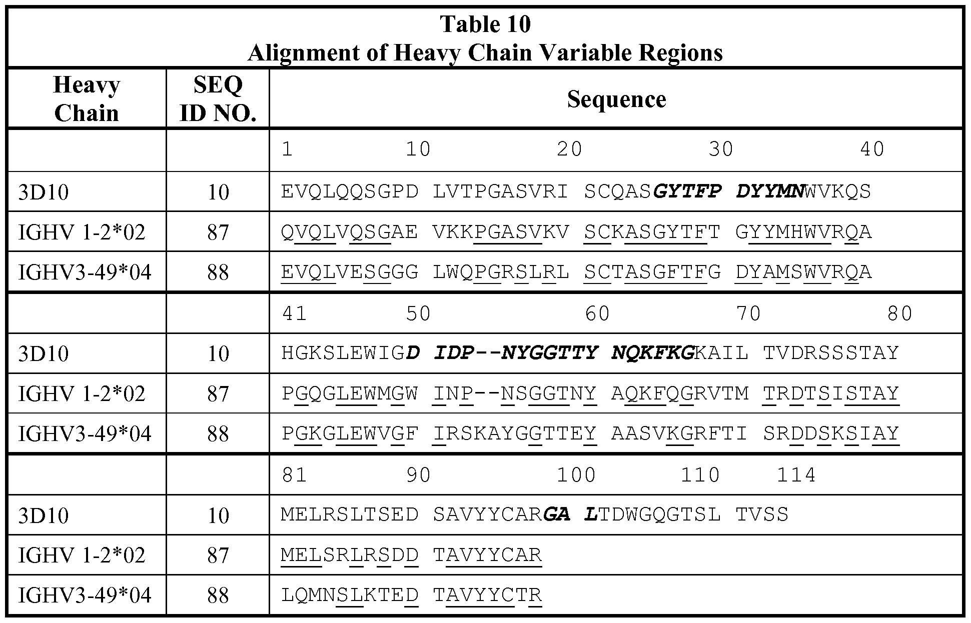


Claims
Priority Applications (19)
| Application Number | Priority Date | Filing Date | Title |
|---|---|---|---|
| RS20180683A RS57324B1 (en) | 2011-04-20 | 2012-04-19 | Antibodies and other molecules that bind b7-h1 and pd-1 |
| RU2013151455A RU2625034C2 (en) | 2011-04-20 | 2012-04-19 | Antibodies and other molecules binding b7-h1 and pd-1 |
| US14/111,402 US9205148B2 (en) | 2011-04-20 | 2012-04-19 | Antibodies and other molecules that bind B7-H1 and PD-1 |
| SI201231320T SI2699264T1 (en) | 2011-04-20 | 2012-04-19 | Antibodies and other molecules that bind b7-h1 and pd-1 |
| MX2013012088A MX338353B (en) | 2011-04-20 | 2012-04-19 | Antibodies and other molecules that bind b7-h1 and pd-1. |
| CA2833636A CA2833636A1 (en) | 2011-04-20 | 2012-04-19 | Antibodies and other molecules that bind b7-h1 and pd-1 |
| AU2012245477A AU2012245477C1 (en) | 2011-04-20 | 2012-04-19 | Antibodies and other molecules that bind B7-H1 and PD-1 |
| DK12774724.4T DK2699264T3 (en) | 2011-04-20 | 2012-04-19 | ANTIBODIES AND OTHER MOLECULES BINDING B7-H1 AND PD-1 |
| KR1020137030691A KR101970025B1 (en) | 2011-04-20 | 2012-04-19 | Antibodies and other molecules that bind b7-h1 and pd-1 |
| EP12774724.4A EP2699264B1 (en) | 2011-04-20 | 2012-04-19 | Antibodies and other molecules that bind b7-h1 and pd-1 |
| LTEP12774724.4T LT2699264T (en) | 2011-04-20 | 2012-04-19 | Antibodies and other molecules that bind b7-h1 and pd-1 |
| JP2014506544A JP6072771B2 (en) | 2011-04-20 | 2012-04-19 | Antibodies and other molecules that bind to B7-H1 and PD-1 |
| EP18161596.4A EP3403672A1 (en) | 2011-04-20 | 2012-04-19 | Antibodies and other molecules that bind b7-h1 and pd-1 |
| ES12774724.4T ES2669310T3 (en) | 2011-04-20 | 2012-04-19 | Antibodies and other molecules that bind with B7-H1 and PD-1 |
| PL12774724T PL2699264T3 (en) | 2011-04-20 | 2012-04-19 | Antibodies and other molecules that bind b7-h1 and pd-1 |
| CN201280030691.9A CN103608040B (en) | 2011-04-20 | 2012-04-19 | Antibody in conjunction with B7 H1 and PD 1 and other molecules |
| US14/926,732 US20160130348A1 (en) | 2011-04-20 | 2015-10-29 | Antibodies and other molecules that bind b7-h1 and pd-1 |
| HRP20180893TT HRP20180893T1 (en) | 2011-04-20 | 2018-06-07 | Antibodies and other molecules that bind b7-h1 and pd-1 |
| CY181100615T CY1120626T1 (en) | 2011-04-20 | 2018-06-13 | ANTIBODIES AND OTHER MOLECULES BIND B7-H1 AND PD-1 |
Applications Claiming Priority (2)
| Application Number | Priority Date | Filing Date | Title |
|---|---|---|---|
| US201161477414P | 2011-04-20 | 2011-04-20 | |
| US61/477,414 | 2011-04-20 |
Related Child Applications (2)
| Application Number | Title | Priority Date | Filing Date |
|---|---|---|---|
| US14/111,402 A-371-Of-International US9205148B2 (en) | 2011-04-20 | 2012-04-19 | Antibodies and other molecules that bind B7-H1 and PD-1 |
| US14/926,732 Division US20160130348A1 (en) | 2011-04-20 | 2015-10-29 | Antibodies and other molecules that bind b7-h1 and pd-1 |
Publications (1)
| Publication Number | Publication Date |
|---|---|
| WO2012145493A1 true WO2012145493A1 (en) | 2012-10-26 |
Family
ID=47041916
Family Applications (1)
| Application Number | Title | Priority Date | Filing Date |
|---|---|---|---|
| PCT/US2012/034223 WO2012145493A1 (en) | 2011-04-20 | 2012-04-19 | Antibodies and other molecules that bind b7-h1 and pd-1 |
Country Status (20)
| Country | Link |
|---|---|
| US (2) | US9205148B2 (en) |
| EP (2) | EP2699264B1 (en) |
| JP (2) | JP6072771B2 (en) |
| KR (1) | KR101970025B1 (en) |
| CN (1) | CN103608040B (en) |
| AU (1) | AU2012245477C1 (en) |
| CA (1) | CA2833636A1 (en) |
| CY (1) | CY1120626T1 (en) |
| DK (1) | DK2699264T3 (en) |
| ES (1) | ES2669310T3 (en) |
| HR (1) | HRP20180893T1 (en) |
| HU (1) | HUE037651T2 (en) |
| LT (1) | LT2699264T (en) |
| MX (1) | MX338353B (en) |
| PL (1) | PL2699264T3 (en) |
| PT (1) | PT2699264T (en) |
| RS (1) | RS57324B1 (en) |
| RU (1) | RU2625034C2 (en) |
| SI (1) | SI2699264T1 (en) |
| WO (1) | WO2012145493A1 (en) |
Cited By (595)
| Publication number | Priority date | Publication date | Assignee | Title |
|---|---|---|---|---|
| WO2014055648A1 (en) | 2012-10-02 | 2014-04-10 | Bristol-Myers Squibb Company | Combination of anti-kir antibodies and anti-pd-1 antibodies to treat cancer |
| WO2014100483A1 (en) * | 2012-12-19 | 2014-06-26 | Amplimmune, Inc. | Anti-human b7-h4 antibodies and their uses |
| WO2014135876A1 (en) | 2013-03-06 | 2014-09-12 | Astrazeneca Ab | Quinazoline inhibitors of activating mutant forms of epidermal growth factor receptor |
| WO2014194293A1 (en) * | 2013-05-30 | 2014-12-04 | Amplimmune, Inc. | Improved methods for the selection of patients for pd-1 or b7-h4 targeted therapies, and combination therapies thereof |
| WO2015042246A1 (en) | 2013-09-20 | 2015-03-26 | Bristol-Myers Squibb Company | Combination of anti-lag-3 antibodies and anti-pd-1 antibodies to treat tumors |
| WO2015048312A1 (en) * | 2013-09-26 | 2015-04-02 | Costim Pharmaceuticals Inc. | Methods for treating hematologic cancers |
| WO2015100282A1 (en) | 2013-12-24 | 2015-07-02 | Bristol-Myers Squibb Company | Tricyclic compounds as anticancer agents |
| WO2015112800A1 (en) | 2014-01-23 | 2015-07-30 | Regeneron Pharmaceuticals, Inc. | Human antibodies to pd-1 |
| CN104968679A (en) * | 2012-12-21 | 2015-10-07 | 艾普利穆恩公司 | Anti-H7CR antibodies |
| WO2015187835A2 (en) | 2014-06-06 | 2015-12-10 | Bristol-Myers Squibb Company | Antibodies against glucocorticoid-induced tumor necrosis factor receptor (gitr) and uses thereof |
| US9212224B2 (en) | 2012-05-15 | 2015-12-15 | Bristol-Myers Squibb Company | Antibodies that bind PD-L1 and uses thereof |
| CN105209919A (en) * | 2013-03-15 | 2015-12-30 | 豪夫迈·罗氏有限公司 | Biomarkers and methods of treating PD-1 and PD-L1 related conditions |
| WO2016020856A2 (en) | 2014-08-05 | 2016-02-11 | MabQuest SA | Immunological reagents |
| WO2016057898A1 (en) | 2014-10-10 | 2016-04-14 | Idera Pharmaceuticals, Inc. | Treatment of cancer using tlr9 agonist with checkpoint inhibitors |
| WO2016069727A1 (en) | 2014-10-29 | 2016-05-06 | Five Prime Therapeutics, Inc. | Combination therapy for cancer |
| WO2016081748A2 (en) | 2014-11-21 | 2016-05-26 | Bristol-Myers Squibb Company | Antibodies against cd73 and uses thereof |
| WO2016090070A1 (en) | 2014-12-04 | 2016-06-09 | Bristol-Myers Squibb Company | Combination of anti-cs1 and anti-pd1 antibodies to treat cancer (myeloma) |
| WO2016106266A1 (en) | 2014-12-22 | 2016-06-30 | Bristol-Myers Squibb Company | TGFβ RECEPTOR ANTAGONISTS |
| WO2016127052A1 (en) | 2015-02-05 | 2016-08-11 | Bristol-Myers Squibb Company | Cxcl11 and smica as predictive biomarkers for efficacy of anti-ctla4 immunotherapy |
| WO2016140884A1 (en) | 2015-03-02 | 2016-09-09 | Rigel Pharmaceuticals, Inc. | TGF-β INHIBITORS |
| WO2016149201A2 (en) | 2015-03-13 | 2016-09-22 | Cytomx Therapeutics, Inc. | Anti-pdl1 antibodies, activatable anti-pdl1 antibodies, and methods of use thereof |
| WO2016161279A1 (en) | 2015-04-03 | 2016-10-06 | Bristol-Myers Squibb Company | Inhibitors of indoleamine 2,3-dioxygenase for the treatment of cancer |
| WO2016162505A1 (en) | 2015-04-08 | 2016-10-13 | F-Star Biotechnology Limited | Her2 binding agent therapies |
| WO2016176503A1 (en) | 2015-04-28 | 2016-11-03 | Bristol-Myers Squibb Company | Treatment of pd-l1-negative melanoma using an anti-pd-1 antibody and an anti-ctla-4 antibody |
| WO2016176504A1 (en) | 2015-04-28 | 2016-11-03 | Bristol-Myers Squibb Company | Treatment of pd-l1-positive melanoma using an anti-pd-1 antibody |
| WO2016183115A1 (en) | 2015-05-12 | 2016-11-17 | Bristol-Myers Squibb Company | 5h-pyrido[3,2-b]indole compounds as anticancer agents |
| WO2016183118A1 (en) | 2015-05-12 | 2016-11-17 | Bristol-Myers Squibb Company | Tricyclic compounds as anticancer agents |
| WO2016183114A1 (en) | 2015-05-11 | 2016-11-17 | Bristol-Myers Squibb Company | Tricyclic compounds as anticancer agents |
| WO2016191751A1 (en) | 2015-05-28 | 2016-12-01 | Bristol-Myers Squibb Company | Treatment of pd-l1 positive lung cancer using an anti-pd-1 antibody |
| WO2016196228A1 (en) | 2015-05-29 | 2016-12-08 | Bristol-Myers Squibb Company | Antibodies against ox40 and uses thereof |
| WO2016196389A1 (en) | 2015-05-29 | 2016-12-08 | Bristol-Myers Squibb Company | Treatment of renal cell carcinoma |
| WO2016201425A1 (en) | 2015-06-12 | 2016-12-15 | Bristol-Myers Squibb Company | Treatment of cancer by combined blockade of the pd-1 and cxcr4 signaling pathways |
| WO2017004016A1 (en) | 2015-06-29 | 2017-01-05 | The Rockefeller University | Antibodies to cd40 with enhanced agonist activity |
| WO2017011666A1 (en) | 2015-07-14 | 2017-01-19 | Bristol-Myers Squibb Company | Method of treating cancer using immune checkpoint inhibitor |
| WO2017019757A1 (en) | 2015-07-28 | 2017-02-02 | Bristol-Myers Squibb Company | Tgf beta receptor antagonists |
| WO2017025498A1 (en) | 2015-08-07 | 2017-02-16 | Pieris Pharmaceuticals Gmbh | Novel fusion polypeptide specific for lag-3 and pd-1 |
| WO2017035118A1 (en) | 2015-08-25 | 2017-03-02 | Bristol-Myers Squibb Company | Tgf beta receptor antagonists |
| US9598422B2 (en) | 2014-11-05 | 2017-03-21 | Flexus Biosciences, Inc. | Immunoregulatory agents |
| US9605070B2 (en) | 2014-01-31 | 2017-03-28 | Novartis Ag | Antibody molecules to TIM-3 and uses thereof |
| WO2017055404A1 (en) | 2015-10-02 | 2017-04-06 | F. Hoffmann-La Roche Ag | Bispecific antibodies specific for pd1 and tim3 |
| WO2017055443A1 (en) | 2015-10-02 | 2017-04-06 | F. Hoffmann-La Roche Ag | Anti-pd1 antibodies and methods of use |
| WO2017064043A1 (en) | 2015-10-12 | 2017-04-20 | Innate Pharma | Cd73 blocking agents |
| WO2017069291A1 (en) | 2015-10-23 | 2017-04-27 | Canbas Co., Ltd. | Peptides and peptidomimetics in combination with t cell activating and/or checkpoint inhibiting agents for cancer treatment |
| US9643972B2 (en) | 2014-11-05 | 2017-05-09 | Flexus Biosciences, Inc. | Immunoregulatory agents |
| WO2017079116A2 (en) | 2015-11-03 | 2017-05-11 | Janssen Biotech, Inc. | Antibodies specifically binding pd-1 and tim-3 and their uses |
| WO2017077382A1 (en) | 2015-11-06 | 2017-05-11 | Orionis Biosciences Nv | Bi-functional chimeric proteins and uses thereof |
| WO2017079117A1 (en) | 2015-11-02 | 2017-05-11 | Five Prime Therapeutics, Inc. | Cd80 extracellular domain polypeptides and their use in cancer treatment |
| WO2017087678A2 (en) | 2015-11-19 | 2017-05-26 | Bristol-Myers Squibb Company | Antibodies against glucocorticoid-induced tumor necrosis factor receptor (gitr) and uses thereof |
| WO2017087599A1 (en) | 2015-11-18 | 2017-05-26 | Lyvgen Biopharma Holdings Limited | Anti-pd-1 antibodies and therapeutic uses thereof |
| WO2017087870A1 (en) | 2015-11-18 | 2017-05-26 | Bristol-Myers Squibb Company | Treatment of lung cancer using a combination of an anti-pd-1 antibody and an anti-ctla-4 antibody |
| WO2017091580A1 (en) | 2015-11-23 | 2017-06-01 | Five Prime Therapeutics, Inc. | Predicting response to cancer treatment with fgfr2 inhibitors |
| US9683048B2 (en) | 2014-01-24 | 2017-06-20 | Novartis Ag | Antibody molecules to PD-1 and uses thereof |
| WO2017106291A1 (en) | 2015-12-15 | 2017-06-22 | Bristol-Myers Squibb Company | Cxcr4 receptor antagonists |
| EP3044323A4 (en) * | 2013-09-13 | 2017-07-05 | F. Hoffmann-La Roche AG | Compositions and methods for detecting and quantifying host cell protein in cell lines and recombinant polypeptide products |
| WO2017125532A1 (en) | 2016-01-21 | 2017-07-27 | Innate Pharma | Neutralization of inhibitory pathways in lymphocytes |
| WO2017125815A2 (en) | 2016-01-22 | 2017-07-27 | MabQuest SA | Immunological reagents |
| EP3044234A4 (en) * | 2013-09-13 | 2017-08-09 | Beigene, Ltd. | Anti-pd1 antibodies and their use as therapeutics and diagnostics |
| WO2017134305A1 (en) | 2016-02-05 | 2017-08-10 | Orionis Biosciences Nv | Bispecific signaling agents and uses thereof |
| WO2017141208A1 (en) | 2016-02-17 | 2017-08-24 | Novartis Ag | Tgfbeta 2 antibodies |
| JP2017525354A (en) * | 2014-08-08 | 2017-09-07 | ザ ボード オブ トラスティーズ オブ ザ レランド スタンフォード ジュニア ユニバーシティー | High affinity PD-1 drug and method of use |
| WO2017152085A1 (en) | 2016-03-04 | 2017-09-08 | Bristol-Myers Squibb Company | Combination therapy with anti-cd73 antibodies |
| WO2017153433A1 (en) | 2016-03-08 | 2017-09-14 | Innate Pharma | Siglec neutralizing antibodies |
| WO2017160754A1 (en) | 2016-03-15 | 2017-09-21 | Mersana Therapeutics,Inc. | Napi2b-targeted antibody-drug conjugates and methods of use thereof |
| US9771425B2 (en) | 2014-10-27 | 2017-09-26 | Agency For Science, Technology And Research | Anti-PD-1 antibodies |
| WO2017167921A1 (en) | 2016-03-30 | 2017-10-05 | Centre Léon-Bérard | Lymphocytes expressing cd73 in cancerous patient dictates therapy |
| WO2017176925A1 (en) | 2016-04-05 | 2017-10-12 | Bristol-Myers Squibb Company | Cytokine profiling analysis for predicting prognosis of a patient in need of an anti-cancer treatment |
| WO2017184619A2 (en) | 2016-04-18 | 2017-10-26 | Celldex Therapeutics, Inc. | Agonistic antibodies that bind human cd40 and uses thereof |
| EP3243832A1 (en) | 2016-05-13 | 2017-11-15 | F. Hoffmann-La Roche AG | Antigen binding molecules comprising a tnf family ligand trimer and pd1 binding moiety |
| WO2017194782A2 (en) | 2016-05-13 | 2017-11-16 | Orionis Biosciences Nv | Therapeutic targeting of non-cellular structures |
| WO2017194783A1 (en) | 2016-05-13 | 2017-11-16 | Orionis Biosciences Nv | Targeted mutant interferon-beta and uses thereof |
| WO2017210637A1 (en) | 2016-06-03 | 2017-12-07 | Bristol-Myers Squibb Company | Use of anti-pd-1 antibody in the treatment of patients with colorectal cancer |
| WO2017210335A1 (en) | 2016-06-01 | 2017-12-07 | Bristol-Myers Squibb Company | Imaging methods using 18f-radiolabeled biologics |
| WO2017210631A1 (en) | 2016-06-03 | 2017-12-07 | Bristol-Myers Squibb Company | Anti-pd-1 antibody for use in a method of treatment of recurrent small cell lung cancer |
| WO2017210624A1 (en) | 2016-06-03 | 2017-12-07 | Bristol-Myers Squibb Company | Anti-pd-1 antibody for use in a method of treating a tumor |
| WO2017210473A1 (en) | 2016-06-02 | 2017-12-07 | Bristol-Myers Squibb Company | Use of an anti-pd-1 antibody in combination with an anti-cd30 antibody in lymphoma treatment |
| WO2017210453A1 (en) | 2016-06-02 | 2017-12-07 | Bristol-Myers Squibb Company | Pd-1 blockade with nivolumab in refractory hodgkin's lymphoma |
| WO2017218533A1 (en) | 2016-06-13 | 2017-12-21 | Torque Therapeutics, Inc. | Methods and compositions for promoting immune cell function |
| WO2017220989A1 (en) | 2016-06-20 | 2017-12-28 | Kymab Limited | Anti-pd-l1 and il-2 cytokines |
| EP3176180A4 (en) * | 2014-07-30 | 2018-01-03 | Livzon Mabpharm Inc. | Anti-pd-1 antibody and use thereof |
| WO2018013818A2 (en) | 2016-07-14 | 2018-01-18 | Bristol-Myers Squibb Company | Antibodies against tim3 and uses thereof |
| WO2018017633A1 (en) | 2016-07-21 | 2018-01-25 | Bristol-Myers Squibb Company | TGF Beta RECEPTOR ANTAGONISTS |
| WO2018029474A2 (en) | 2016-08-09 | 2018-02-15 | Kymab Limited | Anti-icos antibodies |
| US9908936B2 (en) | 2014-03-14 | 2018-03-06 | Novartis Ag | Antibody molecules to LAG-3 and uses thereof |
| WO2018048975A1 (en) | 2016-09-09 | 2018-03-15 | Bristol-Myers Squibb Company | Use of an anti-pd-1 antibody in combination with an anti-mesothelin antibody in cancer treatment |
| US9920120B2 (en) | 2013-09-13 | 2018-03-20 | Genentech, Inc. | Methods and compositions comprising purified recombinant polypeptides |
| US9938345B2 (en) | 2014-01-23 | 2018-04-10 | Regeneron Pharmaceuticals, Inc. | Human antibodies to PD-L1 |
| US9957323B2 (en) | 2016-06-20 | 2018-05-01 | Kymab Limited | Anti-ICOS antibodies |
| WO2018077893A1 (en) | 2016-10-24 | 2018-05-03 | Orionis Biosciences Nv | Targeted mutant interferon-gamma and uses thereof |
| WO2018081621A1 (en) | 2016-10-28 | 2018-05-03 | Bristol-Myers Squibb Company | Methods of treating urothelial carcinoma using an anti-pd-1 antibody |
| WO2018085555A1 (en) | 2016-11-03 | 2018-05-11 | Bristol-Myers Squibb Company | Activatable anti-ctla-4 antibodies and uses thereof |
| WO2018083087A2 (en) | 2016-11-02 | 2018-05-11 | Glaxosmithkline Intellectual Property (No.2) Limited | Binding proteins |
| WO2018098269A2 (en) | 2016-11-23 | 2018-05-31 | Mersana Therapeutics, Inc. | Peptide-containing linkers for antibody-drug conjugates |
| US9988452B2 (en) | 2014-10-14 | 2018-06-05 | Novartis Ag | Antibody molecules to PD-L1 and uses thereof |
| WO2018101448A1 (en) | 2016-11-30 | 2018-06-07 | Kyowa Hakko Kirin Co., Ltd. | Method of treating cancer using anti-ccr4 antibody and anti-pd-1 antibody |
| WO2018112364A1 (en) | 2016-12-16 | 2018-06-21 | Evelo Biosciences, Inc. | Combination therapies for treating melanoma |
| WO2018112360A1 (en) | 2016-12-16 | 2018-06-21 | Evelo Biosciences, Inc. | Combination therapies for treating cancer |
| WO2018119475A1 (en) * | 2016-12-23 | 2018-06-28 | Remd Biotherapeutics, Inc. | Immunotherapy using antibodies that bind programmed death ligand-1 (pd-l1) |
| WO2018127570A1 (en) | 2017-01-05 | 2018-07-12 | Netris Pharma | Combined treatment with netrin-1 interfering drug and immune checkpoint inhibitors drugs |
| CN108289953A (en) * | 2015-09-29 | 2018-07-17 | 细胞基因公司 | PD-1 binding proteins and its application method |
| WO2018132279A1 (en) | 2017-01-05 | 2018-07-19 | Bristol-Myers Squibb Company | Tgf beta receptor antagonists |
| WO2018134279A1 (en) | 2017-01-18 | 2018-07-26 | Pieris Pharmaceuticals Gmbh | Novel fusion polypeptides specific for lag-3 and pd-1 |
| WO2018134784A1 (en) | 2017-01-20 | 2018-07-26 | Novartis Ag | Combination therapy for the treatment of cancer |
| WO2018136700A1 (en) | 2017-01-20 | 2018-07-26 | Arcus Biosciences, Inc. | Azolopyrimidine for the treatment of cancer-related disorders |
| WO2018134681A1 (en) | 2017-01-20 | 2018-07-26 | Sanofi | Anti-tgf-beta antibodies and their use |
| WO2018141964A1 (en) | 2017-02-06 | 2018-08-09 | Orionis Biosciences Nv | Targeted chimeric proteins and uses thereof |
| WO2018144999A1 (en) | 2017-02-06 | 2018-08-09 | Orionis Biosciences, Inc. | Targeted engineered interferon and uses thereof |
| WO2018141959A1 (en) | 2017-02-06 | 2018-08-09 | Innate Pharma | Immunomodulatory antibody drug conjugates binding to a human mica polypeptide |
| WO2018146612A1 (en) | 2017-02-10 | 2018-08-16 | Novartis Ag | 1-(4-amino-5-bromo-6-(1 h-pyrazol-1-yl)pyrimidin-2-yl)-1 h-pyrazol-4-ol and use thereof in the treatment of cancer |
| WO2018152396A1 (en) | 2017-02-17 | 2018-08-23 | Innate Tumor Immunity, Inc. | Substituted imidazo-quinolines as nlrp3 modulators |
| WO2018160540A1 (en) | 2017-02-28 | 2018-09-07 | Sanofi | Therapeutic rna |
| WO2018160538A1 (en) | 2017-02-28 | 2018-09-07 | Mersana Therapeutics, Inc. | Combination therapies of her2-targeted antibody-drug conjugates |
| US10076518B2 (en) | 2015-03-06 | 2018-09-18 | Beyondspring Pharmaceuticals, Inc. | Method of treating a brain tumor |
| WO2018167267A1 (en) | 2017-03-16 | 2018-09-20 | Innate Pharma | Compositions and methods for treating cancer |
| WO2018183928A1 (en) | 2017-03-31 | 2018-10-04 | Bristol-Myers Squibb Company | Methods of treating tumor |
| WO2018183608A1 (en) | 2017-03-31 | 2018-10-04 | Five Prime Therapeutics, Inc. | Combination therapy for cancer using anti-gitr antibodies |
| WO2018187057A1 (en) | 2017-04-06 | 2018-10-11 | Regeneron Pharmaceuticals, Inc. | Stable antibody formulation |
| WO2018185043A1 (en) | 2017-04-05 | 2018-10-11 | F. Hoffmann-La Roche Ag | Bispecific antibodies specifically binding to pd1 and lag3 |
| WO2018187613A2 (en) | 2017-04-07 | 2018-10-11 | Bristol-Myers Squibb Company | Anti-icos agonist antibodies and uses thereof |
| WO2018195397A2 (en) | 2017-04-21 | 2018-10-25 | Kyn Therapeutics | Indole ahr inhibitors and uses thereof |
| WO2018198091A1 (en) | 2017-04-28 | 2018-11-01 | Novartis Ag | Antibody conjugates comprising toll-like receptor agonist and combination therapies |
| WO2018200430A1 (en) | 2017-04-26 | 2018-11-01 | Bristol-Myers Squibb Company | Methods of antibody production that minimize disulfide bond reduction |
| WO2018209049A1 (en) | 2017-05-12 | 2018-11-15 | Bristol-Myers Squibb Company | Inhibitors of indoleamine 2,3-dioxygenase and methods of their use |
| WO2018213297A1 (en) | 2017-05-16 | 2018-11-22 | Bristol-Myers Squibb Company | Treatment of cancer with anti-gitr agonist antibodies |
| WO2018213377A1 (en) | 2017-05-17 | 2018-11-22 | Arcus Biosciences, Inc. | Quinazoline-pyrazole derivatives for the treatment of cancer-related disorders |
| WO2018211453A1 (en) | 2017-05-19 | 2018-11-22 | Novartis Ag | Compositions comprising naphthyridine derivatives and aluminium adjuvant for use in treating solid tumors |
| WO2018218056A1 (en) | 2017-05-25 | 2018-11-29 | Birstol-Myers Squibb Company | Antibodies comprising modified heavy constant regions |
| WO2018222718A1 (en) | 2017-05-30 | 2018-12-06 | Bristol-Myers Squibb Company | Treatment of lag-3 positive tumors |
| WO2018220546A1 (en) | 2017-05-31 | 2018-12-06 | Novartis Ag | Crystalline forms of 5-bromo-2,6-di(1 h-pyrazol-1-yl)pyrimidin-4-amine and new salts |
| WO2018222711A2 (en) | 2017-05-30 | 2018-12-06 | Bristol-Myers Squibb Company | Compositions comprising a combination of an anti-lag-3 antibody, a pd-1 pathway inhibitor, and an immunotherapeutic agent |
| WO2018222722A2 (en) | 2017-05-30 | 2018-12-06 | Bristol-Myers Squibb Company | Compositions comprising an anti-lag-3 antibody or an anti-lag-3 antibody and an anti-pd-1 or anti-pd-l1 antibody |
| WO2018223040A1 (en) | 2017-06-01 | 2018-12-06 | Bristol-Myers Squibb Company | Methods of treating a tumor using an anti-pd-1 antibody |
| US10155037B2 (en) | 2014-12-09 | 2018-12-18 | Rinat Neuroscience Corp. | Anti-PD-1 antibodies and methods of use thereof |
| US10155748B2 (en) | 2015-07-13 | 2018-12-18 | Beyondspring Pharmaceuticals, Inc. | Plinabulin compositions |
| WO2018229715A1 (en) | 2017-06-16 | 2018-12-20 | Novartis Ag | Compositions comprising anti-cd32b antibodies and methods of use thereof |
| US10160806B2 (en) | 2014-06-26 | 2018-12-25 | Macrogenics, Inc. | Covalently bonded diabodies having immunoreactivity with PD-1 and LAG-3, and methods of use thereof |
| WO2018235056A1 (en) | 2017-06-22 | 2018-12-27 | Novartis Ag | Il-1beta binding antibodies for use in treating cancer |
| WO2018234879A1 (en) | 2017-06-22 | 2018-12-27 | Novartis Ag | Il-1beta binding antibodies for use in treating cancer |
| WO2018237173A1 (en) | 2017-06-22 | 2018-12-27 | Novartis Ag | Antibody molecules to cd73 and uses thereof |
| WO2018237157A1 (en) | 2017-06-22 | 2018-12-27 | Novartis Ag | Antibody molecules to cd73 and uses thereof |
| EP3421494A1 (en) | 2017-06-29 | 2019-01-02 | Sanofi | Use of isatuximab in combination with an anti-pd-1 antibody |
| WO2019006283A1 (en) | 2017-06-30 | 2019-01-03 | Bristol-Myers Squibb Company | Amorphous and crystalline forms of ido inhibitors |
| WO2019006007A1 (en) | 2017-06-27 | 2019-01-03 | Novartis Ag | Dosage regimens for anti-tim-3 antibodies and uses thereof |
| WO2019014402A1 (en) | 2017-07-14 | 2019-01-17 | Innate Tumor Immunity, Inc. | Nlrp3 modulators |
| WO2019011855A1 (en) | 2017-07-10 | 2019-01-17 | Innate Pharma | Siglec-9-neutralizing antibodies |
| WO2019018730A1 (en) | 2017-07-20 | 2019-01-24 | Novartis Ag | Dosage regimens of anti-lag-3 antibodies and uses thereof |
| WO2019023624A1 (en) | 2017-07-28 | 2019-01-31 | Bristol-Myers Squibb Company | Predictive peripheral blood biomarker for checkpoint inhibitors |
| WO2019023459A1 (en) | 2017-07-28 | 2019-01-31 | Bristol-Myers Squibb Company | Cyclic dinucleotides as anticancer agents |
| WO2019032431A1 (en) | 2017-08-07 | 2019-02-14 | Amgen Inc. | Treatment of triple negative breast cancer or colorectal cancer with liver metastases with an anti pd-l1 antibody and an oncolytic virus |
| EP3444271A1 (en) | 2013-08-08 | 2019-02-20 | Cytune Pharma | Il-15 and il-15raplha sushi domain based modulokines |
| US10214586B2 (en) | 2015-08-24 | 2019-02-26 | Eli Lilly And Company | PD-L1 antibodies |
| WO2019040780A1 (en) | 2017-08-25 | 2019-02-28 | Five Prime Therapeutics Inc. | B7-h4 antibodies and methods of use thereof |
| WO2019046500A1 (en) | 2017-08-31 | 2019-03-07 | Bristol-Myers Squibb Company | Cyclic dinucleotides as anticancer agents |
| WO2019046496A1 (en) | 2017-08-31 | 2019-03-07 | Bristol-Myers Squibb Company | Cyclic dinucleotides as anticancer agents |
| WO2019046321A1 (en) | 2017-08-28 | 2019-03-07 | Bristol-Myers Squibb Company | Tim-3 antagonists for the treatment and diagnosis of cancers |
| WO2019046498A1 (en) | 2017-08-31 | 2019-03-07 | Bristol-Myers Squibb Company | Cyclic dinucleotides as anticancer agents |
| US10238650B2 (en) | 2015-03-06 | 2019-03-26 | Beyondspring Pharmaceuticals, Inc. | Method of treating cancer associated with a RAS mutation |
| WO2019068907A1 (en) | 2017-10-06 | 2019-04-11 | Innate Pharma | Restoration of t cell activity via the cd39/cd73 axis |
| WO2019074887A1 (en) | 2017-10-10 | 2019-04-18 | Bristol-Myers Squibb Company | Cyclic dinucleotides as anticancer agents |
| WO2019075090A1 (en) | 2017-10-10 | 2019-04-18 | Tilos Therapeutics, Inc. | Anti-lap antibodies and uses thereof |
| WO2019074824A1 (en) | 2017-10-09 | 2019-04-18 | Bristol-Myers Squibb Company | Inhibitors of indoleamine 2,3-dioxygenase and methods of their use |
| WO2019074822A1 (en) | 2017-10-09 | 2019-04-18 | Bristol-Myers Squibb Company | Inhibitors of indoleamine 2,3-dioxygenase and methods of their use |
| WO2019075468A1 (en) | 2017-10-15 | 2019-04-18 | Bristol-Myers Squibb Company | Methods of treating tumor |
| WO2019079261A1 (en) | 2017-10-16 | 2019-04-25 | Bristol-Myers Squibb Company | Cyclic dinucleotides as anticancer agents |
| WO2019081983A1 (en) | 2017-10-25 | 2019-05-02 | Novartis Ag | Antibodies targeting cd32b and methods of use thereof |
| WO2019090263A1 (en) | 2017-11-06 | 2019-05-09 | Genentech, Inc. | Diagnostic and therapeutic methods for cancer |
| WO2019090198A1 (en) | 2017-11-06 | 2019-05-09 | Bristol-Myers Squibb Company | Isofuranone compounds useful as hpk1 inhibitors |
| WO2019089921A1 (en) | 2017-11-01 | 2019-05-09 | Bristol-Myers Squibb Company | Immunostimulatory agonistic antibodies for use in treating cancer |
| WO2019090330A1 (en) | 2017-11-06 | 2019-05-09 | Bristol-Myers Squibb Company | Methods of treating a tumor |
| WO2019099838A1 (en) | 2017-11-16 | 2019-05-23 | Novartis Ag | Combination therapies |
| WO2019104289A1 (en) | 2017-11-27 | 2019-05-31 | Mersana Therapeutics, Inc. | Pyrrolobenzodiazepine antibody conjugates |
| WO2019108900A1 (en) | 2017-11-30 | 2019-06-06 | Novartis Ag | Bcma-targeting chimeric antigen receptor, and uses thereof |
| US10316089B2 (en) | 2015-08-10 | 2019-06-11 | Innovent Biologics (Suzhou) Co. Ltd. | PD-1 antibodies |
| US10323004B2 (en) | 2016-05-04 | 2019-06-18 | Bristol-Myers Squibb Company | Inhibitors of indoleamine 2,3-dioxygenase and methods of their use |
| WO2019122884A1 (en) | 2017-12-19 | 2019-06-27 | Kymab Limited | Antibodies to icos |
| WO2019126691A1 (en) | 2017-12-21 | 2019-06-27 | Mersana Therapeutics, Inc. | Pyrrolobenzodiazepine antibody conjugates |
| WO2019133747A1 (en) | 2017-12-27 | 2019-07-04 | Bristol-Myers Squibb Company | Anti-cd40 antibodies and uses thereof |
| US10344090B2 (en) | 2013-12-12 | 2019-07-09 | Shanghai Hangrui Pharmaceutical Co., Ltd. | PD-1 antibody, antigen-binding fragment thereof, and medical application thereof |
| WO2019136432A1 (en) | 2018-01-08 | 2019-07-11 | Novartis Ag | Immune-enhancing rnas for combination with chimeric antigen receptor therapy |
| WO2019136112A1 (en) | 2018-01-05 | 2019-07-11 | Bristol-Myers Squibb Company | Inhibitors of indoleamine 2,3-dioxygenase and methods of their use |
| WO2019140229A1 (en) | 2018-01-12 | 2019-07-18 | Bristol-Myers Squibb Company | Antibodies against tim3 and uses thereof |
| WO2019140150A1 (en) | 2018-01-12 | 2019-07-18 | Bristol-Myers Squibb Company | Combination therapy with anti-il-8 antibodies and anti-pd-1 antibodies for treating cancer |
| WO2019144098A1 (en) | 2018-01-22 | 2019-07-25 | Bristol-Myers Squibb Company | Compositions and methods of treating cancer |
| WO2019144126A1 (en) | 2018-01-22 | 2019-07-25 | Pascal Biosciences Inc. | Cannabinoids and derivatives for promoting immunogenicity of tumor and infected cells |
| WO2019143607A1 (en) | 2018-01-16 | 2019-07-25 | Bristol-Myers Squibb Company | Methods of treating cancer with antibodies against tim3 |
| WO2019149716A1 (en) | 2018-01-31 | 2019-08-08 | F. Hoffmann-La Roche Ag | Bispecific antibodies comprising an antigen-binding site binding to lag3 |
| WO2019152660A1 (en) | 2018-01-31 | 2019-08-08 | Novartis Ag | Combination therapy using a chimeric antigen receptor |
| WO2019157124A1 (en) | 2018-02-08 | 2019-08-15 | Bristol-Myers Squibb Company | Combination of a tetanus toxoid, anti-ox40 antibody and/or anti-pd-1 antibody to treat tumors |
| WO2019160956A1 (en) | 2018-02-13 | 2019-08-22 | Novartis Ag | Chimeric antigen receptor therapy in combination with il-15r and il15 |
| WO2019160884A1 (en) | 2018-02-13 | 2019-08-22 | Bristol-Myers Squibb Company | Cyclic dinucleotides as anticancer agents |
| US10392442B2 (en) | 2015-12-17 | 2019-08-27 | Bristol-Myers Squibb Company | Use of anti-PD-1 antibody in combination with anti-CD27 antibody in cancer treatment |
| WO2019165315A1 (en) | 2018-02-23 | 2019-08-29 | Syntrix Biosystems Inc. | Method for treating cancer using chemokine antagonists alone or in combination |
| WO2019173587A1 (en) | 2018-03-08 | 2019-09-12 | Bristol-Myers Squibb Company | Cyclic dinucleotides as anticancer agents |
| WO2019183040A1 (en) | 2018-03-21 | 2019-09-26 | Five Prime Therapeutics, Inc. | ANTIBODIES BINDING TO VISTA AT ACIDIC pH |
| US10428146B2 (en) | 2014-07-22 | 2019-10-01 | Cb Therapeutics, Inc. | Anti PD-1 antibodies |
| WO2019191676A1 (en) | 2018-03-30 | 2019-10-03 | Bristol-Myers Squibb Company | Methods of treating tumor |
| US10435470B2 (en) | 2014-08-05 | 2019-10-08 | Cb Therapeutics, Inc. | Anti-PD-L1 antibodies |
| EP3550019A1 (en) | 2014-10-24 | 2019-10-09 | Astrazeneca AB | Combination |
| GB201912107D0 (en) | 2019-08-22 | 2019-10-09 | Amazentis Sa | Combination |
| WO2019195452A1 (en) | 2018-04-04 | 2019-10-10 | Bristol-Myers Squibb Company | Anti-cd27 antibodies and uses thereof |
| WO2019200256A1 (en) | 2018-04-12 | 2019-10-17 | Bristol-Myers Squibb Company | Anticancer combination therapy with cd73 antagonist antibody and pd-1/pd-l1 axis antagonist antibody |
| WO2019200229A1 (en) | 2018-04-13 | 2019-10-17 | Novartis Ag | Dosage regimens for anti-pd-l1 antibodies and uses thereof |
| WO2019204257A1 (en) | 2018-04-16 | 2019-10-24 | Arrys Therapeutics, Inc. | Ep4 inhibitors and use thereof |
| US10457725B2 (en) | 2016-05-13 | 2019-10-29 | Regeneron Pharmaceuticals, Inc. | Methods of treating skin cancer by administering a PD-1 inhibitor |
| WO2019209896A1 (en) | 2018-04-25 | 2019-10-31 | Innate Tumor Immunity, Inc. | Nlrp3 modulators |
| WO2019211492A1 (en) | 2018-05-04 | 2019-11-07 | Tollys | Tlr3 ligands that activate both epithelial and myeloid cells |
| WO2019213340A1 (en) | 2018-05-03 | 2019-11-07 | Bristol-Myers Squibb Company | Uracil derivatives as mer-axl inhibitors |
| EP3572430A2 (en) | 2014-03-05 | 2019-11-27 | Bristol-Myers Squibb Company | Treatment of renal cancer using a combination of an anti-pd-1 antibody and another anti-cancer agent |
| WO2019224275A1 (en) | 2018-05-23 | 2019-11-28 | Adc Therapeutics Sa | Molecular adjuvant |
| WO2019232244A2 (en) | 2018-05-31 | 2019-12-05 | Novartis Ag | Antibody molecules to cd73 and uses thereof |
| WO2019229658A1 (en) | 2018-05-30 | 2019-12-05 | Novartis Ag | Entpd2 antibodies, combination therapies, and methods of using the antibodies and combination therapies |
| WO2019229701A2 (en) | 2018-06-01 | 2019-12-05 | Novartis Ag | Binding molecules against bcma and uses thereof |
| WO2019232319A1 (en) | 2018-05-31 | 2019-12-05 | Peloton Therapeutics, Inc. | Compositions and methods for inhibiting cd73 |
| WO2019241730A2 (en) | 2018-06-15 | 2019-12-19 | Flagship Pioneering Innovations V, Inc. | Increasing immune activity through modulation of postcellular signaling factors |
| US10512689B2 (en) | 2015-04-17 | 2019-12-24 | Bristol-Myers Squibb Company | Compositions comprising a combination of nivolumab and ipilimumab |
| US10513558B2 (en) | 2015-07-13 | 2019-12-24 | Cytomx Therapeutics, Inc. | Anti-PD1 antibodies, activatable anti-PD1 antibodies, and methods of use thereof |
| WO2019243252A1 (en) | 2018-06-18 | 2019-12-26 | Innate Pharma | Compositions and methods for treating cancer |
| WO2019243833A1 (en) | 2018-06-22 | 2019-12-26 | Bicycletx Limited | Bicyclic peptide ligands specific for nectin-4 |
| WO2019246557A1 (en) | 2018-06-23 | 2019-12-26 | Genentech, Inc. | Methods of treating lung cancer with a pd-1 axis binding antagonist, a platinum agent, and a topoisomerase ii inhibitor |
| WO2020006016A1 (en) | 2018-06-27 | 2020-01-02 | Bristol-Myers Squibb Company | Naphthyridinone compounds useful as t cell activators |
| WO2020006018A1 (en) | 2018-06-27 | 2020-01-02 | Bristol-Myers Squibb Company | Substituted naphthyridinone compounds useful as t cell activators |
| WO2020014327A2 (en) | 2018-07-11 | 2020-01-16 | Five Prime Therapeutics, Inc. | Antibodies binding to vista at acidic ph |
| WO2020012334A1 (en) | 2018-07-10 | 2020-01-16 | Novartis Ag | 3-(5-hydroxy-1-oxoisoindolin-2-yl)piperidine-2,6-dione derivatives and their use in the treatment of ikaros family zinc finger 2 (ikzf2)-dependent diseases |
| WO2020014132A2 (en) | 2018-07-09 | 2020-01-16 | Five Prime Therapeutics, Inc. | Antibodies binding to ilt4 |
| WO2020014583A1 (en) | 2018-07-13 | 2020-01-16 | Bristol-Myers Squibb Company | Ox-40 agonist, pd-1 pathway inhibitor and ctla-4 inhibitor combination for use in a mehtod of treating a cancer or a solid tumor |
| WO2020012337A1 (en) | 2018-07-10 | 2020-01-16 | Novartis Ag | 3-(5-amino-1-oxoisoindolin-2-yl)piperidine-2,6-dione derivatives and their use in the treatment of i karos family zinc finger 2 (ikzf2)-dependent diseases |
| WO2020018789A1 (en) | 2018-07-18 | 2020-01-23 | Genentech, Inc. | Methods of treating lung cancer with a pd-1 axis binding antagonist, an antimetabolite, and a platinum agent |
| US10544223B2 (en) | 2017-04-20 | 2020-01-28 | Adc Therapeutics Sa | Combination therapy with an anti-axl antibody-drug conjugate |
| US10544225B2 (en) | 2014-07-03 | 2020-01-28 | Beigene, Ltd. | Anti-PD-L1 antibodies and their use as therapeutics and diagnostics |
| US10544099B2 (en) | 2016-05-04 | 2020-01-28 | Bristol-Myers Squibb Company | Inhibitors of indoleamine 2,3-dioxygenase and methods of their use |
| WO2020023356A1 (en) | 2018-07-23 | 2020-01-30 | Bristol-Myers Squibb Company | Inhibitors of indoleamine 2,3-dioxygenase and methods of their use |
| WO2020021061A1 (en) | 2018-07-26 | 2020-01-30 | Pieris Pharmaceuticals Gmbh | Humanized anti-pd-1 antibodies and uses thereof |
| WO2020023707A1 (en) | 2018-07-26 | 2020-01-30 | Bristol-Myers Squibb Company | Lag-3 combination therapy for the treatment of cancer |
| WO2020023355A1 (en) | 2018-07-23 | 2020-01-30 | Bristol-Myers Squibb Company | Inhibitors of indoleamine 2,3-dioxygenase and methods of their use |
| WO2020021465A1 (en) | 2018-07-25 | 2020-01-30 | Advanced Accelerator Applications (Italy) S.R.L. | Method of treatment of neuroendocrine tumors |
| WO2020037091A1 (en) | 2018-08-16 | 2020-02-20 | Innate Tumor Immunity, Inc. | Imidazo[4,5-c]quinoline derived nlrp3-modulators |
| WO2020037092A1 (en) | 2018-08-16 | 2020-02-20 | Innate Tumor Immunity, Inc. | Imidazo[4,5-c]quinoline derived nlrp3-modulators |
| WO2020037094A1 (en) | 2018-08-16 | 2020-02-20 | Innate Tumor Immunity, Inc. | Substitued 4-amino-1h-imidazo[4,5-c]quinoline compounds and improved methods for their preparation |
| WO2020041655A1 (en) | 2018-08-24 | 2020-02-27 | Sanofi | Therapeutic rna for solid tumor cancers |
| US10577422B2 (en) | 2015-07-30 | 2020-03-03 | Macrogenics, Inc. | PD-1-binding molecules and methods of use thereof |
| WO2020043683A1 (en) | 2018-08-27 | 2020-03-05 | Pieris Pharmaceuticals Gmbh | Combination therapies comprising cd137/her2 bispecific agents and pd-1 axis inhibitors and uses thereof |
| WO2020044252A1 (en) | 2018-08-31 | 2020-03-05 | Novartis Ag | Dosage regimes for anti-m-csf antibodies and uses thereof |
| WO2020051099A1 (en) | 2018-09-03 | 2020-03-12 | Genentech, Inc. | Carboxamide and sulfonamide derivatives useful as tead modulators |
| WO2020051424A1 (en) | 2018-09-07 | 2020-03-12 | Pic Therapeutics | Eif4e inhibitors and uses thereof |
| WO2020049534A1 (en) | 2018-09-07 | 2020-03-12 | Novartis Ag | Sting agonist and combination therapy thereof for the treatment of cancer |
| WO2020061376A2 (en) | 2018-09-19 | 2020-03-26 | Alpine Immune Sciences, Inc. | Methods and uses of variant cd80 fusion proteins and related constructs |
| US10604574B2 (en) | 2016-06-30 | 2020-03-31 | Oncorus, Inc. | Oncolytic viral delivery of therapeutic polypeptides |
| WO2020076799A1 (en) | 2018-10-09 | 2020-04-16 | Bristol-Myers Squibb Company | Anti-mertk antibodies for treating cancer |
| WO2020076969A2 (en) | 2018-10-10 | 2020-04-16 | Tilos Therapeutics, Inc. | Anti-lap antibody variants and uses thereof |
| WO2020079581A1 (en) | 2018-10-16 | 2020-04-23 | Novartis Ag | Tumor mutation burden alone or in combination with immune markers as biomarkers for predicting response to targeted therapy |
| WO2020081928A1 (en) | 2018-10-19 | 2020-04-23 | Bristol-Myers Squibb Company | Combination therapy for melanoma |
| US10633342B2 (en) | 2016-05-04 | 2020-04-28 | Bristol-Myers Squibb Company | Inhibitors of indoleamine 2,3-dioxygenase and methods of their use |
| WO2020086724A1 (en) | 2018-10-23 | 2020-04-30 | Bristol-Myers Squibb Company | Methods of treating tumor |
| WO2020089811A1 (en) | 2018-10-31 | 2020-05-07 | Novartis Ag | Dc-sign antibody drug conjugates |
| WO2020092385A1 (en) | 2018-10-29 | 2020-05-07 | Mersana Therapeutics, Inc. | Cysteine engineered antibody-drug conjugates with peptide-containing linkers |
| WO2020102728A1 (en) | 2018-11-16 | 2020-05-22 | Neoimmunetech, Inc. | Method of treating a tumor with a combination of il-7 protein and an immune checkpoint inhibitor |
| WO2020102501A1 (en) | 2018-11-16 | 2020-05-22 | Bristol-Myers Squibb Company | Anti-nkg2a antibodies and uses thereof |
| US10660909B2 (en) | 2016-11-17 | 2020-05-26 | Syntrix Biosystems Inc. | Method for treating cancer using chemokine antagonists |
| US10662253B2 (en) | 2008-01-31 | 2020-05-26 | Inserm (Institut National De La Sante Et De La Recherche Medicale) | Antibodies against human CD39 and use thereof for inhibiting T regulatory cells activity |
| WO2020106695A1 (en) | 2018-11-19 | 2020-05-28 | Ariagen, Inc. | Methods of treating cancer |
| EP3659622A1 (en) | 2013-08-08 | 2020-06-03 | Cytune Pharma | Combined pharmaceutical composition |
| WO2020112781A1 (en) | 2018-11-28 | 2020-06-04 | Bristol-Myers Squibb Company | Antibodies comprising modified heavy constant regions |
| WO2020117849A1 (en) | 2018-12-04 | 2020-06-11 | Bristol-Myers Squibb Company | Methods of analysis using in-sample calibration curve by multiple isotopologue reaction monitoring |
| WO2020117988A1 (en) | 2018-12-04 | 2020-06-11 | Tolero Pharmaceuticals, Inc. | Cdk9 inhibitors and polymorphs thereof for use as agents for treatment of cancer |
| EP3613768A4 (en) * | 2017-04-20 | 2020-06-17 | Suzhou Stainwei Biotech Inc. | Monoclonal antibody antagonizing and inhibiting binding between human pd-1 antigen and ligand thereof, preparation method therefor and application thereof |
| EP3666905A1 (en) | 2018-12-11 | 2020-06-17 | Sanofi | E. coli positive for pks island as marker of positive response to anti-pd1 therapy in colorectal cancer |
| WO2020123453A2 (en) | 2018-12-11 | 2020-06-18 | Theravance Biopharma R&D Ip, Llc | Alk5 inhibitors |
| US10689445B2 (en) | 2014-07-11 | 2020-06-23 | Ventana Medical Systems, Inc. | Anti-PD-L1 antibodies and diagnostic uses thereof |
| WO2020127965A1 (en) | 2018-12-21 | 2020-06-25 | Onxeo | New conjugated nucleic acid molecules and their uses |
| WO2020128636A1 (en) | 2018-12-21 | 2020-06-25 | Novartis Ag | Use of il-1 beta antibodies in the treatment or prevention of myelodysplastic syndrome |
| WO2020128620A1 (en) | 2018-12-21 | 2020-06-25 | Novartis Ag | Use of il-1beta binding antibodies |
| WO2020128972A1 (en) | 2018-12-20 | 2020-06-25 | Novartis Ag | Dosing regimen and pharmaceutical combination comprising 3-(1-oxoisoindolin-2-yl)piperidine-2,6-dione derivatives |
| WO2020128637A1 (en) | 2018-12-21 | 2020-06-25 | Novartis Ag | Use of il-1 binding antibodies in the treatment of a msi-h cancer |
| WO2020127853A1 (en) | 2018-12-20 | 2020-06-25 | Abivax | Biomarkers, and uses in treatment of viral infections, inflammations, or cancer |
| WO2020128613A1 (en) | 2018-12-21 | 2020-06-25 | Novartis Ag | Use of il-1beta binding antibodies |
| US10696648B2 (en) | 2016-05-04 | 2020-06-30 | Bristol-Myers Squibb Company | Inhibitors of indoleamine 2,3-dioxygenase and methods of their use |
| US10696650B2 (en) | 2017-08-17 | 2020-06-30 | Ikena Oncology, Inc. | AHR inhibitors and uses thereof |
| WO2020136133A1 (en) | 2018-12-23 | 2020-07-02 | F. Hoffmann-La Roche Ag | Tumor classification based on predicted tumor mutational burden |
| WO2020143749A1 (en) | 2019-01-10 | 2020-07-16 | 迈威(上海)生物科技有限公司 | Recombinant anti-human pd-1 antibody and application thereof |
| WO2020150116A1 (en) | 2019-01-14 | 2020-07-23 | Innate Tumor Immunity, Inc. | Nlrp3 modulators |
| WO2020150113A1 (en) | 2019-01-14 | 2020-07-23 | Innate Tumor Immunity, Inc. | Substituted quinazolines as nlrp3 modulators, for use in the treatment of cancer |
| WO2020150152A1 (en) | 2019-01-14 | 2020-07-23 | Genentech, Inc. | Methods of treating cancer with a pd-1 axis binding antagonist and an rna vaccine |
| WO2020150115A1 (en) | 2019-01-14 | 2020-07-23 | Innate Tumor Immunity, Inc. | Nlrp3 modulators |
| WO2020150114A1 (en) | 2019-01-14 | 2020-07-23 | Innate Tumor Immunity, Inc. | Heterocyclic nlrp3 modulators, for use in the treatment of cancer |
| WO2020165834A1 (en) | 2019-02-15 | 2020-08-20 | Novartis Ag | Substituted 3-(1-oxoisoindolin-2-yl)piperidine-2,6-dione derivatives and uses thereof |
| WO2020165733A1 (en) | 2019-02-12 | 2020-08-20 | Novartis Ag | Pharmaceutical combination comprising tno155 and a pd-1 inhibitor |
| WO2020165833A1 (en) | 2019-02-15 | 2020-08-20 | Novartis Ag | 3-(1-oxo-5-(piperidin-4-yl)isoindolin-2-yl)piperidine-2,6-dione derivatives and uses thereof |
| US10766955B2 (en) | 2017-01-20 | 2020-09-08 | Sanofi | Anti-TGF-β antibodies and their use |
| WO2020187998A1 (en) | 2019-03-19 | 2020-09-24 | Fundació Privada Institut D'investigació Oncològica De Vall Hebron | Combination therapy with omomyc and an antibody binding pd-1 or ctla-4 for the treatment of cancer |
| WO2020198676A1 (en) | 2019-03-28 | 2020-10-01 | Bristol-Myers Squibb Company | Methods of treating tumor |
| WO2020198672A1 (en) | 2019-03-28 | 2020-10-01 | Bristol-Myers Squibb Company | Methods of treating tumor |
| WO2020198077A1 (en) | 2019-03-22 | 2020-10-01 | Sumitomo Dainippon Pharma Oncology, Inc. | Compositions comprising pkm2 modulators and methods of treatment using the same |
| US10793563B2 (en) | 2018-01-29 | 2020-10-06 | Merck Patent Gmbh | GCN2 inhibitors and uses thereof |
| WO2020201753A1 (en) | 2019-04-02 | 2020-10-08 | Bicycletx Limited | Bicycle toxin conjugates and uses thereof |
| WO2020214995A1 (en) | 2019-04-19 | 2020-10-22 | Genentech, Inc. | Anti-mertk antibodies and their methods of use |
| EP3036256B1 (en) | 2013-08-20 | 2020-10-28 | Merck Sharp & Dohme Corp. | Modulation of tumor immunity |
| WO2020216697A1 (en) | 2019-04-23 | 2020-10-29 | Innate Pharma | Cd73 blocking antibodies |
| EP3736294A2 (en) | 2014-10-10 | 2020-11-11 | Innate Pharma | Cd73 blockade |
| WO2020226986A2 (en) | 2019-05-03 | 2020-11-12 | Genentech, Inc. | Methods of treating cancer with an anti-pd-l1 antibody |
| WO2020227159A2 (en) | 2019-05-03 | 2020-11-12 | Flagship Pioneering Innovations V, Inc. | Methods of modulating immune activity |
| WO2020231766A1 (en) | 2019-05-13 | 2020-11-19 | Bristol-Myers Squibb Company | AGONISTS OF ROR GAMMAt |
| WO2020231713A1 (en) | 2019-05-13 | 2020-11-19 | Bristol-Myers Squibb Company | AGONISTS OF ROR GAMMAt |
| WO2020232019A1 (en) | 2019-05-13 | 2020-11-19 | Regeneron Pharmaceuticals, Inc. | Combination of pd-1 inhibitors and lag-3 inhibitors for enhanced efficacy in treating cancer |
| US10844115B2 (en) | 2017-01-20 | 2020-11-24 | Genzyme Corporation | Bone-targeting antibodies |
| WO2020243563A1 (en) | 2019-05-30 | 2020-12-03 | Bristol-Myers Squibb Company | Multi-tumor gene signatures for suitability to immuno-oncology therapy |
| WO2020243568A1 (en) | 2019-05-30 | 2020-12-03 | Bristol-Myers Squibb Company | Methods of identifying a subject suitable for an immuno-oncology (i-o) therapy |
| WO2020243423A1 (en) | 2019-05-31 | 2020-12-03 | Ikena Oncology, Inc. | Tead inhibitors and uses thereof |
| WO2020243570A1 (en) | 2019-05-30 | 2020-12-03 | Bristol-Myers Squibb Company | Cell localization signature and combination therapy |
| US10864203B2 (en) | 2016-07-05 | 2020-12-15 | Beigene, Ltd. | Combination of a PD-1 antagonist and a RAF inhibitor for treating cancer |
| US10874743B2 (en) | 2017-12-26 | 2020-12-29 | Kymera Therapeutics, Inc. | IRAK degraders and uses thereof |
| US10875864B2 (en) | 2011-07-21 | 2020-12-29 | Sumitomo Dainippon Pharma Oncology, Inc. | Substituted imidazo[1,2-B]pyridazines as protein kinase inhibitors |
| EP3760229A2 (en) | 2014-05-15 | 2021-01-06 | Bristol-Myers Squibb Company | Treatment of lung cancer using a combination of an anti-pd-1 antibody and another anti-cancer agent |
| EP3608341A4 (en) * | 2017-04-01 | 2021-01-06 | Beijing Hanmi Pharmaceutical Co., Ltd. | Anti-pd-l1/anti-pd-1 natural antibody structure-like heterodimeric bispecific antibody and preparation thereof |
| WO2021003417A1 (en) | 2019-07-03 | 2021-01-07 | Sumitomo Dainippon Pharma Oncology, Inc. | Tyrosine kinase non-receptor 1 (tnk1) inhibitors and uses thereof |
| US10912748B2 (en) | 2016-02-08 | 2021-02-09 | Beyondspring Pharmaceuticals, Inc. | Compositions containing tucaresol or its analogs |
| WO2021024020A1 (en) | 2019-08-06 | 2021-02-11 | Astellas Pharma Inc. | Combination therapy involving antibodies against claudin 18.2 and immune checkpoint inhibitors for treatment of cancer |
| WO2021026179A1 (en) | 2019-08-06 | 2021-02-11 | Bristol-Myers Squibb Company | AGONISTS OF ROR GAMMAt |
| WO2021041588A1 (en) | 2019-08-28 | 2021-03-04 | Bristol-Myers Squibb Company | Substituted pyridopyrimidinonyl compounds useful as t cell activators |
| EP3789399A1 (en) | 2014-11-21 | 2021-03-10 | Bristol-Myers Squibb Company | Antibodies comprising modified heavy constant regions |
| US10954301B2 (en) | 2015-12-14 | 2021-03-23 | Macrogenics, Inc. | Bispecific molecules having immunoreactivity with PD-1 and CTLA-4, and methods of use thereof |
| WO2021053559A1 (en) | 2019-09-18 | 2021-03-25 | Novartis Ag | Entpd2 antibodies, combination therapies, and methods of using the antibodies and combination therapies |
| WO2021055698A1 (en) | 2019-09-19 | 2021-03-25 | Bristol-Myers Squibb Company | Antibodies binding to vista at acidic ph |
| WO2021053560A1 (en) | 2019-09-18 | 2021-03-25 | Novartis Ag | Combination therapy with entpd2 and cd73 antibodies |
| WO2021055994A1 (en) | 2019-09-22 | 2021-03-25 | Bristol-Myers Squibb Company | Quantitative spatial profiling for lag-3 antagonist therapy |
| US10959986B2 (en) | 2018-08-29 | 2021-03-30 | Bristol-Myers Squibb Company | Inhibitors of indoleamine 2,3-dioxygenase and methods of their use |
| WO2021062122A1 (en) | 2019-09-25 | 2021-04-01 | Seagen Inc. | Combination anti-cd30 adc, anti-pd-1 and chemotherapeutic for treatment of hematopoietic cancers |
| WO2021062018A1 (en) | 2019-09-25 | 2021-04-01 | Bristol-Myers Squibb Company | Composite biomarker for cancer therapy |
| EP3799885A1 (en) | 2014-09-16 | 2021-04-07 | Innate Pharma | Neutralization of inhibitory pathways in lymphocytes |
| GB202102396D0 (en) | 2021-02-19 | 2021-04-07 | Adc Therapeutics Sa | Molecular adjuvant |
| WO2021064567A1 (en) | 2019-09-30 | 2021-04-08 | Astrazeneca Ab | Combination treatment for cancer |
| WO2021067644A1 (en) | 2019-10-01 | 2021-04-08 | Silverback Therapeutics, Inc. | Combination therapy with immune stimulatory conjugates |
| US10988477B2 (en) | 2018-01-29 | 2021-04-27 | Merck Patent Gmbh | GCN2 inhibitors and uses thereof |
| US10987322B2 (en) | 2014-06-06 | 2021-04-27 | Flexus Biosciences, Inc. | Immunoregulatory agents |
| WO2021079195A1 (en) | 2019-10-21 | 2021-04-29 | Novartis Ag | Tim-3 inhibitors and uses thereof |
| WO2021079188A1 (en) | 2019-10-21 | 2021-04-29 | Novartis Ag | Combination therapies with venetoclax and tim-3 inhibitors |
| WO2021090146A1 (en) | 2019-11-04 | 2021-05-14 | Astrazeneca Ab | Combination therapy for treating cancer |
| WO2021092220A1 (en) | 2019-11-06 | 2021-05-14 | Bristol-Myers Squibb Company | Methods of identifying a subject with a tumor suitable for a checkpoint inhibitor therapy |
| WO2021092044A1 (en) | 2019-11-05 | 2021-05-14 | Bristol-Myers Squibb Company | M-protein assays and uses thereof |
| WO2021092380A1 (en) | 2019-11-08 | 2021-05-14 | Bristol-Myers Squibb Company | Lag-3 antagonist therapy for melanoma |
| WO2021092221A1 (en) | 2019-11-06 | 2021-05-14 | Bristol-Myers Squibb Company | Methods of identifying a subject with a tumor suitable for a checkpoint inhibitor therapy |
| WO2021091885A2 (en) | 2019-11-04 | 2021-05-14 | Alector Llc | Siglec-9 ecd fusion molecules and methods of use thereof |
| WO2021097256A1 (en) | 2019-11-14 | 2021-05-20 | Cohbar, Inc. | Cxcr4 antagonist peptides |
| WO2021097110A1 (en) | 2019-11-13 | 2021-05-20 | Genentech, Inc. | Therapeutic compounds and methods of use |
| WO2021102343A1 (en) | 2019-11-22 | 2021-05-27 | Sumitomo Dainippon Pharma Oncology, Inc. | Solid dose pharmaceutical composition |
| WO2021102468A1 (en) | 2019-11-22 | 2021-05-27 | Theravance Biopharma R&D Ip, Llc | Substituted 1,5-naphthyridines or quinolines as alk5 inhibitors |
| WO2021101919A1 (en) | 2019-11-19 | 2021-05-27 | Bristol-Myers Squibb Company | Compounds useful as inhibitors of helios protein |
| WO2021104834A1 (en) | 2019-11-27 | 2021-06-03 | Adc Therapeutics Sa | Combination therapy |
| WO2021108528A1 (en) | 2019-11-26 | 2021-06-03 | Ikena Oncology, Inc. | Polymorphic carbazole derivatives and uses thereof |
| WO2021108288A1 (en) | 2019-11-26 | 2021-06-03 | Bristol-Myers Squibb Company | Salts/cocrystals of (r)-n-(4-chlorophenyl)-2-((1s,4s)-4-(6-fluoroquinolin-4-yl)cyclohexyl)propanamide |
| WO2021119105A1 (en) | 2019-12-09 | 2021-06-17 | Seagen Inc. | Combination therapy with liv1-adc and pd-1 antagonist |
| WO2021123902A1 (en) | 2019-12-20 | 2021-06-24 | Novartis Ag | Combination of anti tim-3 antibody mbg453 and anti tgf-beta antibody nis793, with or without decitabine or the anti pd-1 antibody spartalizumab, for treating myelofibrosis and myelodysplastic syndrome |
| WO2021127217A1 (en) | 2019-12-17 | 2021-06-24 | Flagship Pioneering Innovations V, Inc. | Combination anti-cancer therapies with inducers of iron-dependent cellular disassembly |
| WO2021127554A1 (en) | 2019-12-19 | 2021-06-24 | Bristol-Myers Squibb Company | Combinations of dgk inhibitors and checkpoint antagonists |
| WO2021133751A1 (en) | 2019-12-23 | 2021-07-01 | Bristol-Myers Squibb Company | Substituted quinazolinyl compounds useful as t cell activators |
| WO2021133749A1 (en) | 2019-12-23 | 2021-07-01 | Bristol-Myers Squibb Company | Substituted piperazine derivatives useful as t cell activators |
| WO2021133748A1 (en) | 2019-12-23 | 2021-07-01 | Bristol-Myers Squibb Company | Substituted quinolinonyl piperazine compounds useful as t cell activators |
| WO2021133750A1 (en) | 2019-12-23 | 2021-07-01 | Bristol-Myers Squibb Company | Substituted bicyclic piperidine derivatives useful as t cell activators |
| WO2021133752A1 (en) | 2019-12-23 | 2021-07-01 | Bristol-Myers Squibb Company | Substituted heteroaryl compounds useful as t cell activators |
| US11059893B2 (en) | 2015-04-15 | 2021-07-13 | Bergenbio Asa | Humanized anti-AXL antibodies |
| WO2021139682A1 (en) | 2020-01-07 | 2021-07-15 | Hifibio (Hk) Limited | Anti-galectin-9 antibody and uses thereof |
| WO2021141907A1 (en) | 2020-01-06 | 2021-07-15 | Hifibio (Hong Kong) Limited | Anti-tnfr2 antibody and uses thereof |
| WO2021142203A1 (en) | 2020-01-10 | 2021-07-15 | Innate Tumor Immunity, Inc. | Nlrp3 modulators |
| US11066383B2 (en) | 2016-05-04 | 2021-07-20 | Bristol-Myers Squibb Company | Inhibitors of indoleamine 2,3-dioxygenase and methods of their use |
| WO2021144657A1 (en) | 2020-01-17 | 2021-07-22 | Novartis Ag | Combination comprising a tim-3 inhibitor and a hypomethylating agent for use in treating myelodysplastic syndrome or chronic myelomonocytic leukemia |
| WO2021146370A1 (en) | 2020-01-15 | 2021-07-22 | Blueprint Medicines Corporation | Map4k1 inhibitors |
| US11072653B2 (en) | 2015-06-08 | 2021-07-27 | Macrogenics, Inc. | LAG-3-binding molecules and methods of use thereof |
| US11078279B2 (en) | 2015-06-12 | 2021-08-03 | Macrogenics, Inc. | Combination therapy for the treatment of cancer |
| WO2021155042A1 (en) | 2020-01-28 | 2021-08-05 | Genentech, Inc. | Il15/il15r alpha heterodimeric fc-fusion proteins for the treatment of cancer |
| WO2021152548A1 (en) | 2020-01-30 | 2021-08-05 | Benitah Salvador Aznar | Combination therapy for treatment of cancer and cancer metastasis |
| WO2021152005A1 (en) | 2020-01-28 | 2021-08-05 | Universite De Strasbourg | Antisense oligonucleotide targeting linc00518 for treating melanoma |
| WO2021155149A1 (en) | 2020-01-31 | 2021-08-05 | Genentech, Inc. | Methods of inducing neoepitope-specific t cells with a pd-1 axis binding antagonist and an rna vaccine |
| WO2021158938A1 (en) | 2020-02-06 | 2021-08-12 | Bristol-Myers Squibb Company | Il-10 and uses thereof |
| WO2021167964A1 (en) | 2020-02-18 | 2021-08-26 | Alector Llc | Pilra antibodies and methods of use thereof |
| WO2021171260A2 (en) | 2020-02-28 | 2021-09-02 | Novartis Ag | A triple pharmaceutical combination comprising dabrafenib, an erk inhibitor and a raf inhibitor or a pd-1 inhibitor |
| WO2021176424A1 (en) | 2020-03-06 | 2021-09-10 | Ona Therapeutics, S.L. | Anti-cd36 antibodies and their use to treat cancer |
| WO2021178488A1 (en) | 2020-03-03 | 2021-09-10 | PIC Therapeutics, Inc. | Eif4e inhibitors and uses thereof |
| WO2021178807A1 (en) | 2020-03-06 | 2021-09-10 | Celgene Quanticel Research, Inc. | Combination of an lsd-1 inhibitor and nivolumab for use in treating sclc or sqnsclc |
| US11117889B1 (en) | 2018-11-30 | 2021-09-14 | Kymera Therapeutics, Inc. | IRAK degraders and uses thereof |
| WO2021183428A1 (en) | 2020-03-09 | 2021-09-16 | Bristol-Myers Squibb Company | Antibodies to cd40 with enhanced agonist activity |
| WO2021188769A1 (en) | 2020-03-19 | 2021-09-23 | Arcus Biosciences, Inc. | Tetralin and tetrahydroquinoline compounds as inhibitors of hif-2alpha |
| WO2021194942A1 (en) | 2020-03-23 | 2021-09-30 | Bristol-Myers Squibb Company | Anti-ccr8 antibodies for treating cancer |
| WO2021194914A1 (en) | 2020-03-23 | 2021-09-30 | Bristol-Myers Squibb Company | Substituted oxoisoindoline compounds for the treatment of cancer |
| WO2021203131A1 (en) | 2020-03-31 | 2021-10-07 | Theravance Biopharma R&D Ip, Llc | Substituted pyrimidines and methods of use |
| WO2021207449A1 (en) | 2020-04-09 | 2021-10-14 | Merck Sharp & Dohme Corp. | Affinity matured anti-lap antibodies and uses thereof |
| WO2021214623A1 (en) | 2020-04-21 | 2021-10-28 | Novartis Ag | Dosing regimen for treating a disease modulated by csf-1r |
| US11168144B2 (en) | 2017-06-01 | 2021-11-09 | Cytomx Therapeutics, Inc. | Activatable anti-PDL1 antibodies, and methods of use thereof |
| SE2050559A1 (en) * | 2020-05-13 | 2021-11-14 | Khs Biomedinvent Ab | PD-1 as a predictive marker for therapy in cancer |
| US11174315B2 (en) | 2015-10-08 | 2021-11-16 | Macrogenics, Inc. | Combination therapy for the treatment of cancer |
| WO2021231732A1 (en) | 2020-05-15 | 2021-11-18 | Bristol-Myers Squibb Company | Antibodies to garp |
| WO2021247591A1 (en) | 2020-06-02 | 2021-12-09 | Arcus Biosciences, Inc. | Antibodies to tigit |
| WO2021252920A1 (en) | 2020-06-11 | 2021-12-16 | Novartis Ag | Zbtb32 inhibitors and uses thereof |
| WO2021253041A1 (en) | 2020-06-10 | 2021-12-16 | Theravance Biopharma R&D Ip, Llc | Naphthyridine derivatives useful as alk5 inhibitors |
| WO2021255223A1 (en) | 2020-06-19 | 2021-12-23 | Onxeo | New conjugated nucleic acid molecules and their uses |
| WO2021258010A1 (en) | 2020-06-19 | 2021-12-23 | Gossamer Bio Services, Inc. | Oxime compounds useful as t cell activators |
| WO2021260528A1 (en) | 2020-06-23 | 2021-12-30 | Novartis Ag | Dosing regimen comprising 3-(1-oxoisoindolin-2-yl)piperidine-2,6-dione derivatives |
| US11214619B2 (en) | 2018-07-20 | 2022-01-04 | Surface Oncology, Inc. | Anti-CD112R compositions and methods |
| WO2022006179A1 (en) | 2020-06-29 | 2022-01-06 | Flagship Pioneering Innovations V, Inc. | Viruses engineered to promote thanotransmission and their use in treating cancer |
| WO2022003568A1 (en) | 2020-06-30 | 2022-01-06 | Dcprime B.V. | Use of leukemia-derived cells in ovarian cancer vaccines |
| WO2022009157A1 (en) | 2020-07-10 | 2022-01-13 | Novartis Ag | Lhc165 and spartalizumab combinations for treating solid tumors |
| JP2022008601A (en) * | 2016-05-06 | 2022-01-13 | メディミューン,エルエルシー | Bispecific binding proteins and uses thereof |
| WO2022008519A1 (en) | 2020-07-07 | 2022-01-13 | BioNTech SE | Therapeutic rna for hpv-positive cancer |
| US11229642B2 (en) | 2016-06-06 | 2022-01-25 | Beyondspring Pharmaceuticals, Inc. | Composition and method for reducing neutropenia |
| WO2022020716A1 (en) | 2020-07-24 | 2022-01-27 | Genentech, Inc. | Heterocyclic inhibitors of tead for treating cancer |
| US11242393B2 (en) | 2018-03-23 | 2022-02-08 | Bristol-Myers Squibb Company | Antibodies against MICA and/or MICB and uses thereof |
| US11242319B2 (en) | 2014-11-05 | 2022-02-08 | Flexus Biosciences, Inc. | Immunoregulatory agents |
| WO2022029573A1 (en) | 2020-08-03 | 2022-02-10 | Novartis Ag | Heteroaryl substituted 3-(1-oxoisoindolin-2-yl)piperidine-2,6-dione derivatives and uses thereof |
| WO2022033419A2 (en) | 2020-08-10 | 2022-02-17 | Shanghai Xbh Biotechnology Co., Ltd. | Compositions and methods for treating autoimmune diseases and cancers by targeting igsf8 |
| WO2022036079A1 (en) | 2020-08-13 | 2022-02-17 | Bristol-Myers Squibb Company | Methods of redirecting of il-2 to target cells of interest |
| US11253525B2 (en) | 2018-08-29 | 2022-02-22 | Bristol-Myers Squibb Company | Inhibitors of indoleamine 2,3-dioxygenase and methods of their use |
| WO2022038158A1 (en) | 2020-08-17 | 2022-02-24 | Bicycletx Limited | Bicycle conjugates specific for nectin-4 and uses thereof |
| WO2022043558A1 (en) | 2020-08-31 | 2022-03-03 | Advanced Accelerator Applications International Sa | Method of treating psma-expressing cancers |
| WO2022043557A1 (en) | 2020-08-31 | 2022-03-03 | Advanced Accelerator Applications International Sa | Method of treating psma-expressing cancers |
| WO2022046833A1 (en) | 2020-08-26 | 2022-03-03 | Regeneron Pharmaceuticals, Inc. | Methods of treating cancer by administering a pd-1 inhibitor |
| WO2022047189A1 (en) | 2020-08-28 | 2022-03-03 | Bristol-Myers Squibb Company | Lag-3 antagonist therapy for hepatocellular carcinoma |
| WO2022047412A1 (en) | 2020-08-31 | 2022-03-03 | Bristol-Myers Squibb Company | Cell localization signature and immunotherapy |
| US11268109B2 (en) | 2014-10-23 | 2022-03-08 | Regeneron Pharmaceuticals, Inc. | CHO integration sites and uses thereof |
| US11279694B2 (en) | 2016-11-18 | 2022-03-22 | Sumitomo Dainippon Pharma Oncology, Inc. | Alvocidib prodrugs and their use as protein kinase inhibitors |
| US11292792B2 (en) | 2018-07-06 | 2022-04-05 | Kymera Therapeutics, Inc. | Tricyclic CRBN ligands and uses thereof |
| WO2022076596A1 (en) | 2020-10-06 | 2022-04-14 | Codiak Biosciences, Inc. | Extracellular vesicle-aso constructs targeting stat6 |
| WO2022076318A1 (en) | 2020-10-05 | 2022-04-14 | Bristol-Myers Squibb Company | Methods for concentrating proteins |
| WO2022087402A1 (en) | 2020-10-23 | 2022-04-28 | Bristol-Myers Squibb Company | Lag-3 antagonist therapy for lung cancer |
| WO2022086957A1 (en) | 2020-10-20 | 2022-04-28 | Genentech, Inc. | Peg-conjugated anti-mertk antibodies and methods of use |
| US11319378B2 (en) | 2016-11-18 | 2022-05-03 | Beijing Hanmi Pharmaceutical Co., Ltd. | Anti-PD-1/anti-HER2 natural antibody structural heterodimeric bispecific antibody and method of preparing the same |
| US11318211B2 (en) | 2017-06-14 | 2022-05-03 | Adc Therapeutics Sa | Dosage regimes for the administration of an anti-CD19 ADC |
| WO2022094567A1 (en) | 2020-10-28 | 2022-05-05 | Ikena Oncology, Inc. | Combination of an ahr inhibitor with a pdx inhibitor or doxorubicine |
| WO2022093981A1 (en) | 2020-10-28 | 2022-05-05 | Genentech, Inc. | Combination therapy comprising ptpn22 inhibitors and pd-l1 binding antagonists |
| WO2022097060A1 (en) | 2020-11-06 | 2022-05-12 | Novartis Ag | Cd19 binding molecules and uses thereof |
| WO2022098648A2 (en) | 2020-11-04 | 2022-05-12 | Genentech, Inc. | Dosing for treatment with anti-cd20/anti-cd3 bispecific antibodies and anti-cd79b antibody drug conjugates |
| WO2022098638A2 (en) | 2020-11-04 | 2022-05-12 | Genentech, Inc. | Dosing for treatment with anti-cd20/anti-cd3 bispecific antibodies |
| WO2022098628A2 (en) | 2020-11-04 | 2022-05-12 | Genentech, Inc. | Subcutaneous dosing of anti-cd20/anti-cd3 bispecific antibodies |
| WO2022103904A1 (en) | 2020-11-13 | 2022-05-19 | Genentech, Inc. | Methods and compositions comprising a krasg12c inhibitor and a pd-l1 binding antagonist for treating lung cancer |
| US11344620B2 (en) | 2014-09-13 | 2022-05-31 | Novartis Ag | Combination therapies |
| US11351164B2 (en) | 2016-08-26 | 2022-06-07 | Bristol-Myers Squibb Company | Inhibitors of indoleamine 2,3-dioxygenase and methods of their use |
| WO2022120179A1 (en) | 2020-12-03 | 2022-06-09 | Bristol-Myers Squibb Company | Multi-tumor gene signatures and uses thereof |
| WO2022119830A1 (en) | 2020-12-02 | 2022-06-09 | Genentech, Inc. | Methods and compositions for neoadjuvant and adjuvant urothelial carcinoma therapy |
| WO2022120353A1 (en) | 2020-12-02 | 2022-06-09 | Ikena Oncology, Inc. | Tead inhibitors and uses thereof |
| WO2022120354A1 (en) | 2020-12-02 | 2022-06-09 | Ikena Oncology, Inc. | Tead inhibitors and uses thereof |
| US11358948B2 (en) | 2017-09-22 | 2022-06-14 | Kymera Therapeutics, Inc. | CRBN ligands and uses thereof |
| WO2022133083A1 (en) | 2020-12-16 | 2022-06-23 | Gossamer Bio Services, Inc. | Compounds useful as t cell activators |
| WO2022135666A1 (en) | 2020-12-21 | 2022-06-30 | BioNTech SE | Treatment schedule for cytokine proteins |
| WO2022136266A1 (en) | 2020-12-21 | 2022-06-30 | BioNTech SE | Therapeutic rna for treating cancer |
| WO2022135667A1 (en) | 2020-12-21 | 2022-06-30 | BioNTech SE | Therapeutic rna for treating cancer |
| WO2022146948A1 (en) | 2020-12-28 | 2022-07-07 | Bristol-Myers Squibb Company | Subcutaneous administration of pd1/pd-l1 antibodies |
| WO2022146947A1 (en) | 2020-12-28 | 2022-07-07 | Bristol-Myers Squibb Company | Antibody compositions and methods of use thereof |
| WO2022150557A1 (en) | 2021-01-08 | 2022-07-14 | Bristol-Myers Squibb Company | Combination therapy using an anti-fucosyl-gm1 antibody |
| WO2022148979A1 (en) | 2021-01-11 | 2022-07-14 | Bicycletx Limited | Methods for treating cancer |
| US11390621B2 (en) | 2019-04-15 | 2022-07-19 | Ariagen, Inc. | Chiral indole compounds and their use |
| WO2022157715A1 (en) | 2021-01-22 | 2022-07-28 | Dcprime B.V. | Methods of tumor vaccination |
| US11400086B2 (en) | 2017-02-01 | 2022-08-02 | Beyondspring Pharmaceuticals, Inc. | Method of reducing chemotherapy-induced neutropenia |
| WO2022162569A1 (en) | 2021-01-29 | 2022-08-04 | Novartis Ag | Dosage regimes for anti-cd73 and anti-entpd2 antibodies and uses thereof |
| WO2022167457A1 (en) | 2021-02-02 | 2022-08-11 | Liminal Biosciences Limited | Gpr84 antagonists and uses thereof |
| WO2022167445A1 (en) | 2021-02-02 | 2022-08-11 | Liminal Biosciences Limited | Gpr84 antagonists and uses thereof |
| WO2022169921A1 (en) | 2021-02-04 | 2022-08-11 | Bristol-Myers Squibb Company | Benzofuran compounds as sting agonists |
| US11413331B2 (en) | 2017-04-03 | 2022-08-16 | Hoffmann-La Roche Inc. | Immunoconjugates |
| WO2022171745A1 (en) | 2021-02-12 | 2022-08-18 | F. Hoffmann-La Roche Ag | Bicyclic tetrahydroazepine derivatives for the treatment of cancer |
| US11427576B2 (en) | 2017-11-20 | 2022-08-30 | Ariagen, Inc. | Indole compounds and their use |
| US11440960B2 (en) | 2017-06-20 | 2022-09-13 | Kymab Limited | TIGIT antibodies, encoding nucleic acids and methods of using said antibodies in vivo |
| WO2022192145A1 (en) | 2021-03-08 | 2022-09-15 | Blueprint Medicines Corporation | Map4k1 inhibitors |
| WO2022190058A1 (en) | 2021-03-12 | 2022-09-15 | Dcprime B.V. | Methods of vaccination and use of cd47 blockade |
| WO2022195551A1 (en) | 2021-03-18 | 2022-09-22 | Novartis Ag | Biomarkers for cancer and methods of use thereof |
| WO2022197641A1 (en) | 2021-03-15 | 2022-09-22 | Rapt Therapeutics, Inc. | 1h-pyrazolo[3,4-d]pyrimidin-6-yl-amine derivatives as hematopoietic progenitor kinase 1 (hpk1) modulators and/or inhibitors for the treatment of cancer and other diseases |
| WO2022204672A1 (en) | 2021-03-23 | 2022-09-29 | Regeneron Pharmaceuticals, Inc. | Methods of treating cancer in immunosuppressed or immunocompromised patients by administering a pd-1 inhibitor |
| WO2022203090A1 (en) | 2021-03-25 | 2022-09-29 | Astellas Pharma Inc. | Combination therapy involving antibodies against claudin 18.2 for treatment of cancer |
| US11459394B2 (en) | 2017-02-24 | 2022-10-04 | Macrogenics, Inc. | Bispecific binding molecules that are capable of binding CD137 and tumor antigens, and uses thereof |
| WO2022212784A1 (en) | 2021-03-31 | 2022-10-06 | Flagship Pioneering Innovations V, Inc. | Thanotransmission polypeptides and their use in treating cancer |
| WO2022212400A1 (en) | 2021-03-29 | 2022-10-06 | Juno Therapeutics, Inc. | Methods for dosing and treatment with a combination of a checkpoint inhibitor therapy and a car t cell therapy |
| WO2022212876A1 (en) | 2021-04-02 | 2022-10-06 | The Regents Of The University Of California | Antibodies against cleaved cdcp1 and uses thereof |
| WO2022215011A1 (en) | 2021-04-07 | 2022-10-13 | Novartis Ag | USES OF ANTI-TGFβ ANTIBODIES AND OTHER THERAPEUTIC AGENTS FOR THE TREATMENT OF PROLIFERATIVE DISEASES |
| WO2022216573A1 (en) | 2021-04-05 | 2022-10-13 | Bristol-Myers Squibb Company | Pyridinyl substituted oxoisoindoline compounds for the treatment of cancer |
| WO2022216644A1 (en) | 2021-04-06 | 2022-10-13 | Bristol-Myers Squibb Company | Pyridinyl substituted oxoisoindoline compounds |
| US11471456B2 (en) | 2019-02-12 | 2022-10-18 | Sumitomo Pharma Oncology, Inc. | Formulations comprising heterocyclic protein kinase inhibitors |
| WO2022221227A1 (en) | 2021-04-13 | 2022-10-20 | Nuvalent, Inc. | Amino-substituted heterocycles for treating cancers with egfr mutations |
| WO2022221866A1 (en) | 2021-04-16 | 2022-10-20 | Ikena Oncology, Inc. | Mek inhibitors and uses thereof |
| US11479608B2 (en) | 2016-08-23 | 2022-10-25 | Akeso Biopharma, Inc. | Anti-CTLA4 antibodies |
| US11485750B1 (en) | 2019-04-05 | 2022-11-01 | Kymera Therapeutics, Inc. | STAT degraders and uses thereof |
| US11485743B2 (en) | 2018-01-12 | 2022-11-01 | Kymera Therapeutics, Inc. | Protein degraders and uses thereof |
| WO2022228705A1 (en) | 2021-04-30 | 2022-11-03 | F. Hoffmann-La Roche Ag | Dosing for combination treatment with anti-cd20/anti-cd3 bispecific antibody and anti-cd79b antibody drug conjugate |
| WO2022232503A1 (en) | 2021-04-30 | 2022-11-03 | Genentech, Inc. | Therapeutic and diagnostic methods and compositions for cancer |
| US11498977B2 (en) | 2016-09-29 | 2022-11-15 | Beijing Hanmi Pharmaceutical Co., Ltd. | Heterodimeric immunoglobulin constructs and preparation methods thereof |
| US11497756B2 (en) | 2017-09-12 | 2022-11-15 | Sumitomo Pharma Oncology, Inc. | Treatment regimen for cancers that are insensitive to BCL-2 inhibitors using the MCL-1 inhibitor alvocidib |
| WO2022246179A1 (en) | 2021-05-21 | 2022-11-24 | Arcus Biosciences, Inc. | Axl inhibitor compounds |
| WO2022243378A1 (en) | 2021-05-18 | 2022-11-24 | Kymab Limited | Uses of anti-icos antibodies |
| WO2022246177A1 (en) | 2021-05-21 | 2022-11-24 | Arcus Biosciences, Inc. | Axl compounds |
| WO2022243846A1 (en) | 2021-05-18 | 2022-11-24 | Novartis Ag | Combination therapies |
| US11512080B2 (en) | 2018-01-12 | 2022-11-29 | Kymera Therapeutics, Inc. | CRBN ligands and uses thereof |
| WO2022251359A1 (en) | 2021-05-26 | 2022-12-01 | Theravance Biopharma R&D Ip, Llc | Bicyclic inhibitors of alk5 and methods of use |
| WO2022254227A1 (en) | 2021-06-04 | 2022-12-08 | Kymab Limited | Treatment of pd-l1 negative or low expressing cancer with anti-icos antibodies |
| WO2022266510A1 (en) | 2021-06-18 | 2022-12-22 | Genzyme Corporation | Anti-tgf-beta antibody formulations and their use |
| WO2023279092A2 (en) | 2021-07-02 | 2023-01-05 | Genentech, Inc. | Methods and compositions for treating cancer |
| WO2023278641A1 (en) | 2021-06-29 | 2023-01-05 | Flagship Pioneering Innovations V, Inc. | Immune cells engineered to promote thanotransmission and uses thereof |
| US11547698B2 (en) | 2016-12-26 | 2023-01-10 | Ariagen, Inc. | Aryl hydrocarbon receptor modulators |
| US11555038B2 (en) | 2017-01-25 | 2023-01-17 | Beigene, Ltd. | Crystalline forms of (S)-7-(1-(but-2-ynoyl)piperidin-4-yl)-2-(4-phenoxyphenyl)-4,5,6,7-tetrahydropyrazolo[1,5-a]pyrimidine-3-carboxamide, preparation, and uses thereof |
| WO2023288264A1 (en) | 2021-07-15 | 2023-01-19 | Blueprint Medicines Corporation | Map4k1 inhibitors |
| WO2023288254A1 (en) | 2021-07-14 | 2023-01-19 | Blueprint Medicines Corporation | Heterocyclic compounds as map4k1 inhibitors |
| WO2023285552A1 (en) | 2021-07-13 | 2023-01-19 | BioNTech SE | Multispecific binding agents against cd40 and cd137 in combination therapy for cancer |
| US11560425B2 (en) | 2017-06-27 | 2023-01-24 | Neuracle Science Co., Ltd. | Use of anti-FAM19A5 antibodies for treating cancers |
| WO2023010095A1 (en) | 2021-07-28 | 2023-02-02 | F. Hoffmann-La Roche Ag | Methods and compositions for treating cancer |
| WO2023010094A2 (en) | 2021-07-28 | 2023-02-02 | Genentech, Inc. | Methods and compositions for treating cancer |
| WO2023007472A1 (en) | 2021-07-30 | 2023-02-02 | ONA Therapeutics S.L. | Anti-cd36 antibodies and their use to treat cancer |
| WO2023015198A1 (en) | 2021-08-04 | 2023-02-09 | Genentech, Inc. | Il15/il15r alpha heterodimeric fc-fusion proteins for the expansion of nk cells in the treatment of solid tumours |
| WO2023012147A1 (en) | 2021-08-03 | 2023-02-09 | F. Hoffmann-La Roche Ag | Bispecific antibodies and methods of use |
| US11578128B2 (en) | 2016-08-23 | 2023-02-14 | Akeso Pharmaceuticals, Inc. | Anti-CTLA4 and anti-PD-1 bifunctional antibody, pharmaceutical composition thereof and use thereof |
| US11591332B2 (en) | 2019-12-17 | 2023-02-28 | Kymera Therapeutics, Inc. | IRAK degraders and uses thereof |
| WO2023028235A1 (en) | 2021-08-25 | 2023-03-02 | PIC Therapeutics, Inc. | Eif4e inhibitors and uses thereof |
| WO2023028238A1 (en) | 2021-08-25 | 2023-03-02 | PIC Therapeutics, Inc. | Eif4e inhibitors and uses thereof |
| US11597768B2 (en) | 2017-06-26 | 2023-03-07 | Beigene, Ltd. | Immunotherapy for hepatocellular carcinoma |
| US11607453B2 (en) | 2017-05-12 | 2023-03-21 | Harpoon Therapeutics, Inc. | Mesothelin binding proteins |
| US11613581B2 (en) | 2017-10-27 | 2023-03-28 | Pfizer, Inc. | Antibodies and antibody-drug conjugates specific for CD123 and uses thereof |
| WO2023051926A1 (en) | 2021-09-30 | 2023-04-06 | BioNTech SE | Treatment involving non-immunogenic rna for antigen vaccination and pd-1 axis binding antagonists |
| WO2023056403A1 (en) | 2021-09-30 | 2023-04-06 | Genentech, Inc. | Methods for treatment of hematologic cancers using anti-tigit antibodies, anti-cd38 antibodies, and pd-1 axis binding antagonists |
| US11623958B2 (en) | 2016-05-20 | 2023-04-11 | Harpoon Therapeutics, Inc. | Single chain variable fragment CD3 binding proteins |
| US11623932B2 (en) | 2017-09-22 | 2023-04-11 | Kymera Therapeutics, Inc. | Protein degraders and uses thereof |
| WO2023057534A1 (en) | 2021-10-06 | 2023-04-13 | Genmab A/S | Multispecific binding agents against pd-l1 and cd137 in combination |
| US11629189B2 (en) | 2017-12-19 | 2023-04-18 | Kymab Limited | Bispecific antibody for ICOS and PD-L1 |
| WO2023061930A1 (en) | 2021-10-11 | 2023-04-20 | BioNTech SE | Therapeutic rna for lung cancer |
| US11633393B2 (en) | 2017-01-06 | 2023-04-25 | Beyondspring Pharmaceuticals, Inc. | Tubulin binding compounds and therapeutic use thereof |
| WO2023077090A1 (en) | 2021-10-29 | 2023-05-04 | Bristol-Myers Squibb Company | Lag-3 antagonist therapy for hematological cancer |
| WO2023077034A1 (en) | 2021-10-28 | 2023-05-04 | Lyell Immunopharma, Inc. | Methods for culturing immune cells |
| WO2023077046A1 (en) | 2021-10-29 | 2023-05-04 | Arcus Biosciences, Inc. | Inhibitors of hif-2alpha and methods of use thereof |
| WO2023080900A1 (en) | 2021-11-05 | 2023-05-11 | Genentech, Inc. | Methods and compositions for classifying and treating kidney cancer |
| WO2023083439A1 (en) | 2021-11-09 | 2023-05-19 | BioNTech SE | Tlr7 agonist and combinations for cancer treatment |
| WO2023084445A1 (en) | 2021-11-12 | 2023-05-19 | Novartis Ag | Combination therapy for treating lung cancer |
| WO2023097195A1 (en) | 2021-11-24 | 2023-06-01 | Genentech, Inc. | Therapeutic indazole compounds and methods of use in the treatment of cancer |
| WO2023097194A2 (en) | 2021-11-24 | 2023-06-01 | Genentech, Inc. | Therapeutic compounds and methods of use |
| US11679109B2 (en) | 2019-12-23 | 2023-06-20 | Kymera Therapeutics, Inc. | SMARCA degraders and uses thereof |
| WO2023114984A1 (en) | 2021-12-17 | 2023-06-22 | Ikena Oncology, Inc. | Tead inhibitors and uses thereof |
| WO2023111203A1 (en) | 2021-12-16 | 2023-06-22 | Onxeo | New conjugated nucleic acid molecules and their uses |
| US11685750B2 (en) | 2020-06-03 | 2023-06-27 | Kymera Therapeutics, Inc. | Crystalline forms of IRAK degraders |
| WO2023122777A1 (en) | 2021-12-22 | 2023-06-29 | Gossamer Bio Services, Inc. | Oxime derivatives useful as t cell activators |
| WO2023122772A1 (en) | 2021-12-22 | 2023-06-29 | Gossamer Bio Services, Inc. | Oxime derivatives useful as t cell activators |
| WO2023122778A1 (en) | 2021-12-22 | 2023-06-29 | Gossamer Bio Services, Inc. | Pyridazinone derivatives useful as t cell activators |
| US11701357B2 (en) | 2016-08-19 | 2023-07-18 | Beigene Switzerland Gmbh | Treatment of B cell cancers using a combination comprising Btk inhibitors |
| WO2023137161A1 (en) | 2022-01-14 | 2023-07-20 | Amgen Inc. | Triple blockade of tigit, cd112r, and pd-l1 |
| US11707457B2 (en) | 2019-12-17 | 2023-07-25 | Kymera Therapeutics, Inc. | IRAK degraders and uses thereof |
| WO2023147371A1 (en) | 2022-01-26 | 2023-08-03 | Bristol-Myers Squibb Company | Combination therapy for hepatocellular carcinoma |
| WO2023150186A1 (en) | 2022-02-01 | 2023-08-10 | Arvinas Operations, Inc. | Dgk targeting compounds and uses thereof |
| US11732043B2 (en) | 2017-07-06 | 2023-08-22 | Merus N.V. | Antibodies that modulate a biological activity expressed by a cell |
| WO2023159102A1 (en) | 2022-02-17 | 2023-08-24 | Regeneron Pharmaceuticals, Inc. | Combinations of checkpoint inhibitors and oncolytic virus for treating cancer |
| WO2023161453A1 (en) | 2022-02-24 | 2023-08-31 | Amazentis Sa | Uses of urolithins |
| WO2023164638A1 (en) | 2022-02-25 | 2023-08-31 | Bristol-Myers Squibb Company | Combination therapy for colorectal carcinoma |
| US11746103B2 (en) | 2020-12-10 | 2023-09-05 | Sumitomo Pharma Oncology, Inc. | ALK-5 inhibitors and uses thereof |
| WO2023168404A1 (en) | 2022-03-04 | 2023-09-07 | Bristol-Myers Squibb Company | Methods of treating a tumor |
| US11753471B2 (en) | 2018-02-08 | 2023-09-12 | Beijing Hanmi Pharmaceutical Co., Ltd. | Anti-PD-1/anti-HER2 natural antibody structural heterodimeric bispecific antibody and method of preparing same |
| US11753479B2 (en) | 2014-03-04 | 2023-09-12 | Kymab Limited | Nucleic acids encoding anti-OX40L antibodies |
| WO2023173053A1 (en) | 2022-03-10 | 2023-09-14 | Ikena Oncology, Inc. | Mek inhibitors and uses thereof |
| WO2023173057A1 (en) | 2022-03-10 | 2023-09-14 | Ikena Oncology, Inc. | Mek inhibitors and uses thereof |
| WO2023170606A1 (en) | 2022-03-08 | 2023-09-14 | Alentis Therapeutics Ag | Use of anti-claudin-1 antibodies to increase t cell availability |
| WO2023178329A1 (en) | 2022-03-18 | 2023-09-21 | Bristol-Myers Squibb Company | Methods of isolating polypeptides |
| WO2023174210A1 (en) | 2022-03-14 | 2023-09-21 | Laekna Limited | Combination treatment for cancer |
| EP4249066A2 (en) | 2014-12-23 | 2023-09-27 | Bristol-Myers Squibb Company | Antibodies to tigit |
| US11773103B2 (en) | 2021-02-15 | 2023-10-03 | Kymera Therapeutics, Inc. | IRAK4 degraders and uses thereof |
| WO2023192478A1 (en) | 2022-04-01 | 2023-10-05 | Bristol-Myers Squibb Company | Combination therapy with anti-il-8 antibodies and anti-pd-1 antibodies for treating cancer |
| WO2023191816A1 (en) | 2022-04-01 | 2023-10-05 | Genentech, Inc. | Dosing for treatment with anti-fcrh5/anti-cd3 bispecific antibodies |
| US11779604B2 (en) | 2016-11-03 | 2023-10-10 | Kymab Limited | Antibodies, combinations comprising antibodies, biomarkers, uses and methods |
| WO2023196964A1 (en) | 2022-04-08 | 2023-10-12 | Bristol-Myers Squibb Company | Machine learning identification, classification, and quantification of tertiary lymphoid structures |
| WO2023196987A1 (en) | 2022-04-07 | 2023-10-12 | Bristol-Myers Squibb Company | Methods of treating tumor |
| US11786529B2 (en) | 2017-11-29 | 2023-10-17 | Beigene Switzerland Gmbh | Treatment of indolent or aggressive B-cell lymphomas using a combination comprising BTK inhibitors |
| US11786523B2 (en) | 2018-01-24 | 2023-10-17 | Beyondspring Pharmaceuticals, Inc. | Composition and method for reducing thrombocytopenia |
| US11793802B2 (en) | 2019-03-20 | 2023-10-24 | Sumitomo Pharma Oncology, Inc. | Treatment of acute myeloid leukemia (AML) with venetoclax failure |
| WO2023211889A1 (en) | 2022-04-25 | 2023-11-02 | Ikena Oncology, Inc. | Polymorphic compounds and uses thereof |
| US11807692B2 (en) | 2018-09-25 | 2023-11-07 | Harpoon Therapeutics, Inc. | DLL3 binding proteins and methods of use |
| WO2023214325A1 (en) | 2022-05-05 | 2023-11-09 | Novartis Ag | Pyrazolopyrimidine derivatives and uses thereof as tet2 inhibitors |
| WO2023215719A1 (en) | 2022-05-02 | 2023-11-09 | Arcus Biosciences, Inc. | Anti-tigit antibodies and uses of the same |
| WO2023219613A1 (en) | 2022-05-11 | 2023-11-16 | Genentech, Inc. | Dosing for treatment with anti-fcrh5/anti-cd3 bispecific antibodies |
| WO2023220703A1 (en) | 2022-05-12 | 2023-11-16 | Genentech, Inc. | Methods and compositions comprising a shp2 inhibitor and a pd-l1 binding antagonist |
| WO2023218046A1 (en) | 2022-05-12 | 2023-11-16 | Genmab A/S | Binding agents capable of binding to cd27 in combination therapy |
| WO2023222854A1 (en) | 2022-05-18 | 2023-11-23 | Kymab Limited | Uses of anti-icos antibodies |
| WO2023230205A1 (en) | 2022-05-25 | 2023-11-30 | Ikena Oncology, Inc. | Mek inhibitors and uses thereof |
| WO2023235847A1 (en) | 2022-06-02 | 2023-12-07 | Bristol-Myers Squibb Company | Antibody compositions and methods of use thereof |
| WO2023240058A2 (en) | 2022-06-07 | 2023-12-14 | Genentech, Inc. | Prognostic and therapeutic methods for cancer |
| US11857535B2 (en) | 2020-07-30 | 2024-01-02 | Kymera Therapeutics, Inc. | Methods of treating mutant lymphomas |
| US11858996B2 (en) | 2016-08-09 | 2024-01-02 | Kymab Limited | Anti-ICOS antibodies |
| US11865081B2 (en) | 2017-12-29 | 2024-01-09 | Virogin Biotech Canada Ltd. | Oncolytic viral delivery of therapeutic polypeptides |
| WO2024015251A1 (en) | 2022-07-15 | 2024-01-18 | Arcus Biosciences, Inc. | Inhibitors of hpk1 and methods of use thereof |
| WO2024015897A1 (en) | 2022-07-13 | 2024-01-18 | Genentech, Inc. | Dosing for treatment with anti-fcrh5/anti-cd3 bispecific antibodies |
| EP4310197A1 (en) | 2022-07-21 | 2024-01-24 | Fundación para la Investigación Biomédica del Hospital Universitario Puerta de Hierro Majadahonda | Method for identifying lung cancer patients for a combination treatment of immuno- and chemotherapy |
| WO2024020034A1 (en) | 2022-07-20 | 2024-01-25 | Arcus Biosciences, Inc. | Cbl-b inhibitors and methods of use thereof |
| WO2024020432A1 (en) | 2022-07-19 | 2024-01-25 | Genentech, Inc. | Dosing for treatment with anti-fcrh5/anti-cd3 bispecific antibodies |
| WO2024023740A1 (en) | 2022-07-27 | 2024-02-01 | Astrazeneca Ab | Combinations of recombinant virus expressing interleukin-12 with pd-1/pd-l1 inhibitors |
| WO2024028364A1 (en) | 2022-08-02 | 2024-02-08 | Liminal Biosciences Limited | Aryl-triazolyl and related gpr84 antagonists and uses thereof |
| WO2024028363A1 (en) | 2022-08-02 | 2024-02-08 | Liminal Biosciences Limited | Heteroaryl carboxamide and related gpr84 antagonists and uses thereof |
| WO2024028365A1 (en) | 2022-08-02 | 2024-02-08 | Liminal Biosciences Limited | Substituted pyridone gpr84 antagonists and uses thereof |
| US11896643B2 (en) | 2018-02-05 | 2024-02-13 | Orionis Biosciences, Inc. | Fibroblast binding agents and use thereof |
| WO2024033458A1 (en) | 2022-08-11 | 2024-02-15 | F. Hoffmann-La Roche Ag | Bicyclic tetrahydroazepine derivatives |
| WO2024033388A1 (en) | 2022-08-11 | 2024-02-15 | F. Hoffmann-La Roche Ag | Bicyclic tetrahydrothiazepine derivatives |
| WO2024033457A1 (en) | 2022-08-11 | 2024-02-15 | F. Hoffmann-La Roche Ag | Bicyclic tetrahydrothiazepine derivatives |
| WO2024036100A1 (en) | 2022-08-08 | 2024-02-15 | Bristol-Myers Squibb Company | Substituted tetrazolyl compounds useful as t cell activators |
| WO2024036101A1 (en) | 2022-08-09 | 2024-02-15 | Bristol-Myers Squibb Company | Tertiary amine substituted bicyclic compounds useful as t cell activators |
| WO2024033389A1 (en) | 2022-08-11 | 2024-02-15 | F. Hoffmann-La Roche Ag | Bicyclic tetrahydrothiazepine derivatives |
| WO2024040175A1 (en) | 2022-08-18 | 2024-02-22 | Pulmatrix Operating Company, Inc. | Methods for treating cancer using inhaled angiogenesis inhibitor |
| WO2024049949A1 (en) | 2022-09-01 | 2024-03-07 | Genentech, Inc. | Therapeutic and diagnostic methods for bladder cancer |
| US11926625B2 (en) | 2021-03-05 | 2024-03-12 | Nimbus Saturn, Inc. | HPK1 antagonists and uses thereof |
| US11932624B2 (en) | 2020-03-19 | 2024-03-19 | Kymera Therapeutics, Inc. | MDM2 degraders and uses thereof |
| WO2024059142A1 (en) | 2022-09-14 | 2024-03-21 | Arcus Biosciences, Inc. | Dispersions of etrumadenant |
| US11939383B2 (en) | 2018-03-02 | 2024-03-26 | Five Prime Therapeutics, Inc. | B7-H4 antibodies and methods and use thereof |
| WO2024069009A1 (en) | 2022-09-30 | 2024-04-04 | Alentis Therapeutics Ag | Treatment of drug-resistant hepatocellular carcinoma |
Families Citing this family (83)
| Publication number | Priority date | Publication date | Assignee | Title |
|---|---|---|---|---|
| CN102245640B (en) | 2008-12-09 | 2014-12-31 | 霍夫曼-拉罗奇有限公司 | Anti-PD-L1 antibodies and their use to enhance T-cell function |
| US9163087B2 (en) * | 2010-06-18 | 2015-10-20 | The Brigham And Women's Hospital, Inc. | Bi-specific antibodies against TIM-3 and PD-1 for immunotherapy in chronic immune conditions |
| CN112587671A (en) | 2012-07-18 | 2021-04-02 | 博笛生物科技有限公司 | Targeted immunotherapy for cancer |
| US9657082B2 (en) | 2013-01-31 | 2017-05-23 | Thomas Jefferson University | PD-L1 and PD-L2-based fusion proteins and uses thereof |
| US20160145355A1 (en) * | 2013-06-24 | 2016-05-26 | Biomed Valley Discoveries, Inc. | Bispecific antibodies |
| WO2015103989A1 (en) | 2014-01-10 | 2015-07-16 | Shanghai Birdie Biotech, Inc. | Compounds and compositions for immunotherapy |
| US10519237B2 (en) | 2014-03-12 | 2019-12-31 | Yeda Research And Development Co. Ltd | Reducing systemic regulatory T cell levels or activity for treatment of disease and injury of the CNS |
| US9394365B1 (en) | 2014-03-12 | 2016-07-19 | Yeda Research And Development Co., Ltd | Reducing systemic regulatory T cell levels or activity for treatment of alzheimer's disease |
| KR20230097209A (en) | 2014-03-12 | 2023-06-30 | 예다 리서치 앤드 디벨럽먼트 캄파니 리미티드 | Reducing systemic regulatory t cell levels or activity for treatment of disease and injury of the cns |
| US10618963B2 (en) | 2014-03-12 | 2020-04-14 | Yeda Research And Development Co. Ltd | Reducing systemic regulatory T cell levels or activity for treatment of disease and injury of the CNS |
| DK3166976T3 (en) | 2014-07-09 | 2022-04-11 | Birdie Biopharmaceuticals Inc | ANTI-PD-L1 COMBINATIONS FOR TREATMENT OF TUMORS |
| US9982052B2 (en) | 2014-08-05 | 2018-05-29 | MabQuest, SA | Immunological reagents |
| CN112587672A (en) | 2014-09-01 | 2021-04-02 | 博笛生物科技有限公司 | anti-PD-L1 conjugates for the treatment of tumors |
| CN113855806A (en) | 2014-10-21 | 2021-12-31 | 赛生药品股份国际有限公司 | Treatment of cancer with immunostimulants |
| HUE050750T2 (en) | 2015-05-29 | 2021-01-28 | Agenus Inc | Anti-ctla-4 antibodies and methods of use thereof |
| WO2016196218A1 (en) | 2015-05-31 | 2016-12-08 | Curegenix Corporation | Combination compositions for immunotherapy |
| PE20181090A1 (en) | 2015-06-24 | 2018-07-09 | Janssen Biotech Inc | IMMUNE MODULATION AND TREATMENT OF SOLID TUMORS WITH ANTIBODIES THAT SPECIFICALLY BIND CD38 |
| KR102055396B1 (en) | 2015-08-11 | 2019-12-12 | 우시 바이올로직스 (케이만) 인코포레이티드 | Novel Anti-PD-1 Antibodies |
| CN106432494B9 (en) * | 2015-08-11 | 2022-02-15 | 广州誉衡生物科技有限公司 | Novel anti-PD-1 antibodies |
| WO2017040790A1 (en) | 2015-09-01 | 2017-03-09 | Agenus Inc. | Anti-pd-1 antibodies and methods of use thereof |
| BR112018006360A2 (en) | 2015-09-30 | 2018-10-09 | Janssen Biotech Inc | agonistic antibodies that specifically bind to human cd40 and methods of use |
| EP3365062A4 (en) | 2015-10-19 | 2019-07-17 | Cold Genesys, Inc. | Methods of treating solid or lymphatic tumors by combination therapy |
| JP6983371B2 (en) * | 2015-11-17 | 2021-12-17 | スーヂョウ サンケイディア バイオファーマスーティカルズ カンパニー リミテッド | Anti-PD-L1 antibody, its antigen-binding fragment and its medical use |
| CA3006058A1 (en) | 2015-11-25 | 2017-06-01 | Visterra, Inc. | Antibody molecules to april and uses thereof |
| CN115350279A (en) | 2016-01-07 | 2022-11-18 | 博笛生物科技有限公司 | anti-HER 2 combinations for the treatment of tumors |
| CN106943596A (en) | 2016-01-07 | 2017-07-14 | 博笛生物科技(北京)有限公司 | Anti-CD 20 for treating tumour is combined |
| CN115252792A (en) | 2016-01-07 | 2022-11-01 | 博笛生物科技有限公司 | anti-EGFR combinations for the treatment of tumors |
| WO2017122203A1 (en) | 2016-01-11 | 2017-07-20 | Technion Research & Development Foundation Limited | Methods of determining prognosis of sepsis and treating same |
| US11214617B2 (en) | 2016-01-22 | 2022-01-04 | MabQuest SA | Immunological reagents |
| WO2017156349A1 (en) | 2016-03-10 | 2017-09-14 | Cold Genesys, Inc. | Methods of treating solid or lymphatic tumors by combination therapy |
| WO2017165778A1 (en) | 2016-03-24 | 2017-09-28 | Millennium Pharmaceuticals, Inc. | Methods of treating gastrointestinal immune-related adverse events in immune oncology treatments |
| WO2017165742A1 (en) | 2016-03-24 | 2017-09-28 | Millennium Pharmaceuticals, Inc. | Methods of treating gastrointestinal immune-related adverse events in anti-ctla4 anti-pd-1 combination treatments |
| CN105968200B (en) * | 2016-05-20 | 2019-03-15 | 瑞阳(苏州)生物科技有限公司 | Anti human PD-L 1 Humanized monoclonal antibodies and its application |
| CN106008714B (en) | 2016-05-24 | 2019-03-15 | 瑞阳(苏州)生物科技有限公司 | Anti-human PD-1 Humanized monoclonal antibodies and its application |
| KR102366813B1 (en) | 2016-05-27 | 2022-02-24 | 아게누스 인코포레이티드 | Anti-TIM-3 Antibodies and Methods of Using Same |
| KR102590454B1 (en) | 2016-07-07 | 2023-10-17 | 더 보드 어브 트러스티스 어브 더 리랜드 스탠포드 주니어 유니버시티 | Antibody-Adjuvant Conjugate |
| CN106977602B (en) * | 2016-08-23 | 2018-09-25 | 中山康方生物医药有限公司 | A kind of anti-PD1 monoclonal antibodies, its medical composition and its use |
| JP2019534859A (en) | 2016-09-19 | 2019-12-05 | セルジーン コーポレイション | Method for treating vitiligo using PD-1 binding protein |
| CN109952317A (en) | 2016-09-19 | 2019-06-28 | 细胞基因公司 | Use the method for PD-1 binding protein treatment immune disorders |
| EP3515478B1 (en) | 2016-09-21 | 2024-02-28 | Nextcure, Inc. | Antibodies for siglec-15 and methods of use thereof |
| IL265800B2 (en) | 2016-10-11 | 2023-10-01 | Agenus Inc | Anti-lag-3 antibodies and methods of use thereof |
| WO2018098352A2 (en) | 2016-11-22 | 2018-05-31 | Jun Oishi | Targeting kras induced immune checkpoint expression |
| KR102603681B1 (en) | 2016-12-07 | 2023-11-17 | 아게누스 인코포레이티드 | Antibodies and methods of using them |
| MA50949B1 (en) | 2016-12-07 | 2023-12-29 | Memorial Sloan Kettering Cancer Center | ANTI-CTLA-4 ANTIBODIES AND METHODS OF USE THEREOF |
| EP3568412A2 (en) | 2017-01-13 | 2019-11-20 | Agenus Inc. | T cell receptors that bind to ny-eso-1 and methods of use thereof |
| EP3571228A1 (en) * | 2017-01-18 | 2019-11-27 | H. Hoffnabb-La Roche Ag | Idiotypic antibodies against anti-pd-l1 antibodies and uses thereof |
| TW201841942A (en) | 2017-04-13 | 2018-12-01 | 美商艾吉納斯公司 | Anti-CD137 antibodies and methods of use thereof |
| CN108728444A (en) | 2017-04-18 | 2018-11-02 | 长春华普生物技术股份有限公司 | Immunoregulation polynucleotide and its application |
| CA3057744A1 (en) | 2017-04-20 | 2018-10-25 | Adc Therapeutics Sa | Combination therapy with an anti-cd25 antibody-drug conjugate |
| CN108794467A (en) | 2017-04-27 | 2018-11-13 | 博笛生物科技有限公司 | 2- amino-quinoline derivatives |
| MA50957A (en) | 2017-05-01 | 2020-10-14 | Agenus Inc | ANTI-TIGIT ANTIBODIES AND THEIR METHODS OF USE |
| CN110997725A (en) * | 2017-06-12 | 2020-04-10 | 蓝鳍生物医药公司 | anti-IL 1RAP antibodies and antibody drug conjugates |
| CA3067268A1 (en) | 2017-06-23 | 2018-12-27 | Birdie Biopharmaceuticals, Inc. | Crystalline resiquimod monosulfate anhydrate and its preparation and uses |
| CA3070519A1 (en) | 2017-07-21 | 2019-01-24 | Novimmune Sa | Generating multispecific antibody mixtures and methods of uses thereof |
| AU2018326875A1 (en) | 2017-09-04 | 2020-03-19 | Agenus Inc. | T cell receptors that bind to mixed lineage leukemia (MLL)-specific phosphopeptides and methods of use thereof |
| EP3679070A1 (en) | 2017-09-07 | 2020-07-15 | Augusta University Research Institute, Inc. | Antibodies to programmed cell death protein 1 |
| CN117343193A (en) * | 2017-11-02 | 2024-01-05 | 西雅图免疫公司 | Bispecific antibodies and methods of making and using the same |
| CN115925943A (en) * | 2017-12-27 | 2023-04-07 | 信达生物制药(苏州)有限公司 | Anti-PD-L1 antibodies and uses thereof |
| US11324774B2 (en) | 2018-01-05 | 2022-05-10 | Augusta University Research Institute, Inc. | Compositions of oral alkaline salts and metabolic acid inducers and uses thereof |
| WO2019169229A1 (en) | 2018-03-01 | 2019-09-06 | Nextcure, Inc. | Klrg1 binding compositions and methods of use thereof |
| MA52363A (en) | 2018-04-26 | 2021-03-03 | Agenus Inc | THERMAL SHOCK PROTEIN (HSP) PEPTIDIC COMPOSITIONS AND THEIR METHODS OF USE |
| CN112399973B (en) * | 2018-06-05 | 2022-12-16 | 江苏康宁杰瑞生物制药有限公司 | Dimers and uses thereof |
| TW202028222A (en) | 2018-11-14 | 2020-08-01 | 美商Ionis製藥公司 | Modulators of foxp3 expression |
| CN113645960A (en) | 2019-01-17 | 2021-11-12 | 佐治亚技术研究公司 | Drug delivery system containing oxidized cholesterol |
| WO2020154189A1 (en) | 2019-01-21 | 2020-07-30 | Sanofi | Therapeutic rna and anti-pd1 antibodies for advanced stage solid tumor cancers |
| EP3924521A4 (en) | 2019-02-15 | 2023-03-29 | IncellDx, Inc. | Assaying bladder-associated samples, identifying and treating bladder-associated neoplasia, and kits for use therein |
| CN113993549A (en) | 2019-03-15 | 2022-01-28 | 博尔特生物治疗药物有限公司 | Immunoconjugates targeting HER2 |
| EP3725370A1 (en) | 2019-04-19 | 2020-10-21 | ImmunoBrain Checkpoint, Inc. | Modified anti-pd-l1 antibodies and methods and uses for treating a neurodegenerative disease |
| US11246906B2 (en) | 2019-06-11 | 2022-02-15 | Alkermes Pharma Ireland Limited | Compositions and methods for subcutaneous administration of cancer immunotherapy |
| CN114630675A (en) | 2019-06-18 | 2022-06-14 | 爱尔兰詹森科学公司 | Combination of Hepatitis B Virus (HBV) vaccine and anti-PD-1 or anti-PD-L1 antibody |
| EP3986460A2 (en) | 2019-06-18 | 2022-04-27 | Janssen Sciences Ireland Unlimited Company | Combination of hepatitis b virus (hbv) vaccines and anti-pd-1 antibody |
| WO2021042019A1 (en) | 2019-08-30 | 2021-03-04 | Agenus Inc. | Anti-cd96 antibodies and methods of use thereof |
| CN114761386A (en) | 2019-09-17 | 2022-07-15 | 比亚尔R&D投资股份公司 | Substituted N-heterocyclic carboxamides as acid ceramidase inhibitors and their use as pharmaceuticals |
| US20220411388A1 (en) | 2019-09-17 | 2022-12-29 | Bial - R&D Investments, S.A. | Substituted imidazole carboxamides and their use in the treatment of medical disorders |
| KR20220100860A (en) | 2019-09-17 | 2022-07-18 | 비알 - 알&디 인베스트먼츠, 에스.에이. | Substituted, saturated and unsaturated N-heterocyclic carboxamides and related compounds for use in the treatment of diseases |
| US11897950B2 (en) | 2019-12-06 | 2024-02-13 | Augusta University Research Institute, Inc. | Osteopontin monoclonal antibodies |
| AU2021357520A1 (en) | 2020-03-05 | 2022-09-29 | Neotx Therapeutics Ltd. | Methods and compositions for treating cancer with immune cells |
| US20220218614A1 (en) | 2020-12-04 | 2022-07-14 | Tidal Therapeutics, Inc. | Ionizable cationic lipids and lipid nanoparticles, and methods of synthesis and use thereof |
| EP4284919A1 (en) | 2021-01-29 | 2023-12-06 | Iovance Biotherapeutics, Inc. | Methods of making modified tumor infiltrating lymphocytes and their use in adoptive cell therapy |
| WO2023020315A1 (en) * | 2021-08-19 | 2023-02-23 | 南京吉盛澳玛生物医药有限公司 | Antibody targeting pd-l1/pd-1 and use thereof |
| CA3229448A1 (en) | 2021-08-23 | 2023-03-02 | Immunitas Therapeutics, Inc. | Anti-cd161 antibodies and uses thereof |
| WO2023147488A1 (en) | 2022-01-28 | 2023-08-03 | Iovance Biotherapeutics, Inc. | Cytokine associated tumor infiltrating lymphocytes compositions and methods |
| WO2023240156A1 (en) | 2022-06-08 | 2023-12-14 | Tidal Therapeutics, Inc. | Ionizable cationic lipids and lipid nanoparticles, and methods of synthesis and use thereof |
Citations (135)
| Publication number | Priority date | Publication date | Assignee | Title |
|---|---|---|---|---|
| US529054A (en) | 1894-11-13 | Alfred zempliner | ||
| US598520A (en) | 1898-02-08 | Marker s record for laundrymen | ||
| US4179337A (en) | 1973-07-20 | 1979-12-18 | Davis Frank F | Non-immunogenic polypeptides |
| US4444887A (en) | 1979-12-10 | 1984-04-24 | Sloan-Kettering Institute | Process for making human antibody producing B-lymphocytes |
| US4485045A (en) | 1981-07-06 | 1984-11-27 | Research Corporation | Synthetic phosphatidyl cholines useful in forming liposomes |
| US4526938A (en) | 1982-04-22 | 1985-07-02 | Imperial Chemical Industries Plc | Continuous release formulations |
| US4544545A (en) | 1983-06-20 | 1985-10-01 | Trustees University Of Massachusetts | Liposomes containing modified cholesterol for organ targeting |
| US4676980A (en) | 1985-09-23 | 1987-06-30 | The United States Of America As Represented By The Secretary Of The Department Of Health And Human Services | Target specific cross-linked heteroantibodies |
| EP0239400A2 (en) | 1986-03-27 | 1987-09-30 | Medical Research Council | Recombinant antibodies and methods for their production |
| US4716111A (en) | 1982-08-11 | 1987-12-29 | Trustees Of Boston University | Process for producing human antibodies |
| US4741900A (en) | 1982-11-16 | 1988-05-03 | Cytogen Corporation | Antibody-metal ion complexes |
| WO1988007089A1 (en) | 1987-03-18 | 1988-09-22 | Medical Research Council | Altered antibodies |
| US4816567A (en) | 1983-04-08 | 1989-03-28 | Genentech, Inc. | Recombinant immunoglobin preparations |
| US4816397A (en) | 1983-03-25 | 1989-03-28 | Celltech, Limited | Multichain polypeptides or proteins and processes for their production |
| WO1989007142A1 (en) | 1988-02-05 | 1989-08-10 | Morrison Sherie L | Domain-modified constant region antibodies |
| US4880078A (en) | 1987-06-29 | 1989-11-14 | Honda Giken Kogyo Kabushiki Kaisha | Exhaust muffler |
| WO1990002809A1 (en) | 1988-09-02 | 1990-03-22 | Protein Engineering Corporation | Generation and selection of recombinant varied binding proteins |
| US4946778A (en) | 1987-09-21 | 1990-08-07 | Genex Corporation | Single polypeptide chain binding molecules |
| US4980286A (en) | 1985-07-05 | 1990-12-25 | Whitehead Institute For Biomedical Research | In vivo introduction and expression of foreign genetic material in epithelial cells |
| WO1991005548A1 (en) | 1989-10-10 | 1991-05-02 | Pitman-Moore, Inc. | Sustained release composition for macromolecular proteins |
| US5013556A (en) | 1989-10-20 | 1991-05-07 | Liposome Technology, Inc. | Liposomes with enhanced circulation time |
| WO1991009967A1 (en) | 1989-12-21 | 1991-07-11 | Celltech Limited | Humanised antibodies |
| WO1991010741A1 (en) | 1990-01-12 | 1991-07-25 | Cell Genesys, Inc. | Generation of xenogeneic antibodies |
| WO1991010737A1 (en) | 1990-01-11 | 1991-07-25 | Molecular Affinities Corporation | Production of antibodies using gene libraries |
| WO1992001047A1 (en) | 1990-07-10 | 1992-01-23 | Cambridge Antibody Technology Limited | Methods for producing members of specific binding pairs |
| US5128326A (en) | 1984-12-06 | 1992-07-07 | Biomatrix, Inc. | Drug delivery systems based on hyaluronans derivatives thereof and their salts and methods of producing same |
| WO1992018619A1 (en) | 1991-04-10 | 1992-10-29 | The Scripps Research Institute | Heterodimeric receptor libraries using phagemids |
| WO1992019244A2 (en) | 1991-05-01 | 1992-11-12 | Henry M. Jackson Foundation For The Advancement Of Military Medicine | A method for treating infectious respiratory diseases |
| WO1992022324A1 (en) | 1991-06-14 | 1992-12-23 | Xoma Corporation | Microbially-produced antibody fragments and their conjugates |
| EP0519596A1 (en) | 1991-05-17 | 1992-12-23 | Merck & Co. Inc. | A method for reducing the immunogenicity of antibody variable domains |
| WO1993011236A1 (en) | 1991-12-02 | 1993-06-10 | Medical Research Council | Production of anti-self antibodies from antibody segment repertoires and displayed on phage |
| US5223409A (en) | 1988-09-02 | 1993-06-29 | Protein Engineering Corp. | Directed evolution of novel binding proteins |
| US5225539A (en) | 1986-03-27 | 1993-07-06 | Medical Research Council | Recombinant altered antibodies and methods of making altered antibodies |
| WO1993017105A1 (en) | 1992-02-19 | 1993-09-02 | Scotgen Limited | Altered antibodies, products and processes relating thereto |
| US5258498A (en) | 1987-05-21 | 1993-11-02 | Creative Biomolecules, Inc. | Polypeptide linkers for production of biosynthetic proteins |
| WO1994004678A1 (en) | 1992-08-21 | 1994-03-03 | Casterman Cecile | Immunoglobulins devoid of light chains |
| EP0592106A1 (en) | 1992-09-09 | 1994-04-13 | Immunogen Inc | Resurfacing of rodent antibodies |
| WO1994025591A1 (en) | 1993-04-29 | 1994-11-10 | Unilever N.V. | PRODUCTION OF ANTIBODIES OR (FUNCTIONALIZED) FRAGMENTS THEREOF DERIVED FROM HEAVY CHAIN IMMUNOGLOBULINS OF $i(CAMELIDAE) |
| US5413923A (en) | 1989-07-25 | 1995-05-09 | Cell Genesys, Inc. | Homologous recombination for universal donor cells and chimeric mammalian hosts |
| WO1995015982A2 (en) | 1993-12-08 | 1995-06-15 | Genzyme Corporation | Process for generating specific antibodies |
| US5427908A (en) | 1990-05-01 | 1995-06-27 | Affymax Technologies N.V. | Recombinant library screening methods |
| WO1995020401A1 (en) | 1994-01-31 | 1995-08-03 | Trustees Of Boston University | Polyclonal antibody libraries |
| US5441050A (en) | 1992-12-18 | 1995-08-15 | Neoprobe Corporation | Radiation responsive surgical instrument |
| US5516637A (en) | 1994-06-10 | 1996-05-14 | Dade International Inc. | Method involving display of protein binding pairs on the surface of bacterial pili and bacteriophage |
| US5530101A (en) | 1988-12-28 | 1996-06-25 | Protein Design Labs, Inc. | Humanized immunoglobulins |
| WO1996020698A2 (en) | 1995-01-05 | 1996-07-11 | The Board Of Regents Acting For And On Behalf Of The University Of Michigan | Surface-modified nanoparticles and method of making and using same |
| US5545806A (en) | 1990-08-29 | 1996-08-13 | Genpharm International, Inc. | Ransgenic non-human animals for producing heterologous antibodies |
| US5565332A (en) | 1991-09-23 | 1996-10-15 | Medical Research Council | Production of chimeric antibodies - a combinatorial approach |
| US5569825A (en) | 1990-08-29 | 1996-10-29 | Genpharm International | Transgenic non-human animals capable of producing heterologous antibodies of various isotypes |
| WO1996034096A1 (en) | 1995-04-28 | 1996-10-31 | Abgenix, Inc. | Human antibodies derived from immunized xenomice |
| WO1996033735A1 (en) | 1995-04-27 | 1996-10-31 | Abgenix, Inc. | Human antibodies derived from immunized xenomice |
| US5625126A (en) | 1990-08-29 | 1997-04-29 | Genpharm International, Inc. | Transgenic non-human animals for producing heterologous antibodies |
| US5633425A (en) | 1990-08-29 | 1997-05-27 | Genpharm International, Inc. | Transgenic non-human animals capable of producing heterologous antibodies |
| US5642821A (en) | 1992-10-06 | 1997-07-01 | Haefliger; Werner | Mobile crane with improved boom construction |
| US5661016A (en) | 1990-08-29 | 1997-08-26 | Genpharm International Inc. | Transgenic non-human animals capable of producing heterologous antibodies of various isotypes |
| WO1997032572A2 (en) | 1996-03-04 | 1997-09-12 | The Penn State Research Foundation | Materials and methods for enhancing cellular internalization |
| US5679377A (en) | 1989-11-06 | 1997-10-21 | Alkermes Controlled Therapeutics, Inc. | Protein microspheres and methods of using them |
| WO1997038731A1 (en) | 1996-04-18 | 1997-10-23 | The Regents Of The University Of California | Immunoliposomes that optimize internalization into target cells |
| WO1997044013A1 (en) | 1996-05-24 | 1997-11-27 | Massachusetts Institute Of Technology | Aerodynamically light particles for pulmonary drug delivery |
| US5698426A (en) | 1990-09-28 | 1997-12-16 | Ixsys, Incorporated | Surface expression libraries of heteromeric receptors |
| US5733743A (en) | 1992-03-24 | 1998-03-31 | Cambridge Antibody Technology Limited | Methods for producing members of specific binding pairs |
| WO1998016654A1 (en) | 1996-10-11 | 1998-04-23 | Japan Tobacco, Inc. | Production of a multimeric protein by cell fusion method |
| US5750753A (en) | 1996-01-24 | 1998-05-12 | Chisso Corporation | Method for manufacturing acryloxypropysilane |
| WO1998023289A1 (en) | 1996-11-27 | 1998-06-04 | The General Hospital Corporation | MODULATION OF IgG BINDING TO FcRn |
| WO1998024893A2 (en) | 1996-12-03 | 1998-06-11 | Abgenix, Inc. | TRANSGENIC MAMMALS HAVING HUMAN IG LOCI INCLUDING PLURAL VH AND Vλ REGIONS AND ANTIBODIES PRODUCED THEREFROM |
| US5766886A (en) | 1991-12-13 | 1998-06-16 | Xoma Corporation | Modified antibody variable domains |
| US5780225A (en) | 1990-01-12 | 1998-07-14 | Stratagene | Method for generating libaries of antibody genes comprising amplification of diverse antibody DNAs and methods for using these libraries for the production of diverse antigen combining molecules |
| WO1998031346A1 (en) | 1997-01-16 | 1998-07-23 | Massachusetts Institute Of Technology | Preparation of particles for inhalation |
| US5807715A (en) | 1984-08-27 | 1998-09-15 | The Board Of Trustees Of The Leland Stanford Junior University | Methods and transformed mammalian lymphocyte cells for producing functional antigen-binding protein including chimeric immunoglobulin |
| US5814318A (en) | 1990-08-29 | 1998-09-29 | Genpharm International Inc. | Transgenic non-human animals for producing heterologous antibodies |
| US5821047A (en) | 1990-12-03 | 1998-10-13 | Genentech, Inc. | Monovalent phage display |
| WO1998046645A2 (en) | 1997-04-14 | 1998-10-22 | Micromet Gesellschaft Für Biomedizinische Forschung Mbh | Method for the production of antihuman antigen receptors and uses thereof |
| WO1998050433A2 (en) | 1997-05-05 | 1998-11-12 | Abgenix, Inc. | Human monoclonal antibodies to epidermal growth factor receptor |
| US5843597A (en) | 1997-12-01 | 1998-12-01 | Eveready Battery Company, Inc. | Ribbed gasket for miniature galvanic cell |
| US5855913A (en) | 1997-01-16 | 1999-01-05 | Massachusetts Instite Of Technology | Particles incorporating surfactants for pulmonary drug delivery |
| US5888533A (en) | 1995-10-27 | 1999-03-30 | Atrix Laboratories, Inc. | Non-polymeric sustained release delivery system |
| WO1999015154A1 (en) | 1997-09-24 | 1999-04-01 | Alkermes Controlled Therapeutics, Inc. | Methods for fabricating polymer-based controlled release preparations |
| WO1999020253A1 (en) | 1997-10-23 | 1999-04-29 | Bioglan Therapeutics Ab | Encapsulation method |
| US5912015A (en) | 1992-03-12 | 1999-06-15 | Alkermes Controlled Therapeutics, Inc. | Modulated release from biocompatible polymers |
| US5916597A (en) | 1995-08-31 | 1999-06-29 | Alkermes Controlled Therapeutics, Inc. | Composition and method using solid-phase particles for sustained in vivo release of a biologically active agent |
| US5934272A (en) | 1993-01-29 | 1999-08-10 | Aradigm Corporation | Device and method of creating aerosolized mist of respiratory drug |
| US5945155A (en) | 1996-02-29 | 1999-08-31 | International Business Machines Corporation | Low dielectric constant amorphous fluorinated carbon and method of preparation |
| WO1999051642A1 (en) | 1998-04-02 | 1999-10-14 | Genentech, Inc. | Antibody variants and fragments thereof |
| US5985309A (en) | 1996-05-24 | 1999-11-16 | Massachusetts Institute Of Technology | Preparation of particles for inhalation |
| WO1999058572A1 (en) | 1998-05-08 | 1999-11-18 | Cambridge University Technical Services Limited | Binding molecules derived from immunoglobulins which do not trigger complement mediated lysis |
| US6005079A (en) | 1992-08-21 | 1999-12-21 | Vrije Universiteit Brussels | Immunoglobulins devoid of light chains |
| WO1999066903A2 (en) | 1998-06-24 | 1999-12-29 | Advanced Inhalation Research, Inc. | Large porous particles emitted from an inhaler |
| US6019968A (en) | 1995-04-14 | 2000-02-01 | Inhale Therapeutic Systems, Inc. | Dispersible antibody compositions and methods for their preparation and use |
| WO2000042072A2 (en) | 1999-01-15 | 2000-07-20 | Genentech, Inc. | Polypeptide variants with altered effector function |
| US6194551B1 (en) | 1998-04-02 | 2001-02-27 | Genentech, Inc. | Polypeptide variants |
| WO2001014557A1 (en) | 1999-08-23 | 2001-03-01 | Dana-Farber Cancer Institute, Inc. | Pd-1, a receptor for b7-4, and uses therefor |
| WO2001039722A2 (en) | 1999-11-30 | 2001-06-07 | Mayo Foundation For Medical Education And Research | B7-h1, a novel immunoregulatory molecule |
| US6277375B1 (en) | 1997-03-03 | 2001-08-21 | Board Of Regents, The University Of Texas System | Immunoglobulin-like domains with increased half-lives |
| US6311415B1 (en) | 1998-09-14 | 2001-11-06 | Lind Shoe Company | Bowling shoe with replaceable tip |
| US6350861B1 (en) | 1992-03-09 | 2002-02-26 | Protein Design Labs, Inc. | Antibodies with increased binding affinity |
| US6407213B1 (en) | 1991-06-14 | 2002-06-18 | Genentech, Inc. | Method for making humanized antibodies |
| WO2002086083A2 (en) | 2001-04-20 | 2002-10-31 | Mayo Foundation For Medical Education And Research | Methods of enhancing cell responsiveness |
| US20030044407A1 (en) | 1999-02-22 | 2003-03-06 | Chang Esther H. | Simplified and improved method for preparing an antibody or an antibody fragment targeted immunoliposome or polyplex for systemic administration of a therapeutic or diagnostic agent |
| US20030229208A1 (en) | 1988-12-28 | 2003-12-11 | Protein Design Labs, Inc. | Humanized immunoglobulins |
| WO2004029092A2 (en) | 2002-09-13 | 2004-04-08 | Laboratoire Francais Du Fractionnement Et Des Biotechnologies | Antibody for adcc and inducing cytokine production |
| WO2004029207A2 (en) | 2002-09-27 | 2004-04-08 | Xencor Inc. | Optimized fc variants and methods for their generation |
| WO2004028564A2 (en) | 2002-09-13 | 2004-04-08 | Laboratoire Francais Du Fractionnement Et Des Biotechnologies | Treatment of pathologies which escape the immune response, using optimised antibodies |
| WO2004056875A1 (en) | 2002-12-23 | 2004-07-08 | Wyeth | Antibodies against pd-1 and uses therefor |
| US6784981B1 (en) | 2000-06-02 | 2004-08-31 | Idexx Laboratories, Inc. | Flow cytometry-based hematology system |
| US6787153B1 (en) | 1991-06-28 | 2004-09-07 | Mitsubishi Chemical Corporation | Human monoclonal antibody specifically binding to surface antigen of cancer cell membrane |
| US6794152B2 (en) | 2000-12-22 | 2004-09-21 | Streck Laboratories Inc. | Flow cytometry reagent and system |
| US6803192B1 (en) | 1999-11-30 | 2004-10-12 | Mayo Foundation For Medical Education And Research | B7-H1, a novel immunoregulatory molecule |
| US20040241745A1 (en) | 2001-07-31 | 2004-12-02 | Tasuku Honjo | Substance specific to pd-1 |
| US20050037000A1 (en) | 2003-01-09 | 2005-02-17 | Macrogenics, Inc. | Identification and engineering of antibodies with variant Fc regions and methods of using same |
| US20050064514A1 (en) | 2003-01-09 | 2005-03-24 | Macrogenics, Inc. | Identification and engineering of antibodies with variant Fc regions and methods of using same |
| US20050074403A1 (en) | 1994-10-12 | 2005-04-07 | Jon F. Kayyem | Cell-specific gene delivery vehicles |
| US6982323B1 (en) | 1997-12-23 | 2006-01-03 | Alexion Pharmaceuticals, Inc. | Chimeric proteins for diagnosis and treatment of diabetes |
| US7029674B2 (en) | 2001-04-02 | 2006-04-18 | Wyeth | Methods for downmodulating immune cells using an antibody to PD-1 |
| WO2006133396A2 (en) | 2005-06-08 | 2006-12-14 | Dana-Farber Cancer Institute | Methods and compositions for the treatment of persistent infections and cancer by inhibiting the programmed cell death 1 (pd-1) pathway |
| US7354773B2 (en) | 2003-05-14 | 2008-04-08 | Beckman Coulter, Inc. | Method and apparatus for preparing cell samples for intracellular antigen detection using flow cytometry |
| WO2008083174A2 (en) | 2006-12-27 | 2008-07-10 | Emory University | Compositions and methods for the treatment of infections and tumors |
| US7465543B1 (en) | 2004-05-21 | 2008-12-16 | The United States Of America, As Represented By The Secretary Of Agriculture | Multiplex DNA identification of clinical yeasts using flow cytometry |
| US7479630B2 (en) | 2004-03-25 | 2009-01-20 | Bandura Dmitry R | Method and apparatus for flow cytometry linked with elemental analysis |
| US7486387B2 (en) | 2001-06-29 | 2009-02-03 | Honeywell International Inc. | Optical detection system for flow cytometry |
| US7491502B2 (en) | 2004-12-17 | 2009-02-17 | The General Hospital Corporation | In vivo flow cytometry system and method |
| US20090055944A1 (en) | 2005-07-01 | 2009-02-26 | Medarex, Inc. | Human monoclonal antibodies to be programmed death ligand 1 (pd-l1) |
| US7507548B2 (en) | 2003-03-04 | 2009-03-24 | University Of Salamanca | Multidimensional detection of aberrant phenotypes in neoplastic cells to be used to monitor minimal disease levels using flow cytometry measurements |
| US20090110667A1 (en) | 2007-10-01 | 2009-04-30 | Children's Hospital And Reginonal Medical Center | Detection and treatment of autoimmune disorders |
| US7527978B2 (en) | 2004-10-07 | 2009-05-05 | Children's Hospital & Research Center At Oakland | Flow cytometry based micronucleus assays and kits |
| US7563869B2 (en) | 2003-01-23 | 2009-07-21 | Ono Pharmaceutical Co., Ltd. | Substance specific to human PD-1 |
| US20090217401A1 (en) | 2005-05-09 | 2009-08-27 | Medarex, Inc | Human Monoclonal Antibodies To Programmed Death 1(PD-1) And Methods For Treating Cancer Using Anti-PD-1 Antibodies Alone or in Combination with Other Immunotherapeutics |
| US7590500B2 (en) | 2004-03-03 | 2009-09-15 | Beckman Coulter, Inc. | System for high dynamic range analysis in flow cytometry |
| US7595048B2 (en) | 2002-07-03 | 2009-09-29 | Ono Pharmaceutical Co., Ltd. | Method for treatment of cancer by inhibiting the immunosuppressive signal induced by PD-1 |
| US20090313687A1 (en) | 2004-10-15 | 2009-12-17 | Nicolas Popp | One time password |
| US7635757B2 (en) | 1999-08-23 | 2009-12-22 | Dana-Farber Cancer Institute, Inc. | B7-4 Antibodies and uses therefor |
| US7722868B2 (en) | 2001-11-13 | 2010-05-25 | Dana-Farber Cancer Institute, Inc. | Agents that modulate the interaction of B7-1 polypeptide with PD-L1 and methods of use thereof |
| US7746466B2 (en) | 2007-05-14 | 2010-06-29 | The Regents Of The University Of California | System and method for flow cytometry |
| US7842244B2 (en) | 1999-09-30 | 2010-11-30 | Stc.Unm | Flow cytometry for high throughput screening |
| US7847923B2 (en) | 2006-09-29 | 2010-12-07 | Millipore Corporation | Differentiation of flow cytometry pulses and applications |
| US7876436B2 (en) | 2007-02-02 | 2011-01-25 | Shenzhen Mindray Bio-Medical Electronics Co., Ltd. | Irradiation unit for a flow-cytometry-based analytical instrument and analytical instrument including the same |
Family Cites Families (9)
| Publication number | Priority date | Publication date | Assignee | Title |
|---|---|---|---|---|
| CA2589171A1 (en) * | 2004-11-29 | 2006-06-01 | Pharmacia & Upjohn Company Llc | Thiazepine oxazolidinones as antibacterial agents |
| KR20100054780A (en) * | 2007-06-18 | 2010-05-25 | 엔.브이.오가논 | Antibodies to human programmed death receptor pd-1 |
| JP2011512332A (en) * | 2008-02-11 | 2011-04-21 | キュアー テック リミテッド | Monoclonal antibodies for tumor therapy |
| US8168757B2 (en) * | 2008-03-12 | 2012-05-01 | Merck Sharp & Dohme Corp. | PD-1 binding proteins |
| JP5794917B2 (en) * | 2008-09-12 | 2015-10-14 | アイシス・イノベーション・リミテッドIsis Innovationlimited | PD-1-specific antibodies and uses thereof |
| CA2736829C (en) * | 2008-09-12 | 2018-02-27 | Isis Innovation Limited | Pd-1 specific antibodies and uses thereof |
| EP3530672A1 (en) | 2008-09-26 | 2019-08-28 | Dana Farber Cancer Institute, Inc. | Human anti-pd-1, pd-l1, and pd-l2 antibodies and uses thereof |
| JP5844159B2 (en) * | 2009-02-09 | 2016-01-13 | ユニヴェルシテ デクス−マルセイユUniversite D’Aix−Marseille | PD-1 antibody and PD-L1 antibody and use thereof |
| TW201134488A (en) | 2010-03-11 | 2011-10-16 | Ucb Pharma Sa | PD-1 antibodies |
-
2012
- 2012-04-19 EP EP12774724.4A patent/EP2699264B1/en active Active
- 2012-04-19 RU RU2013151455A patent/RU2625034C2/en not_active IP Right Cessation
- 2012-04-19 KR KR1020137030691A patent/KR101970025B1/en active IP Right Grant
- 2012-04-19 SI SI201231320T patent/SI2699264T1/en unknown
- 2012-04-19 LT LTEP12774724.4T patent/LT2699264T/en unknown
- 2012-04-19 PL PL12774724T patent/PL2699264T3/en unknown
- 2012-04-19 ES ES12774724.4T patent/ES2669310T3/en active Active
- 2012-04-19 EP EP18161596.4A patent/EP3403672A1/en not_active Withdrawn
- 2012-04-19 CN CN201280030691.9A patent/CN103608040B/en not_active Expired - Fee Related
- 2012-04-19 JP JP2014506544A patent/JP6072771B2/en not_active Expired - Fee Related
- 2012-04-19 AU AU2012245477A patent/AU2012245477C1/en not_active Ceased
- 2012-04-19 MX MX2013012088A patent/MX338353B/en active IP Right Grant
- 2012-04-19 WO PCT/US2012/034223 patent/WO2012145493A1/en active Application Filing
- 2012-04-19 RS RS20180683A patent/RS57324B1/en unknown
- 2012-04-19 US US14/111,402 patent/US9205148B2/en not_active Expired - Fee Related
- 2012-04-19 CA CA2833636A patent/CA2833636A1/en not_active Abandoned
- 2012-04-19 HU HUE12774724A patent/HUE037651T2/en unknown
- 2012-04-19 DK DK12774724.4T patent/DK2699264T3/en active
- 2012-04-19 PT PT127747244T patent/PT2699264T/en unknown
-
2015
- 2015-10-29 US US14/926,732 patent/US20160130348A1/en not_active Abandoned
-
2016
- 2016-12-28 JP JP2016254558A patent/JP2017101042A/en active Pending
-
2018
- 2018-06-07 HR HRP20180893TT patent/HRP20180893T1/en unknown
- 2018-06-13 CY CY181100615T patent/CY1120626T1/en unknown
Patent Citations (168)
| Publication number | Priority date | Publication date | Assignee | Title |
|---|---|---|---|---|
| US598520A (en) | 1898-02-08 | Marker s record for laundrymen | ||
| US529054A (en) | 1894-11-13 | Alfred zempliner | ||
| US4179337A (en) | 1973-07-20 | 1979-12-18 | Davis Frank F | Non-immunogenic polypeptides |
| US4444887A (en) | 1979-12-10 | 1984-04-24 | Sloan-Kettering Institute | Process for making human antibody producing B-lymphocytes |
| US4485045A (en) | 1981-07-06 | 1984-11-27 | Research Corporation | Synthetic phosphatidyl cholines useful in forming liposomes |
| US4526938A (en) | 1982-04-22 | 1985-07-02 | Imperial Chemical Industries Plc | Continuous release formulations |
| US4716111A (en) | 1982-08-11 | 1987-12-29 | Trustees Of Boston University | Process for producing human antibodies |
| US4741900A (en) | 1982-11-16 | 1988-05-03 | Cytogen Corporation | Antibody-metal ion complexes |
| US4816397A (en) | 1983-03-25 | 1989-03-28 | Celltech, Limited | Multichain polypeptides or proteins and processes for their production |
| US6331415B1 (en) | 1983-04-08 | 2001-12-18 | Genentech, Inc. | Methods of producing immunoglobulins, vectors and transformed host cells for use therein |
| US4816567A (en) | 1983-04-08 | 1989-03-28 | Genentech, Inc. | Recombinant immunoglobin preparations |
| US4544545A (en) | 1983-06-20 | 1985-10-01 | Trustees University Of Massachusetts | Liposomes containing modified cholesterol for organ targeting |
| US5807715A (en) | 1984-08-27 | 1998-09-15 | The Board Of Trustees Of The Leland Stanford Junior University | Methods and transformed mammalian lymphocyte cells for producing functional antigen-binding protein including chimeric immunoglobulin |
| US5128326A (en) | 1984-12-06 | 1992-07-07 | Biomatrix, Inc. | Drug delivery systems based on hyaluronans derivatives thereof and their salts and methods of producing same |
| US4980286A (en) | 1985-07-05 | 1990-12-25 | Whitehead Institute For Biomedical Research | In vivo introduction and expression of foreign genetic material in epithelial cells |
| US4676980A (en) | 1985-09-23 | 1987-06-30 | The United States Of America As Represented By The Secretary Of The Department Of Health And Human Services | Target specific cross-linked heteroantibodies |
| GB2188638A (en) | 1986-03-27 | 1987-10-07 | Gregory Paul Winter | Chimeric antibodies |
| EP0239400A2 (en) | 1986-03-27 | 1987-09-30 | Medical Research Council | Recombinant antibodies and methods for their production |
| US5225539A (en) | 1986-03-27 | 1993-07-06 | Medical Research Council | Recombinant altered antibodies and methods of making altered antibodies |
| WO1988007089A1 (en) | 1987-03-18 | 1988-09-22 | Medical Research Council | Altered antibodies |
| GB2209757A (en) | 1987-03-18 | 1989-05-24 | Medical Res Council | Altered antibodies |
| US5258498A (en) | 1987-05-21 | 1993-11-02 | Creative Biomolecules, Inc. | Polypeptide linkers for production of biosynthetic proteins |
| US4880078A (en) | 1987-06-29 | 1989-11-14 | Honda Giken Kogyo Kabushiki Kaisha | Exhaust muffler |
| US4946778A (en) | 1987-09-21 | 1990-08-07 | Genex Corporation | Single polypeptide chain binding molecules |
| WO1989007142A1 (en) | 1988-02-05 | 1989-08-10 | Morrison Sherie L | Domain-modified constant region antibodies |
| US5223409A (en) | 1988-09-02 | 1993-06-29 | Protein Engineering Corp. | Directed evolution of novel binding proteins |
| US5571698A (en) | 1988-09-02 | 1996-11-05 | Protein Engineering Corporation | Directed evolution of novel binding proteins |
| US5403484A (en) | 1988-09-02 | 1995-04-04 | Protein Engineering Corporation | Viruses expressing chimeric binding proteins |
| WO1990002809A1 (en) | 1988-09-02 | 1990-03-22 | Protein Engineering Corporation | Generation and selection of recombinant varied binding proteins |
| US20040049014A1 (en) | 1988-12-28 | 2004-03-11 | Protein Design Labs, Inc. | Humanized immunoglobulins |
| US5693761A (en) | 1988-12-28 | 1997-12-02 | Protein Design Labs, Inc. | Polynucleotides encoding improved humanized immunoglobulins |
| US20030229208A1 (en) | 1988-12-28 | 2003-12-11 | Protein Design Labs, Inc. | Humanized immunoglobulins |
| US5530101A (en) | 1988-12-28 | 1996-06-25 | Protein Design Labs, Inc. | Humanized immunoglobulins |
| US5693762A (en) | 1988-12-28 | 1997-12-02 | Protein Design Labs, Inc. | Humanized immunoglobulins |
| US6180370B1 (en) | 1988-12-28 | 2001-01-30 | Protein Design Labs, Inc. | Humanized immunoglobulins and methods of making the same |
| US5585089A (en) | 1988-12-28 | 1996-12-17 | Protein Design Labs, Inc. | Humanized immunoglobulins |
| US5413923A (en) | 1989-07-25 | 1995-05-09 | Cell Genesys, Inc. | Homologous recombination for universal donor cells and chimeric mammalian hosts |
| WO1991005548A1 (en) | 1989-10-10 | 1991-05-02 | Pitman-Moore, Inc. | Sustained release composition for macromolecular proteins |
| US5013556A (en) | 1989-10-20 | 1991-05-07 | Liposome Technology, Inc. | Liposomes with enhanced circulation time |
| US5679377A (en) | 1989-11-06 | 1997-10-21 | Alkermes Controlled Therapeutics, Inc. | Protein microspheres and methods of using them |
| WO1991009967A1 (en) | 1989-12-21 | 1991-07-11 | Celltech Limited | Humanised antibodies |
| WO1991010737A1 (en) | 1990-01-11 | 1991-07-25 | Molecular Affinities Corporation | Production of antibodies using gene libraries |
| US5780225A (en) | 1990-01-12 | 1998-07-14 | Stratagene | Method for generating libaries of antibody genes comprising amplification of diverse antibody DNAs and methods for using these libraries for the production of diverse antigen combining molecules |
| WO1991010741A1 (en) | 1990-01-12 | 1991-07-25 | Cell Genesys, Inc. | Generation of xenogeneic antibodies |
| US5939598A (en) | 1990-01-12 | 1999-08-17 | Abgenix, Inc. | Method of making transgenic mice lacking endogenous heavy chains |
| US5580717A (en) | 1990-05-01 | 1996-12-03 | Affymax Technologies N.V. | Recombinant library screening methods |
| US5427908A (en) | 1990-05-01 | 1995-06-27 | Affymax Technologies N.V. | Recombinant library screening methods |
| US5969108A (en) | 1990-07-10 | 1999-10-19 | Medical Research Council | Methods for producing members of specific binding pairs |
| WO1992001047A1 (en) | 1990-07-10 | 1992-01-23 | Cambridge Antibody Technology Limited | Methods for producing members of specific binding pairs |
| US5814318A (en) | 1990-08-29 | 1998-09-29 | Genpharm International Inc. | Transgenic non-human animals for producing heterologous antibodies |
| US5661016A (en) | 1990-08-29 | 1997-08-26 | Genpharm International Inc. | Transgenic non-human animals capable of producing heterologous antibodies of various isotypes |
| US5545806A (en) | 1990-08-29 | 1996-08-13 | Genpharm International, Inc. | Ransgenic non-human animals for producing heterologous antibodies |
| US5633425A (en) | 1990-08-29 | 1997-05-27 | Genpharm International, Inc. | Transgenic non-human animals capable of producing heterologous antibodies |
| US5569825A (en) | 1990-08-29 | 1996-10-29 | Genpharm International | Transgenic non-human animals capable of producing heterologous antibodies of various isotypes |
| US5625126A (en) | 1990-08-29 | 1997-04-29 | Genpharm International, Inc. | Transgenic non-human animals for producing heterologous antibodies |
| US5698426A (en) | 1990-09-28 | 1997-12-16 | Ixsys, Incorporated | Surface expression libraries of heteromeric receptors |
| US5821047A (en) | 1990-12-03 | 1998-10-13 | Genentech, Inc. | Monovalent phage display |
| WO1992018619A1 (en) | 1991-04-10 | 1992-10-29 | The Scripps Research Institute | Heterodimeric receptor libraries using phagemids |
| US5658727A (en) | 1991-04-10 | 1997-08-19 | The Scripps Research Institute | Heterodimeric receptor libraries using phagemids |
| WO1992019244A2 (en) | 1991-05-01 | 1992-11-12 | Henry M. Jackson Foundation For The Advancement Of Military Medicine | A method for treating infectious respiratory diseases |
| EP0519596A1 (en) | 1991-05-17 | 1992-12-23 | Merck & Co. Inc. | A method for reducing the immunogenicity of antibody variable domains |
| WO1992022324A1 (en) | 1991-06-14 | 1992-12-23 | Xoma Corporation | Microbially-produced antibody fragments and their conjugates |
| US6407213B1 (en) | 1991-06-14 | 2002-06-18 | Genentech, Inc. | Method for making humanized antibodies |
| US6787153B1 (en) | 1991-06-28 | 2004-09-07 | Mitsubishi Chemical Corporation | Human monoclonal antibody specifically binding to surface antigen of cancer cell membrane |
| US5565332A (en) | 1991-09-23 | 1996-10-15 | Medical Research Council | Production of chimeric antibodies - a combinatorial approach |
| WO1993011236A1 (en) | 1991-12-02 | 1993-06-10 | Medical Research Council | Production of anti-self antibodies from antibody segment repertoires and displayed on phage |
| US5766886A (en) | 1991-12-13 | 1998-06-16 | Xoma Corporation | Modified antibody variable domains |
| WO1993017105A1 (en) | 1992-02-19 | 1993-09-02 | Scotgen Limited | Altered antibodies, products and processes relating thereto |
| US6350861B1 (en) | 1992-03-09 | 2002-02-26 | Protein Design Labs, Inc. | Antibodies with increased binding affinity |
| US5912015A (en) | 1992-03-12 | 1999-06-15 | Alkermes Controlled Therapeutics, Inc. | Modulated release from biocompatible polymers |
| US5733743A (en) | 1992-03-24 | 1998-03-31 | Cambridge Antibody Technology Limited | Methods for producing members of specific binding pairs |
| US6005079A (en) | 1992-08-21 | 1999-12-21 | Vrije Universiteit Brussels | Immunoglobulins devoid of light chains |
| WO1994004678A1 (en) | 1992-08-21 | 1994-03-03 | Casterman Cecile | Immunoglobulins devoid of light chains |
| EP0592106A1 (en) | 1992-09-09 | 1994-04-13 | Immunogen Inc | Resurfacing of rodent antibodies |
| US5642821A (en) | 1992-10-06 | 1997-07-01 | Haefliger; Werner | Mobile crane with improved boom construction |
| US5441050A (en) | 1992-12-18 | 1995-08-15 | Neoprobe Corporation | Radiation responsive surgical instrument |
| US5934272A (en) | 1993-01-29 | 1999-08-10 | Aradigm Corporation | Device and method of creating aerosolized mist of respiratory drug |
| WO1994025591A1 (en) | 1993-04-29 | 1994-11-10 | Unilever N.V. | PRODUCTION OF ANTIBODIES OR (FUNCTIONALIZED) FRAGMENTS THEREOF DERIVED FROM HEAVY CHAIN IMMUNOGLOBULINS OF $i(CAMELIDAE) |
| WO1995015982A2 (en) | 1993-12-08 | 1995-06-15 | Genzyme Corporation | Process for generating specific antibodies |
| WO1995020401A1 (en) | 1994-01-31 | 1995-08-03 | Trustees Of Boston University | Polyclonal antibody libraries |
| US5516637A (en) | 1994-06-10 | 1996-05-14 | Dade International Inc. | Method involving display of protein binding pairs on the surface of bacterial pili and bacteriophage |
| US20050074403A1 (en) | 1994-10-12 | 2005-04-07 | Jon F. Kayyem | Cell-specific gene delivery vehicles |
| WO1996020698A2 (en) | 1995-01-05 | 1996-07-11 | The Board Of Regents Acting For And On Behalf Of The University Of Michigan | Surface-modified nanoparticles and method of making and using same |
| US6019968A (en) | 1995-04-14 | 2000-02-01 | Inhale Therapeutic Systems, Inc. | Dispersible antibody compositions and methods for their preparation and use |
| WO1996033735A1 (en) | 1995-04-27 | 1996-10-31 | Abgenix, Inc. | Human antibodies derived from immunized xenomice |
| WO1996034096A1 (en) | 1995-04-28 | 1996-10-31 | Abgenix, Inc. | Human antibodies derived from immunized xenomice |
| US5916597A (en) | 1995-08-31 | 1999-06-29 | Alkermes Controlled Therapeutics, Inc. | Composition and method using solid-phase particles for sustained in vivo release of a biologically active agent |
| US5888533A (en) | 1995-10-27 | 1999-03-30 | Atrix Laboratories, Inc. | Non-polymeric sustained release delivery system |
| US5750753A (en) | 1996-01-24 | 1998-05-12 | Chisso Corporation | Method for manufacturing acryloxypropysilane |
| US5945155A (en) | 1996-02-29 | 1999-08-31 | International Business Machines Corporation | Low dielectric constant amorphous fluorinated carbon and method of preparation |
| WO1997032572A2 (en) | 1996-03-04 | 1997-09-12 | The Penn State Research Foundation | Materials and methods for enhancing cellular internalization |
| WO1997038731A1 (en) | 1996-04-18 | 1997-10-23 | The Regents Of The University Of California | Immunoliposomes that optimize internalization into target cells |
| US5874064A (en) | 1996-05-24 | 1999-02-23 | Massachusetts Institute Of Technology | Aerodynamically light particles for pulmonary drug delivery |
| WO1997044013A1 (en) | 1996-05-24 | 1997-11-27 | Massachusetts Institute Of Technology | Aerodynamically light particles for pulmonary drug delivery |
| US5985309A (en) | 1996-05-24 | 1999-11-16 | Massachusetts Institute Of Technology | Preparation of particles for inhalation |
| US5916771A (en) | 1996-10-11 | 1999-06-29 | Abgenix, Inc. | Production of a multimeric protein by cell fusion method |
| WO1998016654A1 (en) | 1996-10-11 | 1998-04-23 | Japan Tobacco, Inc. | Production of a multimeric protein by cell fusion method |
| WO1998023289A1 (en) | 1996-11-27 | 1998-06-04 | The General Hospital Corporation | MODULATION OF IgG BINDING TO FcRn |
| WO1998024893A2 (en) | 1996-12-03 | 1998-06-11 | Abgenix, Inc. | TRANSGENIC MAMMALS HAVING HUMAN IG LOCI INCLUDING PLURAL VH AND Vλ REGIONS AND ANTIBODIES PRODUCED THEREFROM |
| US5855913A (en) | 1997-01-16 | 1999-01-05 | Massachusetts Instite Of Technology | Particles incorporating surfactants for pulmonary drug delivery |
| WO1998031346A1 (en) | 1997-01-16 | 1998-07-23 | Massachusetts Institute Of Technology | Preparation of particles for inhalation |
| US6277375B1 (en) | 1997-03-03 | 2001-08-21 | Board Of Regents, The University Of Texas System | Immunoglobulin-like domains with increased half-lives |
| WO1998046645A2 (en) | 1997-04-14 | 1998-10-22 | Micromet Gesellschaft Für Biomedizinische Forschung Mbh | Method for the production of antihuman antigen receptors and uses thereof |
| WO1998050433A2 (en) | 1997-05-05 | 1998-11-12 | Abgenix, Inc. | Human monoclonal antibodies to epidermal growth factor receptor |
| US5989463A (en) | 1997-09-24 | 1999-11-23 | Alkermes Controlled Therapeutics, Inc. | Methods for fabricating polymer-based controlled release devices |
| WO1999015154A1 (en) | 1997-09-24 | 1999-04-01 | Alkermes Controlled Therapeutics, Inc. | Methods for fabricating polymer-based controlled release preparations |
| WO1999020253A1 (en) | 1997-10-23 | 1999-04-29 | Bioglan Therapeutics Ab | Encapsulation method |
| US5843597A (en) | 1997-12-01 | 1998-12-01 | Eveready Battery Company, Inc. | Ribbed gasket for miniature galvanic cell |
| US6982323B1 (en) | 1997-12-23 | 2006-01-03 | Alexion Pharmaceuticals, Inc. | Chimeric proteins for diagnosis and treatment of diabetes |
| US6194551B1 (en) | 1998-04-02 | 2001-02-27 | Genentech, Inc. | Polypeptide variants |
| WO1999051642A1 (en) | 1998-04-02 | 1999-10-14 | Genentech, Inc. | Antibody variants and fragments thereof |
| WO1999058572A1 (en) | 1998-05-08 | 1999-11-18 | Cambridge University Technical Services Limited | Binding molecules derived from immunoglobulins which do not trigger complement mediated lysis |
| WO1999066903A2 (en) | 1998-06-24 | 1999-12-29 | Advanced Inhalation Research, Inc. | Large porous particles emitted from an inhaler |
| US6311415B1 (en) | 1998-09-14 | 2001-11-06 | Lind Shoe Company | Bowling shoe with replaceable tip |
| WO2000042072A2 (en) | 1999-01-15 | 2000-07-20 | Genentech, Inc. | Polypeptide variants with altered effector function |
| US20030044407A1 (en) | 1999-02-22 | 2003-03-06 | Chang Esther H. | Simplified and improved method for preparing an antibody or an antibody fragment targeted immunoliposome or polyplex for systemic administration of a therapeutic or diagnostic agent |
| US7101550B2 (en) | 1999-08-23 | 2006-09-05 | Dana-Farber Cancer Institute, Inc. | PD-1, a receptor for B7-4, and uses therefor |
| US7635757B2 (en) | 1999-08-23 | 2009-12-22 | Dana-Farber Cancer Institute, Inc. | B7-4 Antibodies and uses therefor |
| US20070202100A1 (en) | 1999-08-23 | 2007-08-30 | Genetics Institute, Llc | PD-1, a receptor for B7-4, and uses therefor |
| WO2001014557A1 (en) | 1999-08-23 | 2001-03-01 | Dana-Farber Cancer Institute, Inc. | Pd-1, a receptor for b7-4, and uses therefor |
| US6808710B1 (en) | 1999-08-23 | 2004-10-26 | Genetics Institute, Inc. | Downmodulating an immune response with multivalent antibodies to PD-1 |
| US7842244B2 (en) | 1999-09-30 | 2010-11-30 | Stc.Unm | Flow cytometry for high throughput screening |
| US6803192B1 (en) | 1999-11-30 | 2004-10-12 | Mayo Foundation For Medical Education And Research | B7-H1, a novel immunoregulatory molecule |
| US20050059051A1 (en) | 1999-11-30 | 2005-03-17 | Mayo Foundation For Medical Education And Research | B7-H1, a novel immunoregulatory molecule |
| US20090274666A1 (en) | 1999-11-30 | 2009-11-05 | Lieping Chen | B7-h1, a novel immunoregulatory molecule |
| WO2001039722A2 (en) | 1999-11-30 | 2001-06-07 | Mayo Foundation For Medical Education And Research | B7-h1, a novel immunoregulatory molecule |
| US20090317368A1 (en) | 1999-11-30 | 2009-12-24 | Lieping Chen | B7-h1, a novel immunoregulatory molecule |
| US6784981B1 (en) | 2000-06-02 | 2004-08-31 | Idexx Laboratories, Inc. | Flow cytometry-based hematology system |
| US6794152B2 (en) | 2000-12-22 | 2004-09-21 | Streck Laboratories Inc. | Flow cytometry reagent and system |
| US7029674B2 (en) | 2001-04-02 | 2006-04-18 | Wyeth | Methods for downmodulating immune cells using an antibody to PD-1 |
| WO2002086083A2 (en) | 2001-04-20 | 2002-10-31 | Mayo Foundation For Medical Education And Research | Methods of enhancing cell responsiveness |
| US7794710B2 (en) | 2001-04-20 | 2010-09-14 | Mayo Foundation For Medical Education And Research | Methods of enhancing T cell responsiveness |
| US7486387B2 (en) | 2001-06-29 | 2009-02-03 | Honeywell International Inc. | Optical detection system for flow cytometry |
| US20040241745A1 (en) | 2001-07-31 | 2004-12-02 | Tasuku Honjo | Substance specific to pd-1 |
| US20090076250A1 (en) | 2001-07-31 | 2009-03-19 | Tasuku Honjo | Substance that Specifically Recognizes PD-1 |
| US7722868B2 (en) | 2001-11-13 | 2010-05-25 | Dana-Farber Cancer Institute, Inc. | Agents that modulate the interaction of B7-1 polypeptide with PD-L1 and methods of use thereof |
| US7595048B2 (en) | 2002-07-03 | 2009-09-29 | Ono Pharmaceutical Co., Ltd. | Method for treatment of cancer by inhibiting the immunosuppressive signal induced by PD-1 |
| US20090297518A1 (en) | 2002-07-03 | 2009-12-03 | Tasuku Honjo | Immunopotentiative composition |
| WO2004028564A2 (en) | 2002-09-13 | 2004-04-08 | Laboratoire Francais Du Fractionnement Et Des Biotechnologies | Treatment of pathologies which escape the immune response, using optimised antibodies |
| WO2004029092A2 (en) | 2002-09-13 | 2004-04-08 | Laboratoire Francais Du Fractionnement Et Des Biotechnologies | Antibody for adcc and inducing cytokine production |
| WO2004029207A2 (en) | 2002-09-27 | 2004-04-08 | Xencor Inc. | Optimized fc variants and methods for their generation |
| WO2004056875A1 (en) | 2002-12-23 | 2004-07-08 | Wyeth | Antibodies against pd-1 and uses therefor |
| US7488802B2 (en) | 2002-12-23 | 2009-02-10 | Wyeth | Antibodies against PD-1 |
| US20080311117A1 (en) | 2002-12-23 | 2008-12-18 | Mary Collins | Antibodies against PD-1 and uses therefor |
| US20100028330A1 (en) | 2002-12-23 | 2010-02-04 | Medimmune Limited | Methods of upmodulating adaptive immune response using anti-pd1 antibodies |
| US7521051B2 (en) | 2002-12-23 | 2009-04-21 | Medimmune Limited | Methods of upmodulating adaptive immune response using anti-PD-1 antibodies |
| US20050064514A1 (en) | 2003-01-09 | 2005-03-24 | Macrogenics, Inc. | Identification and engineering of antibodies with variant Fc regions and methods of using same |
| US20050037000A1 (en) | 2003-01-09 | 2005-02-17 | Macrogenics, Inc. | Identification and engineering of antibodies with variant Fc regions and methods of using same |
| US20090263865A1 (en) | 2003-01-23 | 2009-10-22 | Tasuku Honjo | Substance Specific to Human PD-1 |
| US7563869B2 (en) | 2003-01-23 | 2009-07-21 | Ono Pharmaceutical Co., Ltd. | Substance specific to human PD-1 |
| US7507548B2 (en) | 2003-03-04 | 2009-03-24 | University Of Salamanca | Multidimensional detection of aberrant phenotypes in neoplastic cells to be used to monitor minimal disease levels using flow cytometry measurements |
| US7354773B2 (en) | 2003-05-14 | 2008-04-08 | Beckman Coulter, Inc. | Method and apparatus for preparing cell samples for intracellular antigen detection using flow cytometry |
| US7590500B2 (en) | 2004-03-03 | 2009-09-15 | Beckman Coulter, Inc. | System for high dynamic range analysis in flow cytometry |
| US7479630B2 (en) | 2004-03-25 | 2009-01-20 | Bandura Dmitry R | Method and apparatus for flow cytometry linked with elemental analysis |
| US7465543B1 (en) | 2004-05-21 | 2008-12-16 | The United States Of America, As Represented By The Secretary Of Agriculture | Multiplex DNA identification of clinical yeasts using flow cytometry |
| US7527978B2 (en) | 2004-10-07 | 2009-05-05 | Children's Hospital & Research Center At Oakland | Flow cytometry based micronucleus assays and kits |
| US20090313687A1 (en) | 2004-10-15 | 2009-12-17 | Nicolas Popp | One time password |
| US7491502B2 (en) | 2004-12-17 | 2009-02-17 | The General Hospital Corporation | In vivo flow cytometry system and method |
| US20090217401A1 (en) | 2005-05-09 | 2009-08-27 | Medarex, Inc | Human Monoclonal Antibodies To Programmed Death 1(PD-1) And Methods For Treating Cancer Using Anti-PD-1 Antibodies Alone or in Combination with Other Immunotherapeutics |
| US20070122378A1 (en) | 2005-06-08 | 2007-05-31 | Gordon Freeman | Methods and compositions for the treatment of persistent infections |
| WO2006133396A2 (en) | 2005-06-08 | 2006-12-14 | Dana-Farber Cancer Institute | Methods and compositions for the treatment of persistent infections and cancer by inhibiting the programmed cell death 1 (pd-1) pathway |
| US20090055944A1 (en) | 2005-07-01 | 2009-02-26 | Medarex, Inc. | Human monoclonal antibodies to be programmed death ligand 1 (pd-l1) |
| US7847923B2 (en) | 2006-09-29 | 2010-12-07 | Millipore Corporation | Differentiation of flow cytometry pulses and applications |
| WO2008083174A2 (en) | 2006-12-27 | 2008-07-10 | Emory University | Compositions and methods for the treatment of infections and tumors |
| US20100040614A1 (en) | 2006-12-27 | 2010-02-18 | Rafi Ahmed | Compositions and methods for the treatment of infections and tumors |
| US7876436B2 (en) | 2007-02-02 | 2011-01-25 | Shenzhen Mindray Bio-Medical Electronics Co., Ltd. | Irradiation unit for a flow-cytometry-based analytical instrument and analytical instrument including the same |
| US7746466B2 (en) | 2007-05-14 | 2010-06-29 | The Regents Of The University Of California | System and method for flow cytometry |
| US20090110667A1 (en) | 2007-10-01 | 2009-04-30 | Children's Hospital And Reginonal Medical Center | Detection and treatment of autoimmune disorders |
Non-Patent Citations (246)
| Title |
|---|
| "Antibody-Drug Conjugates: Targeted Drug Delivery For Cancer", CURR. OPIN. CHEM. BIOL., vol. 14, no. 4, 2010, pages 529 - 537 |
| "Physician's Desk Reference, 56th ed.", 2002, . |
| ; KORMAN, A.J. ET AL.: "Checkpoint Blockade in Cancer Immunotherapy", ADV. IMMUNOL, vol. 90, 2007, pages 297 - 339, XP009101810, DOI: doi:10.1016/S0065-2776(06)90008-X |
| ABRA ET AL., JOURNAL OF LIPOSOME RESEARCH, vol. 12, no. 1&2, 2002, pages 1 - 3 |
| AGARWAL, A. ET AL.: "The Role Of Positive Costimulatory Molecules In Transplantation And Tolerance", CURR. OPIN. ORGAN TRANSPLANT., vol. 13, 2008, pages 366 - 372 |
| AGATA, T. ET AL.: "Expression Of The PD-1 Antigen On The Surface Of Stimulated Mouse T And B Lymphocytes", INT. IMMUNOL., vol. 8, no. 5, 1996, pages 765 - 772 |
| AGATA, Y. ET AL.: "Expression Of The PD-1 Antigen On The Surface Of Stimulated Mouse T And B Lymphocytes", INT. IMMUNOL., vol. 8, no. 5, 1996, pages 765 - 772 |
| AL-LAZINIKI, B. ET AL.: "Standard Conformations For The Canonical Structures Of Immunoglobulins", J. MOLEC. BIOL, vol. 273, 1997, pages 927 - 948, XP004461383, DOI: doi:10.1006/jmbi.1997.1354 |
| ALLEN ET AL., ADV. DRUG DELIV. REV, vol. 13, 1994, pages 285 - 309 |
| ALLEN ET AL., FEBS LETT, vol. 223, 1987, pages 42 - 6 |
| ALLEN ET AL.: "Stealth Liposomes", 1995, CRC PRESS, pages: 233 - 44 |
| AMES, R.S. ET AL.: "Conversion Of Murine Fabs Isolated From A Combinatorial Phage Display Library To Full Length Immunoglobulins", J. IMMUNOL. METHODS, vol. 184, 1995, pages 177 - 186, XP004020985, DOI: doi:10.1016/0022-1759(95)00086-P |
| ANGAL, S. ET AL.: "A Single Amino Acid Substitution Abolishes The Heterogeneity Of Chimeric Mouse/Human (IgG4) Antibody", MOLEC. IMMUNOL., vol. 30, no. 1, 1993, pages 105 - 108, XP023683005, DOI: doi:10.1016/0161-5890(93)90432-B |
| BACA ET AL., J. BIOL. CHEM., vol. 272, 1997, pages 10678 - 84 |
| BALDWIN ET AL.: "MONOCLONAL ANTIBODIES FOR CANCER DETECTION AND THERAPY", ACADEMIC PRESS, article "Analysis, Results, And Future Prospective Of The Therapeutic Use Of Radiolabeled Antibody In Cancer Therapy", pages: 303 - 16 |
| BARBER, D.L. ET AL.: "CD4 T Cells Promote Rather than Control Tuberculosis in the Absence of PD-1-Mediated Inhibition", J. IMMUNOL., vol. 186, 2011, pages 1598 - 1607 |
| BARDERAS, R. ET AL.: "Affinity maturation of antibodies assisted by in silico modeling", PROC. NATL. ACAD. SCI. (USA, vol. 105, no. 26, 2008, pages 9029 - 9034, XP002592575, DOI: doi:10.1073/PNAS.0801221105 |
| BEBBINGTON, C.R. ET AL.: "High-Level Expression Of A Recombinant Antibody From Myeloma Cells Using A Glutamine Synthetase Gene As An Amplifiable Selectable Marker", BIOTECHNOLOGY (NY, vol. 10, no. 2, 1992, pages 169 - 175, XP002135792, DOI: doi:10.1038/nbt0292-169 |
| BENDAS ET AL., BIODRUGS, vol. 14, no. 4, 2001, pages 215 - 224 |
| BENDAS ET AL., BIODRUGS, vol. 15, no. 4, 2001, pages 215 - 224 |
| BETTER, M. ET AL.: "Escherichia coli Secretion Of An Active Chimeric Antibody Fragnzent", SCIENCE, vol. 240, 1988, pages 1041 - 1043, XP002006896, DOI: doi:10.1126/science.3285471 |
| BLANK, C. ET AL.: "Contribution Of The PD-L1/PD-1Pathway To T-Cell Exhaustion: An Update On Implications For Chronic Infections And Tumor Evasion Cancer", IMMUNOL. IMMUNOTHER., vol. 56, no. 5, 29 December 2006 (2006-12-29), pages 739 - 745, XP002497527, DOI: doi:10.1007/s00262-006-0272-1 |
| BLUM ET AL., BIOCHIM. BIOPHYS. ACTA, vol. 1029, 1990, pages 91 - 7 |
| BOLLENBACH, T. ET AL.: "Evolution And Multilevel Optimization Of The Genetic Code", GENOME RES., vol. 17, no. 4, 2007, pages 401 - 404 |
| BORREBACK: "Antibody Engineering", 1992, W. H. FREEMAN |
| BOSTROM, J. ET AL.: "Improving Antibody Binding Affinity And Specificity For Therapeutic Development", METHODS MOL. BIOL, vol. 525, 2009, pages 353 - 376 |
| BRINKMAN, U. ET AL.: "Phage Display Of Disulfide-Stabilized Fv Fragments", J. IMMUNOL. METHODS, vol. 182, 1995, pages 41 - 50 |
| BROWN, J.A. ET AL.: "Blockade Of Programmed Death-1 Ligands On Dendritic Cells Enhances T Cell Activation And Cytokine Production", J. IMMUNOL., vol. 170, 2003, pages 1257 - 1266, XP002973918 |
| BUCHWALD ET AL., SURGERY, vol. 88, 1980, pages 507 |
| BURTON, D.R. ET AL.: "Human Antibodies From Combinatorial Libraries", ADV. IMMUNOL., vol. 57, 1994, pages 191 - 280, XP009164108 |
| BUTTE, M.J. ET AL.: "Interaction of PD-L1 and B7-1", MOLECULAR IMMUNOL., vol. 45, 2008, pages 3567 - 3572, XP022849591, DOI: doi:10.1016/j.molimm.2008.05.014 |
| BUTTE, M.J. ET AL.: "Interaction of PD-Ll and B7-1", MOLECULAR IMMUNOL., vol. 45, 2008, pages 3567 - 3572 |
| CALDAS ET AL., PROTEIN ENG, vol. 13, 2000, pages 353 - 60 |
| CARTER, C. A. ET AL.: "Cell Biology Of HIV-1 Infection Of Macrophages", ANN. REV. MICROBIOL., vol. 62, 2008, pages 425 - 443 |
| CARTER, L. ET AL.: "PD-1:PD-L inhibitory pathway affects both CD4(+) and CD8(+) T cells and is overcome by IL-2", EUR. J. IMMUNOL., vol. 32, no. 3, 2002, pages 634 - 643, XP002980901, DOI: doi:10.1002/1521-4141(200203)32:3<634::AID-IMMU634>3.0.CO;2-9 |
| CARTER, P. ET AL.: "Designer Antibody-Based Therapeutics For Oncology", 2005, AMER. ASSOC. CANCER RES. EDUC. BOOK., pages: 147 - 154 |
| CARTER, P.J. ET AL.: "Antibody-Drug Conjugates for Cancer Therapy", CANCER J, vol. 14, no. 3, 2008, pages 154 - 169, XP009146579, DOI: doi:10.1097/PPO.0b013e318172d704 |
| CARTER, P.J.: "Antibody-Drug Conjugates For Cancer Therapy", CANCER J., vol. 14, no. 3, 2008, pages 154 - 169, XP009146579, DOI: doi:10.1097/PPO.0b013e318172d704 |
| CHARI, R.V.J.: "Targeted Cancer Therapy: Conferring Specificity To Cytotoxic Drugs", ACC. CHEM RES, vol. 41, no. 1, 2008, pages 98 - 107, XP008146241, DOI: doi:10.1021/ar700108g |
| CHATTOPADHYAY, P.K. ET AL.: "Good Cell, Bad Cell: Flow CytometryReveals T-Cell Subsets Important In HIV Disease", CYTOMETRY A., vol. 77, no. 7, 2010, pages 614 - 622, XP055045480, DOI: doi:10.1002/cyto.a.20905 |
| CHEN, Y. ET AL.: "Expression of B7-H1 in Inflammatory Renal Tubular Epithelial Cells", NEPHRON. EXP. NEPHROL., vol. 102, 2005, pages E81 - E92 |
| CHEN, Y. ET AL.: "Expression Of B7-Hl In Inflammatory Renal Tubular Epithelial Cells", NEPHRON. EXP. NEPHROL., vol. 102, no. 3-4, 11 November 2005 (2005-11-11), pages E81 - E92 |
| CHOTHIA ET AL., J. MOL. BIOL., vol. 278, 1998, pages 457 - 479 |
| CHOTHIA, C. ET AL.: "Canonical Structures For The Hypervariable Regions Of Immunoglobulins", J. MOL. BIOL., vol. 196, 1987, pages 901 - 917, XP024010426, DOI: doi:10.1016/0022-2836(87)90412-8 |
| CLEEK ET AL., PRO. INT'L. SYMP. CONTROL. REL. BIOACT. MATER, vol. 24, 1997, pages 853 - 854 |
| CLEEK ET AL.: "Biodegradable Polymeric Carriers for a bFGF Antibody for Cardiovascular Application", PRO. INT'L. SYMP. CONTROL. REL. BIOACT. MATER., vol. 24, 1997, pages 853 - 854 |
| COLLINS, M. ET AL.: "The B7 Family Of Immune-Regulatory Ligands", GENOME BIOL., vol. 6, 2005, pages 223.1 - 223.7 |
| COLLINS, M. ET AL.: "The B7 Family Of Immune-RegulatoryLigands", GENOME BIOL., vol. 6, 2005, pages 223.1 - 223.7 |
| COUTO ET AL., CANCER RES., vol. 55, 1995, pages 1717 - 22 |
| COUTO ET AL., CANCER RES., vol. 55, no. 23, 1995, pages 5973S - 5977S |
| COYLE, A.J. ET AL.: "The Expanding B7 Superfamily: Increasing Complexity In Costimulatory Signals Regulating T Cell Function", NATURE IMMUNOL., vol. 2, no. 3, 2001, pages 203 - 209, XP002336024, DOI: doi:10.1038/85251 |
| DAVIES J. ET AL.: "Expression Of GnTIII In A Recombinant Anti-CD20 CHO Production Cell Line: Expression Of Antibodies With Altered Glycoforms Leads To An Increase In ADCC Through Higher Affinity For FC Gamma RIIl", BIOTECHNOLOGY & BIOENGINEERING, vol. 74, no. 4, 2001, pages 288 - 294, XP002285964, DOI: doi:10.1002/bit.1119 |
| DE HAIJ, S. ET AL.: "Renal Tubular Epithelial Cells Modulate T-Cell Responses Via ICOS-L And B7-H1", KIDNEY INT., vol. 68, 2005, pages 2091 - 2102 |
| DE KEERSMAECKER, B. ET AL.: "Fighting with the Enemy's Weapons? The Role of Costimulatory Molecules in HIV", CURR. MOLEC. MED., vol. 566-5240, 2011, pages 1 - 25 |
| DIETRICH ET AL., BIOCHEMISTRY, vol. 35, 1996, pages 1100 - 1105 |
| DONG ET AL.: "Tumor-associated B7-H1 promotes T-cell apoptosis: a potential mechanism of immune evasion.", NATURE MEDICINE, vol. 8, no. 9, August 2002 (2002-08-01), pages 793 - 800, XP002397368 * |
| DONG, C. ET AL.: "Immune Regulation by Novel Costimulatory Molecules", IMMUNOLOG. RES, vol. 28, no. 1, 2003, pages 39 - 48, XP055354901 |
| DONG, C. ET AL.: "Immune Regulation by Novel Costimulatory Molecules", IMMUNOLOG. RES., vol. 28, no. 1, 2003, pages 39 - 48, XP055354901 |
| DONG, C. ET AL.: "Immune Regulation by Novel Costimulatory Molecules", IMMUNOLOG. RES., vol. 28, no. L, 2003, pages 39 - 48, XP055354901 |
| DONG, H. ET AL.: "Tumor-Associated B7-Hl Promotes T-Cell Apoptosis: A Potential Mechanism Of Immune Evasion", NATURE MED., vol. 8, no. 8, 2002, pages 793 - 800, XP002397368 |
| DONG, H.: "B7-H1Pathway And Its Role In The Evasion Of Tumor Immunity", J. MOL. MED., vol. 81, 2003, pages 281 - 287 |
| DONG, H.: "B7-Hl Pathway And Its Role In The Evasion Of Tumor Immunity", J. MOL. MED., vol. 81, 2003, pages 281 - 287 |
| DORFMAN, D.M. ET AL.: "Programmed Death-1 (PD-1) Is A Marker Of Germinal Center-Associated T Cells And Angioimmunoblastic T-Cell Lymphoma", AM. J. SURG. PATHOL., vol. 30, no. 7, 2006, pages 802 - 810, XP055343182, DOI: doi:10.1097/01.pas.0000209855.28282.ce |
| DORONINA, S.O. ET AL.: "Development Of Potent Monoclonal Antibody Auristatin Conjugates For Cancer Therapy", NAT. BIOTECHNOL., vol. 21, no. 7, 2003, pages 778 - 784, XP002524202, DOI: doi:10.1038/nbt832 |
| DUCRY, L ET AL.: "Antibody-Drug Conjugates: Linking Cytotoxic Payloads To Monoclonal Antibodies", BIOCONJUG CHEM, vol. 21, no. 1, 2010, pages 5 - 13, XP055000588, DOI: doi:10.1021/bc9002019 |
| DURING ET AL., ANN. NEUROL., vol. 25, 1989, pages 351 |
| EICHBAUM, Q.: "PD-1 Signaling In HIV And Chronic Viral Infection Potential For Therapeutic Intervention?", CURR. MED. CHEM., vol. 18, no. 26, 2011, pages 3971 - 3980 |
| ELLIOTT, S. ET AL.: "Enhancement Of Therapeutic Protein In Vivo Activities Through Glycoengineering", NATURE BIOTECLMOL, vol. 21, 2003, pages 414 - 21, XP002354911, DOI: doi:10.1038/nbt799 |
| ELLOSO, M.M. ET AL.: "Expression and Contribution of B7-1 (CD80) and B7-2 (CD86) in the Early Immune Response to Leishmania major Infection", J. IMMUNOL., vol. 162, 1999, pages 6708 - 6715 |
| EPSTEIN, PROC. NATL. ACAD. SCI. USA, vol. 82, 1985, pages 3688 |
| FINLAY, W.J. ET AL.: "Affinity Maturation Of A Humanized Rat Antibody For Anti-RAGE Therapy: Comprehensive Mutagenesis Reveals A High Level Of Mutational Plasticity Both Inside And Outside The Complementarity-Determining Regions", J. MOL. BIOL., vol. 388, no. 3, 2009, pages 541 - 558, XP026046499, DOI: doi:10.1016/j.jmb.2009.03.019 |
| FISHMAN ET AL.: "Medicine, 2nd ed.", 1985, J.B. LIPPINCOTT CO. |
| FLAJNILC, M.F. ET AL.: "Evolution Of The B7 Family: Co-Evolution Of B7H6 And Nkp30, Identification Of A New B7 Family Member, B7H7, And Of B7's Historical Relationship With The MHC", IMMUNOGENETICS, 2012 |
| FLIES, D.B. ET AL.: "The New B7s: Playing a Pivotal Role in Tumor Immunity", J. IMMUNOTHER, vol. 30, no. 3, 2007, pages 251 - 260, XP002679870, DOI: doi:10.1097/CJI.0b013e31802e085a |
| FOOTE, J. ET AL.: "Antibody Framework Residues Affecting The Conformation Of The Hypervariable Loops", J. MOLEC. BIOL., vol. 224, 1992, pages 487 - 499, XP024011188, DOI: doi:10.1016/0022-2836(92)91010-M |
| FREEMAN, G.J. ET AL.: "Engagement Of The PD-1 ImmunoinhibitoryReceptor By A Novel B7 Family Member Leads To Negative Regulation Of Lymphocyte Activation", J. EXP. MED., vol. 192, 2000, pages 1 - 9 |
| GILLIES ET AL., J. IMMUNOL. METHODS, vol. 125, 1989, pages 191 - 202 |
| GLASER, S.M. ET AL.: "Antibody Engineering By Codon-Based Mutagenesis In A Filamentous Phage Vector System", J. IMMUNOL., vol. 149, 1992, pages 3903 - 3913, XP002358561 |
| GOEDDEL ET AL.: "Gene Expression Technology Methods in Enzymology", vol. 185, 1991, ACADEMIC PRESS |
| GOODSON, MEDICAL APPLICATIONS OF CONTROLLED RELEASE, vol. 2, 1984, pages 115 - 138 |
| GRABMEIER-PFISTERSHAMMER, K. ET AL.: "Identification of PD-1 as a Unique Marker for Failing Immune Reconstitution in HIV-1-Infected Patients on Treatment", J ACQUIR. IMMUNE DEFIC. SYNDR, vol. 56, no. 2, 2011, pages 118 - 124 |
| GRABMEIER-PFISTERSHAMMER, K. ET AL.: "Identification of PD-1 as a Unique Marker for Failing Immune Reconstitution in HIV-l-Infected Patients on Treatment", J ACQUIR. IMMUNE DEFIC. SYNDR, vol. 56, no. 2, 2011, pages 118 - 124 |
| GRABMEIER-PFISTERSHAMMER, K. ET AL.: "Identification of PD-l as a Unique Marker for Failing Immune Reconstitution in HIV-1-In-fecte Patients on Treatment", J ACQUIR. IMMUNE DEFIC. SYNDR., vol. 56, no. 2, 2011, pages 118 - 124 |
| GREENSPAN, N.S. ET AL.: "Idiotypes: Structure And Immunogenicity", FASEB J., vol. 7, 1989, pages 437 - 444, XP002988854 |
| GREENWALD, R.J. ET AL.: "The B7 Family Revisited", ANN. REV. IMMUNOL, vol. 23, 2005, pages 515 - 548, XP002481408, DOI: doi:10.1146/annurev.immunol.23.021704.115611 |
| GROSJEAN, H. ET AL.: "Preferential Codon Usage In Prokaryotic Genes: The Optimal Codon-Anticodon Interaction Energy And The Selective Codon Usage In Efficiently Expressed Genes", GENE, vol. 18, no. 3, 1982, pages 199 - 209, XP023539435, DOI: doi:10.1016/0378-1119(82)90157-3 |
| GROSS, J. ET AL.: "Identification And Distribution Of The Costimulatory Receptor CD28 In The Mouse", J. IMMUNOL., vol. 149, 1992, pages 380 - 388 |
| GUSTCHINA, E. ET AL.: "Affinity Maturation By Targeted Diversification Of The CDR-H2 Loop Of A Monoclonal Fab Derived From A Synthetic Naive Human Antibody Library And Directed Against The Internal Trimeric Coiled-Coil Of Gp41 Yields A Set Of Fabs With Improved HIV-1 Neutralization Potency And Breadth,", VIROLOGY, vol. 393, no. 1, 2009, pages 112 - 119, XP026641691, DOI: doi:10.1016/j.virol.2009.07.019 |
| HACKEL, B.J. ET AL.: "Stability And CDR Composition Biases Enrich Binder Functionality Landscapes", J. MOL. BIOL., vol. 401, no. 1, 2010, pages 84 - 96, XP027143129, DOI: doi:10.1016/j.jmb.2010.06.004 |
| HALLETT, W.H. ET AL.: "Immunosuppressive Effects Of Multiple Myeloma Are Overcome By PD-Ll Blockade", BIOL BLOOD MARROW TRANSPLANT., vol. 17, no. 8, 2011, pages 1133 - 1145, XP028380126, DOI: doi:10.1016/j.bbmt.2011.03.011 |
| HANSEN ET AL., BIOCHIM. BIOPHYS. ACTA, vol. 1239, 1995, pages 133 - 144 |
| HIRANO F ET AL., CANCER RES., vol. 65, no. 3, 1 February 2005 (2005-02-01), pages 1089 - 96 |
| HONEGGER, A. ET AL.: "Yet Another Numbering Scheme For Immunoglobulin Variable Domains: An Automatic Modeling And Analysis Tool", J. MOLEC. BIOL, vol. 309, no. 3, 2001, pages 657 - 670, XP004626893, DOI: doi:10.1006/jmbi.2001.4662 |
| HOWARD, J. NEUROSURG., vol. 7 1, 1989, pages 105 |
| HUSTON, J.S. ET AL.: "Protein Engineering Of Single-Chain Fv Analogs And Fusion Proteins", METHODS IN ENZYMOLOGY, vol. 203, 1991, pages 46 - 88, XP008120115 |
| HWANG ET AL., PROC. NATL. ACAD. SCI. USA, vol. 77, 1980, pages 4030 - 4 |
| INOZUME, T. ET AL.: "Selection Of CD8+PD-1+ Lymphocytes In Fresh Human Melanomas Enriches For Tumor-Reactive T Cells", J. IMMUNOTHER, vol. 33, no. 9, 2010, pages 956 - 964, XP008165261, DOI: doi:10.1097/CJI.0b013e3181fad2b0 |
| ISHIDA, Y. ET AL.: "Induced Expression Of PD-1, A Novel Member Of The Immunoglobulin Gene Superfamily, Upon Programmed Cell Death", EMBO J, vol. 11, 1992, pages 3887 - 3895, XP002070368 |
| ISHIDA, Y. ET AL.: "Induced Expression Of PD-1, A Novel Member Of The Immunoglobulin Gene Superfamily, Upon Programmed Cell Death", EMBO J., vol. 11, 1992, pages 3887 - 3895, XP002070368 |
| ISHIWATA, K. ET AL.: "Costimulator Responses Induced by Nippostrongylus brasiliensis", J. IMMUNOL., vol. 184, 10 January 2010 (2010-01-10), pages 2086 - 2094 |
| J. THOMAS AUGUST: "Gene Therapy: Advances in Pharmacology", vol. 40, 1997, ACADEMIC PRESS, pages: 399 - 435 |
| JESTIN, J.L. ET AL.: "Optimization Models And The Structure Of The Genetic Code", J. MOL. EVOL., vol. 69, no. 5, 2009, pages 452 - 457, XP019778179, DOI: doi:10.1007/s00239-009-9287-5 |
| JIN, H.T. ET AL.: "Cooperation Of Tim-3 And PD-1 In CD8 T-Cell Exhaustion During Chronic Viral Infection", PROC. NATL. ACAD. SCI. (USA, vol. 107, no. 33, 2010, pages 14733 - 14738, XP055254340, DOI: doi:10.1073/pnas.1009731107 |
| JOLIOT ET AL., PROC. NATL. ACAD. SCI. USA, vol. 88, 1991, pages 1864 - 1868 |
| JONES ET AL., NATURE, vol. 321, 1986, pages 522 - 525 |
| KABAT ET AL.: "Sequences of Proteins of Immunological Interest, 5th ed.", 1991, NATIONAL INSTITUTES OF HEALTH |
| KABAT, E.A. ET AL.: "Sequences of Proteins of Immunological Interest, 5th ed.", 1991, NIH PUBLICATION NO. 91-3242 |
| KATOH, K. ET AL.: "MAFFT: A Novel Method For Rapid Multiple Sequence Alignment Based On Fast Fourier Transform", NUCLEIC ACIDS RES., vol. 30, 2002, pages 3059 - 3066, XP055388470, DOI: doi:10.1093/nar/gkf436 |
| KETTLEBOROUGH, C.A. ET AL.: "Isolation Of Tumor Cell-Specific Single-Chain Fv From Immunized Mice Using Phage-Antibody Libraries And The ReConstruction Of Whole Antibodies From These Antibody Fragments", EUR. J. IMMUNOL., vol. 24, 1994, pages 952 - 958, XP000570464, DOI: doi:10.1002/eji.1830240426 |
| KHAITAN, A. ET AL.: "Revisiting Immune Exhaustion During HIV Infection", CURR. HIV/AIDS REP, vol. 8, 2011, pages 4 - 11 |
| KILLIAN, M.S. ET AL.: "Natural Suppression of Human Immunodeficiency Virus Type 1 Replication Is Mediated by Memory CD8+ T Cells", J. VIROL., vol. 85, no. 4, 2011, pages 1696 - 1705 |
| KLIBANOV ET AL., BIOCHIM BIOPHYS ACTA, vol. 1062, 1991, pages 142 - 8 |
| KLIBANOV, FEBS LETT., vol. 268, 1990, pages 235 - 7 |
| KNAPPIK, A. ET AL.: "An Improved Affinity Tag Based On The FLAG Peptide For The Detection And Purification Of Recombinant Antibody Fragments", BIOTECHNIQUES, vol. 17, no. 4, 1994, pages 754 - 761, XP008037118 |
| KORMAN, A.J. ET AL.: "Checkpoint Blockade in Cancer Immunotherapy", ADV. IMMUNOL, vol. 90, 2007, pages 297 - 339, XP009101810, DOI: doi:10.1016/S0065-2776(06)90008-X |
| KRAUSE, J.C. ET AL.: "An Insertion Mutation That Distorts Antibody Binding Site Architecture Enhances Function Of A Human Antibody,", MBIO, vol. 2, no. 1, 2011 |
| KUAN, C.T. ET AL.: "Affinity-Matured Anti-Glycoprotein NMB Recombinant Immunotoxins Targeting Malignant Gliomas And Melanomas", INT. J. CANCER, 2010 |
| KURLAND, C.G. ET AL.: "Optimization Of Translation Accuracy", PROG. NUCLEIC ACID RES. MOL. BIOL, vol. 31, 1984, pages 191 - 219 |
| LAM ET AL., PROC. INT'L. SYMP. CONTROL REL. BIOACT. MATER., vol. 24, 1997, pages 759 - 760 |
| LAM ET AL.: "Microencapsulation of Recombinant Humanized Monoclonal Antibody for Local Delivery", PROC. INT'L. SYMP. CONTROL REL. BIOACT. MATER, vol. 24, 1997, pages 759 - 760 |
| LANGER AND WISE: "Medical Applications of Controlled Release", 1974, CRC PRES. |
| LANGER, SCIENCE, vol. 249, 1990, pages 1527 - 1533 |
| LATCHMAN, Y. ET AL.: "PD-L2 Is A Second Ligand For PD-1 And Inhibits T Cell Activation", NAT. IMMUNOL, vol. 2, 2001, pages 261 - 268, XP001064842, DOI: doi:10.1038/85330 |
| LATCHMAN, Y. ET AL.: "PD-L2 Is A Second Ligand For PD-1 And Inhibits T Cell Activation", NATURE IMMUNOL., vol. 2, 2001, pages 261 - 268, XP001064842, DOI: doi:10.1038/85330 |
| LAZAR-MOLNAR, E. ET AL.: "Crystal Structure Of The Complex Between Programmed Death-1 (PD-1) And Its Ligand PD-L2", PROC. NATL. ACAD. SCI. (USA, vol. 105, no. 30, 2008, pages 10483 - 10488, XP055198268 |
| LAZAR-MOLNAR, E. ET AL.: "Programmed Death-l (PD-l)-Deficient Mice Are Extraordinarily Sensitive To Tuberculosis", PROC. NATL. ACAD. SCI. (USA, vol. 107, no. 30, 2010, pages 13402 - 13407 |
| LENSCHOW, D.J. ET AL.: "CD28/B7 System of T Cell Costimulation", ANN. REV. IMMUNOL, vol. 14, 1996, pages 233 - 258, XP000916005, DOI: doi:10.1146/annurev.immunol.14.1.233 |
| LEPENIES, B. ET AL.: "The Role Of Negative Costimulators During Parasitic Infections", ENDOCRINE, METABOLIC & IMMUNE DISORDERS - DRUG TARGETS, vol. 8, 2008, pages 279 - 288 |
| LEVY ET AL., SCIENCE, vol. 228, 1985, pages 190 |
| LINDLEY, P.S. ET AL.: "The Clinical Utility Of Inhibiting CD28-Mediated Costimulation", IMMUNOL. REV., vol. 229, 2009, pages 307 - 321 |
| LINSLEY, P. ET AL.: "Intracellular Trafficking Of CTLA4 And Focal Localization Towards Sites Of TCR Engagement,", IMMUNITY, vol. 4, 1996, pages 535 - 543 |
| LITZINGER ET AL., BIOCHIM. BIOPHYS. ACTA, vol. 1190, 1994, pages 99 - 107 |
| LOIARRO, M. ET AL.: "Targeting TLR/IL-1R Signalling In Human Diseases", MEDIATORS INFLAMM., 8 April 2010 (2010-04-08), pages 674363 |
| LOKE, P. ET AL.: "Emerging Mechanisms Of Immune Regulation: The Extended B7 Family And Regulatory T Cells", ARTHRITIS RES. THER, vol. 6, 2004, pages 208 - 214, XP002594943, DOI: doi:10.1186/AR1225 |
| LONBERG; HUSZAR, INT. REV. IMMUNOL., vol. 13, 1995, pages 65 - 93 |
| LOUGHREY ET AL., BIOCHIM. BIOPHYS. ACTA, vol. 901, 1987, pages 157 - 160 |
| MACH, W.J. ET AL.: "Flow Cytometry And Laser Scanning Cytometry, A Comparison Of Techniques", J. CLIN. MONIT. COMPUT., vol. 24, no. 4, 13 July 2010 (2010-07-13), pages 251 - 259, XP019822568 |
| MARCHLER-BAUER ET AL.: "COD: A Conserved Domain Database For The Functional Annotation Of Proteins", NUCLEIC ACIDS RES., vol. 39, 2011, pages D225 - D229 |
| MARTIN ET AL., BIOCHEMISTRY, vol. 20, 1981, pages 4429 - 38 |
| MARTIN ET AL., J. BIOL. CHEM., vol. 257, 1982, pages 286 - 288 |
| MARTIN, A.C. ET AL.: "Structural Families In Loops Of Homologous Proteins: Automatic Classification, Modelling And Application To Antibodies", J. MOLEC. BIOL, vol. 263, 1996, pages 800 - 815 |
| MARTIN-OROZCO, N. ET AL.: "Inhibitory Costimulation And Anti-Tumor Immuni", SEMIN. CANCER BIOL., vol. 17, no. 4, 2007, pages 288 - 298, XP022190778, DOI: doi:10.1016/j.semcancer.2007.06.003 |
| MARTIN-OROZCO, N. ET AL.: "Inhibitory Costimulation And Anti-Tumor Immunity", SEMIN. CANCER BIOL, vol. 17, no. 4, 2007, pages 288 - 298, XP022190778, DOI: doi:10.1016/j.semcancer.2007.06.003 |
| MARTIN-OROZCO, N. ET AL.: "Inhibitory Costimulation And Anti-Tumor Immunity", SEMIN. CANCER BIOL., vol. 17, no. 4, 2007, pages 288 - 298, XP022190778, DOI: doi:10.1016/j.semcancer.2007.06.003 |
| MARUYAMA ET AL., CHEM. PHARM. BULL., vol. 39, 1991, pages 1620 - 2 |
| MARUYAMA, BIOL. PHARM. BULL, vol. 23, no. 7, 2000, pages 791 - 799 |
| MAYFORTH: "Designing Antibodies", 1993, ACADEMIC PRESS |
| MAZANET, M.M. ET AL.: "B7-Hl Is Expressed By Human Endothelial Cells And Suppresses T Cell Cytokine Synthesis,", J. IMMUNOL., vol. 169, 2002, pages 3581 - 3588, XP002311469 |
| MEYERSON, H.J.: "A Practical Approach To The Flow Cytometric Detection And Diagnosis OfT-Cell Lymphoproliferative Disorders", LAB. HEMATOL, vol. 16, no. 3, 2010, pages 32 - 52 |
| MONTGOMERY, D.L. ET AL.: "Affinity Maturation And Characterization Of A Human Monoclonal Antibody Against HIV-1 gp41", MABS, vol. 1, no. 5, 2009, pages 462 - 474 |
| MOREA ET AL., METHODS, vol. 20, 2000, pages 267 - 79 |
| MORRISON, SCIENCE, vol. 229, 1985, pages 1202 |
| MUELLER, J.P. ET AL.: "Humanized Porcine VCAM-Specific Monoclonal Antibodies With Chimeric IgG2/G4 Constant Regions Block Human Leukocyte Binding To Porcine Endothelial Cells", MOL. IMMUN, vol. 34, no. 6, 1997, pages 441 - 452, XP055166865, DOI: doi:10.1016/S0161-5890(97)00042-4 |
| MUELLER, J.P. ET AL.: "Humanized Porcine VCAM-Specific Monoclonal Antibodies With Chimeric Igg2/G4 Constant Regions Block Human Leukocyte Binding To Porcine Endothelial Cells", MOL. IMMUN., vol. 34, no. 6, 1997, pages 441 - 452, XP055166865, DOI: doi:10.1016/S0161-5890(97)00042-4 |
| MULLINAX, R.L. ET AL.: "Expression Of A Heterodimeric Fab Antibody Protein In One Cloning Step", BIOTECHNIQUES, vol. 12, no. 6, 1992, pages 864 - 869, XP002309250 |
| MURPHY: "Informed Decisions: The Complete Book of Cancer Diagnosis, Treatment, and Recovery", 1997, PENGUIN BOOKS U.S.A., INC. |
| MUYLDERMANS ET AL., TRENDS BIOCHEM. SCI., vol. 26, 2001, pages 230 |
| NARDI, M. ET AL.: "Antiidiotype Antibody Against Platelet Anti-Gpiiia Contributes To The Regulation Of Thrombocytopenia In HIV-1-ITP Patients", J. EXP. MED., vol. 191, no. 12, 2000, pages 2093 - 2100 |
| NI, L. ET AL.: "PD-1 Modulates Regulatory T Cells And Suppresses T-Cell Responses In HCV-Associated Lymphoma", IMMUNOL. CELL. BIOL, vol. 89, no. 4, 2010, pages 535 - 539 |
| NING ET AL., RADIOTHERAPY & ONCOLOGY, vol. 39, 1996, pages 179 - 189 |
| NING ET AL.: "Intratumoral Radioimmunotheraphy of a Human Colon Cancer Xenograft Using a Sustained-Release Gel", RADIOTHERAPY & ONCOLOGY, vol. 39, 1996, pages 179 - 189 |
| NISHIMURA, H. ET AL.: "Facilitation Of Beta Selection And Modification Of Positive Selection In The Thymus Of PD-1-Deficient Mice", J. EXP. MED, vol. 191, 2000, pages 891 - 898 |
| NISINOFF, A.: "Idiotypes: Concepts And Applications,", J. IMMUNOL., vol. 147, no. 8, 1991, pages 2429 - 2438 |
| NOURSADEGHI, M. ET AL.: "HIV-1 Infection Of Mononuclear Phagocytic Cells: The Case For Bacterial Innate Immune Deficiency In AIDS", LANCET INFECT. DIS, vol. 6, 2006, pages 794 - 804, XP024968667, DOI: doi:10.1016/S1473-3099(06)70656-9 |
| NUTTALL ET AL., CUR. PHARM. BIOTECH., vol. 1, 2000, pages 253 |
| OI ET AL., BIOTECHNIQUES, vol. 4, 1986, pages 214 |
| ORNATSKY, O. ET AL.: "Highly Multiparametric Analysis By Mass Cytomet y", J. IMMUNOL. METH., vol. 361, no. 1-2, 21 July 2010 (2010-07-21), pages 1 - 20, XP027301011 |
| PADLAN, MOLECULAR IMMUNOLOGY, vol. 28, no. 4/5, 1991, pages 489 - 498 |
| PARK, BIOSCIENCE REPORTS, vol. 22, no. 2, 2002, pages 267 - 281 |
| PEDERSEN ET AL., J. MOL. BIOL., vol. 235, 1994, pages 959 - 73 |
| PERSIC, L. ET AL.: "An Integrated Vector System For The Eukaryotic Expression Of Antibodies Or Their Fragments After Selection From Phage Display Libraries", GENE, vol. 187, 1997, pages 9 - 18, XP002057893, DOI: doi:10.1016/S0378-1119(96)00628-2 |
| PETERS, J.M. ET AL.: "Multiparameter Flow Cytometry In The Diagnosis And Management Of Acute Leukemia", ARCH. PATHOL. LAB. MED., vol. 135, no. L, 2011, pages 44 - 54 |
| PETROFF, M.G. ET AL.: "B7 Family Molecules: Novel Immunomodulators At The Maternal-Fetal Interface", PLACENTA, vol. 23, 2002, pages S95 - S101 |
| PINCHERA ET AL.: "MONOCLONAL ANTIBODIES '84: BIOLOGICAL AND CLINICAL APPLICATIONS", 1985, article THORPE ET AL.: "Antibody Carriers Of Cytotoxic Agents In Cancer Therapy: A Review", pages: 475 - 506 |
| PLUCKTHUN: "The Pharmacology of Monoclonal Antibodies", vol. 113, 1994, SPRINGER-VERLAG, pages: 269 - 315 |
| PRESTA, CURR. OP. STRUCT. BIOL, vol. 2, 1992, pages 593 - 596 |
| PRESTA, CURR. OP. STRUCT. BIOL., vol. 2, 1992, pages 593 - 596 |
| PRESTA, L.G.: "Molecular Engineering And Design Of Therapeutic Antibodies", CURR. OPIN. IMMUN, vol. 20, 2008, pages 460 - 470, XP025771205, DOI: doi:10.1016/j.coi.2008.06.012 |
| RANGER; PEPPAS, J., MACROMOL. SCI. REV. MACROMOL. CHEM., vol. 23, 1983, pages 61 |
| REICHMANN ET AL., NATURE, vol. 332, 1988, pages 323 - 329 |
| REICHMANN; MUYLDERMANS, J. IMMUNOL. METH., vol. 231, 1999, pages 25 |
| REISFELD ET AL.: "MONOCLONAL ANTIBODIES AND CANCER THERAPY", 1985, ALAN R. LISS, INC., article ARNON ET AL.: "Monoclonal Antibodies For Immunotargeting Of Drugs In Cancer Therapy", pages: 243 - 256 |
| RIECHMANN ET AL., NATURE, vol. 332, 1988, pages 323 |
| RIECHMANN, L. ET AL.: "Reshaping Human Antibodies For Therapy", NATURE, vol. 332, 1988, pages 323 - 327, XP002007067, DOI: doi:10.1038/332323a0 |
| ROBINSON ET AL.: "CONTROLLED DRUG DELIVERY, 2nd ed.", 1987, MARCEL DEKKER, INC., article HELLSTROM ET AL.: "Antibodies For Drug Delivery", pages: 623 - 53 |
| RODRIQUEZ-GARCIA, M. ET AL.: "Expression Of PD-L1And PD-L2 On Human Macrophages Is Up-Regulated By HIV-1 And Differentially Modulated By IL-10", J. LEUKOCYTE BIOL., vol. 89, 19 November 2010 (2010-11-19) |
| RODRIQUEZ-GARCIA, M. ET AL.: "Expression Of PD-LI And PD-L2 On Human Macrophages Is Up-Regulated By HIV-1 And Differentially Modulated By IL-10", J. LEUKOCYTE BIOL., vol. 89, 19 November 2010 (2010-11-19) |
| ROGUSKA ET AL., PNAS, vol. 91, 1994, pages 969 - 973 |
| ROGUSKA ET AL., PROC. NATL. ACAD. SCI. USA, vol. 91, 1994, pages 969 |
| ROGUSKA ET AL., PROC. NATL. ACAD. SCI., vol. 91, 1994, pages 969 - 973 |
| ROGUSKA ET AL., PROTEIN ENG, vol. 9, 1996, pages 895 - 904 |
| ROGUSKA ET AL., PROTEIN ENG., vol. 9, 1996, pages 895 - 904 |
| ROUTLEDGE, E.G. ET AL.: "The Effect Of Aglycosylation On The Immunogenicity Of A Humanized Therapeutic CD3 Monoclonal Antibody", TRANSPLANTATION, vol. 60, no. 8, 1995, pages 847 - 53, XP009092886, DOI: doi:10.1097/00007890-199510270-00015 |
| S.W. BURCHIEL AND B. A. RHODES: "Tumor Imaging: The Radiochemical Detection of Cancer", 1982, MASSON PUBLISHING INC., article S.W. BURCHIEL ET AL.: "Immunopharmacokinetics of Radiolabeled Antibodies and Their Fragments (chapter 13)" |
| SAKAI, S. ET AL.: "PD-1 - PD-LI pathway impairs T l immune response in the late stage of infection with Mycobacterium bovis bacillus Calmette-Guerin", INTL. IMMUNOL., vol. 22, no. 12, 2010, pages 915 - 925 |
| SALI, A. ET AL.: "Comparative Protein Modelling By Satisfaction Of Spatial Restraints", J. MOLEC. BIOL., vol. 234, 1993, pages 779 - 815, XP024008717, DOI: doi:10.1006/jmbi.1993.1626 |
| SANDHU, GENE, vol. 150, 1994, pages 409 - 10 |
| SARIKONDA, G.: "Immunosuppressive Mechanisms During Viral Infectious Diseases", METHODS IN MOLEC. BIOL, vol. 677, 2011, pages 431 - 447 |
| SAUDEK ET AL., N. ENGL. J. MED., vol. 321, 1989, pages 574 |
| SAWAI ET AL.: "Direct Production Of The Fab Fragment Derived From The Sperm Immobilizing Antibody Using Polymerase Chain Reaction And cDNA Expression Vectors", AM. J. REPROD. IMMUNOL., vol. 34, 1995, pages 26 - 34 |
| SCHIER ET AL.: "Isolation Of Picomolar Affinity Anti-C-Erbb-2 Single-Chain Fv By Molecular Evolution Of The Complementarity Determining Regions In The Center Of The Antibody Binding Site", J. MOL. BIOL., vol. 263, 1996, pages 551 - 567, XP029808944, DOI: doi:10.1006/jmbi.1996.0598 |
| See also references of EP2699264A4 * |
| SEFTON, CRC CRIT. REF. BIOMED. ENG., vol. 14, 1987, pages 20 |
| SENTER, P.D.: "Potent Antibody Drug Conjugates For Cancer Therapy", CURR. OPIN. CHEM. BIOL., vol. 13, no. 3, 2009, pages 235 - 244, XP026285196, DOI: doi:10.1016/j.cbpa.2009.03.023 |
| SHARPE, A.H. ET AL.: "The B7-CD28 Superfamily", NATURE REV. IMMUNOL, vol. 2, 2002, pages 116 - 126, XP008018232, DOI: doi:10.1038/nri727 |
| SHARPE, A.H. ET AL.: "The B7-CD28 Superfamily", NATURE REV. IMMUNOL., vol. 2, 2002, pages 116 - 126, XP008018232, DOI: doi:10.1038/nri727 |
| SHIELDS, R.L. ET AL.: "Lack Of Fucose On Human IgG N-Linked Oligosaccharide Improves Binding To Human Fcgamma RIII And Antibody-Dependent Cellular Toxicity", J. BIOL. CHEM., vol. 277, no. 30, 2002, pages 26733 - 26740 |
| SHIELDS, R.L. ET AL.: "Lack Of Fucose On Human IgG N-Linked Oligosaccharide Improves Binding To Human Fcgamma RIII And Antibody-Dependent Cellular Toxicity.", J. BIOL. CHEM., vol. 277, no. 30, 2002, pages 26733 - 26740 |
| SHU, L. ET AL.: "Secretion Of A Single-Gene-Encoded Immunoglobulin From Myeloma Cells", PROC. NATL. ACAD. SCI. (USA, vol. 90, pages 7995 - 7999, XP002982148, DOI: doi:10.1073/pnas.90.17.7995 |
| SKERRA. A. ET AL.: "Assembly Of A Functional Immunoglobulin Fv Fragment In Escherichia coli", SCIENCE, vol. 240, 1988, pages 1038 - 1040 |
| SMOLEN AND BALL: "Controlled Drug Bioavailability, Drug Product Design and Performance", 1984, WILEY |
| SONG ET AL.: "Antibody Mediated Lung Targeting of Long-Circulating Emulsions", PDA JOURNAL OF PHARMACEUTICAL SCIENCE & TECHNOLOGY, vol. 50, 1995, pages 372 - 397 |
| SONG, PDA JOURNAL OF PHARMACEUTICAL SCIENCE & TECHNOLOGY, vol. 50, 1995, pages 372 - 397 |
| SPAHN, J. ET AL.: "Ineffective CD8(+) T-Cell Immunity To Adeno-Associated Virus Can Result In Prolonged Liver Injury And Fibrogenesis", AMER. J. PATHOL, vol. 179, no. 5, 2011, pages 2370 - 2381 |
| STAVENHAGEN, J.B. ET AL.: "Fc Optimization Of Therapeutic Antibodies Enhances Their Ability To Kill Tumor Cells In Vitro And Controls Tumor Expansion In Vivo Via Low-Affinity Activating Fcgamma Receptors", CANCER RES., vol. 57, no. 18, 2007, pages 8882 - 8890, XP002489883, DOI: doi:10.1158/0008-5472.CAN-07-0696 |
| STEIDL, S. ET AL.: "In Vitro Affinity Maturation Of Human GM-CSF Antibodies By Targeted CDR-Diversification", MOL. IMMUNOL, vol. 46, no. 1, 2008, pages 135 - 144, XP025472134, DOI: doi:10.1016/j.molimm.2008.07.013 |
| STUDNICKA ET AL., PROTEIN ENGINEERING, vol. 7, 1994, pages 805 |
| STUDNICKA ET AL., PROTEIN ENGINEERING, vol. 7, no. 6, 1994, pages 805 - 814 |
| STUDNICKA, PROTEIN ENGINEERING, vol. 7, no. 6, 1994, pages 805 - 814 |
| SUBUDHI, S.K. ET AL.: "The Balance Of Immune Responses: Costimulation Verse Coinhibition", J. MOLEC. MED., vol. 83, 2005, pages 193 - 202, XP019320332, DOI: doi:10.1007/s00109-004-0617-1 |
| SWANN, P.G.: "Considerations For The Development Of Therapeutic Monoclonal Antibodies", CURR. OPIN. IMMUN, vol. 20, 2008, pages 493 - 499, XP025771209, DOI: doi:10.1016/j.coi.2008.05.013 |
| TAN ET AL., J. IMMUNOL., vol. 169, 2002, pages 1119 - 25 |
| TAO, M.H. ET AL.: "Studies Of Aglycosylated Chimeric MouseHuman IgG. Role Of Carbohydrate In The Structure And Effector Functions Mediated By The Human IgG Constant Region", J. IMMUNOL., vol. 143, no. 8, 1989, pages 2595 - 2601 |
| TEICHER, B.A.: "Antibody-Drug Conjugate Targets", CURR CANCER DRUG TARGETS., vol. 9, no. 8, 2009, pages 982 - 1004 |
| THOMPSON ET AL.: "Tumor B7-H1 is associated with poor prognosis in renal cell carcinoma patients with long-term follow-up.", CANCER RES, vol. 66, no. 7, 1 April 2006 (2006-04-01), pages 3381 - 5, XP008091321 * |
| THORPE ET AL.: "The Preparation And Cytotoxic Properties Of Antibody-Toxin Conjugates,", IMMUNOL. REV., vol. 62, 1982, pages 119 - 158, XP001179872, DOI: doi:10.1111/j.1600-065X.1982.tb00392.x |
| TITANJI, K. ET AL.: "Acute Depletion Of Activated Memory B Cells Involves The PD-1 Pathway In Rapidly Progressing SIV-Infected Macaques,", J. CLIN. INVEST., vol. 120, no. 11, 2010, pages 3878 - 3890 |
| TORCHILIN, J. LIPOSOME RES, vol. 6, 1996, pages 99 - 116 |
| VANDEWOESTYNE, M. ET AL.: "Laser Capture Microdissection In Forensic Research: A Review", INT. J. LEGAL. MED, vol. 124, no. 6, 3 August 2010 (2010-08-03), pages 513 - 521, XP019847741 |
| VIGLIETTA, V. ET AL.: "Modulating CoStimulation", NEUROTHERAPEUTICS, vol. 4, 2007, pages 666 - 675 |
| VINGERHOEADS, IMMUNOMETHODS, vol. 4, 1994, pages 259 - 72 |
| WALLICK, S.C. ET AL.: "Glycosylation Of A VH Residue Of A Monoclonal Antibody Against Alpha (1----6) Dextran Increases Its Affinity For Antigen", J. EXP. MED., vol. 168, no. 3, 1988, pages 1099 - 1109, XP000983430, DOI: doi:10.1084/jem.168.3.1099 |
| WALLMANN, J. ET AL.: "Anti-Ids in Allergy: Timeliness of a Classic Concept", WORLD ALLERGY ORGANIZ. J., vol. 3, no. 6, 2010, pages 195 - 201, XP021143371, DOI: doi:10.1097/WOX.0b013e3181e61ebf |
| WANG, C. ET AL.: "Phenotype, Effector Function, And Tissue Localization Of PD-1-Expressing Human Follicular Helper T Cell Subsets", BMC IMMUNOL., vol. 12, no. 53, 2011, pages 1 - 15 |
| WANG, L. ET AL.: "VISTA, A Novel Mouse Ig Superfamily Ligand That Negatively Regulates T Cell Responses", J. EXP. MED., 7 March 2011 (2011-03-07) |
| WANG, S. ET AL.: "Co-Signaling Molecules Of The B7-CD28 Family In Positive And Negative Regulation Of T Lymphocyte Responses", MICROBES INFECT., vol. 6, 2004, pages 759 - 766, XP009142434, DOI: doi:10.1016/j.micinf.2004.03.007 |
| WILSON, I.A. ET AL.: "The Structure Of An Antigenic Determinant In A Protein", CELL, vol. 37, 1984, pages 767 - 778, XP027462592, DOI: doi:10.1016/0092-8674(84)90412-4 |
| WOHLGEMUTH, I. ET AL.: "Evolutionary Optimization Of Speed And Accuracy Of Decoding On The Ribosome", PHILOS. TRANS. R. SOC. LOND. B BIOL. SCI., vol. 366, no. 1580, 2011, pages 2979 - 2986 |
| WU, H. ET AL.: "Stepwise In Vitro Affinity Maturation Of Vitaxin, An Alphav Beta3-Specific Humanized Mab", PROC. NATL. ACAD. SCI. (USA, vol. 95, no. 11, 1998, pages 6037 - 6042 |
| WU, K. ET AL.: "Kupffer Cell Suppression of CD8+ T Cells in Human Hepatocellular Carcinoma Is Mediated by B7-Hl/Programmed Death-1Interactions", CANCER RES, vol. 69, no. 20, 2009, pages 8067 - 8075 |
| WU; WU, J. BIOL. CHEM., vol. 262, 1987, pages 4429 - 4432 |
| XU, HUANBIN ET AL.: "Increased B7-Hl Expression on Dendritic Cells Correlates with Programmed Death 1 Expression on T Cells in Simian Immunodeficiency Virus-Infected Macaques and May Contribute to T Cell Dysfunction and Disease Progression", J. IMMUNOL., vol. 185, 2010, pages 7340 - 7348, XP055178284, DOI: doi:10.4049/jimmunol.1001642 |
| YAMAZAKI, T. ET AL.: "Expression Of Programmed Death 1 Ligands By Murine T Cells And APC", J. IMMUNOL., vol. 169, 2002, pages 5538 - 5545, XP002398158 |
| YELTON, D.E. ET AL.: "Affinity Maturation Of The BR96 Anti-Carcinoma Antibody By Codon-Based Mutagenesis", J. IMMUNOL., vol. 155, 1995, pages 1994 - 2004, XP002096491 |
| YOUNGBLOOD, B.: "Chronic Virus Infection Enforces Demethylation Of The Locus That Encodes PD-1 In Antigen-Specific CD8(+) T Cells", IMMUNITY, vol. 35, no. 3, 2011, pages 400 - 412, XP028298665, DOI: doi:10.1016/j.immuni.2011.06.015 |
| ZANG, Y.C. ET AL.: "Human Anti-Idiotypic T Cells Induced By TCR Peptides Corresponding To A Common CDR3 Sequence Motif In Myelin Basic Protein-Reactive T Cells", INT. IMMUNOL, vol. 15, no. 9, 2003, pages 1073 - 1080 |
Cited By (827)
| Publication number | Priority date | Publication date | Assignee | Title |
|---|---|---|---|---|
| US11685792B2 (en) | 2008-01-31 | 2023-06-27 | Inserm (Institut National De La Sante Et De La Recherche Medicale) | Antibodies against human CD39 and use thereof for inhibiting T regulatory cells activity |
| US10662253B2 (en) | 2008-01-31 | 2020-05-26 | Inserm (Institut National De La Sante Et De La Recherche Medicale) | Antibodies against human CD39 and use thereof for inhibiting T regulatory cells activity |
| US10875864B2 (en) | 2011-07-21 | 2020-12-29 | Sumitomo Dainippon Pharma Oncology, Inc. | Substituted imidazo[1,2-B]pyridazines as protein kinase inhibitors |
| US10577423B2 (en) | 2012-05-15 | 2020-03-03 | Bristol-Myers Squibb Company | Cancer immunotherapy by disrupting PD-1/PD-L1 signaling |
| US10266595B2 (en) | 2012-05-15 | 2019-04-23 | Bristol-Myers Squibb Company | Cancer immunotherapy by disrupting PD-1/PD-L1 signaling |
| US10323092B2 (en) | 2012-05-15 | 2019-06-18 | Bristol-Myers Squibb Company | Cancer immunotherapy by disrupting PD-1/PD-L1 signaling |
| US10323093B2 (en) | 2012-05-15 | 2019-06-18 | Bristol-Myers Squibb Company | Cancer immunotherapy by disrupting PD-1/PD-L1 signaling |
| US9856320B2 (en) | 2012-05-15 | 2018-01-02 | Bristol-Myers Squibb Company | Cancer immunotherapy by disrupting PD-1/PD-L1 signaling |
| US10584170B2 (en) | 2012-05-15 | 2020-03-10 | Bristol-Myers Squibb Company | Cancer immunotherapy by disrupting PD-1/PD-L1 signaling |
| US10316090B2 (en) | 2012-05-15 | 2019-06-11 | Bristol-Myers Squibb Company | Cancer immunotherapy by disrupting PD-1/PD-L1 signaling |
| EP3309175A1 (en) | 2012-05-15 | 2018-04-18 | Bristol-Myers Squibb Company | Cancer immunotherapy by disrupting pd-1/pd-l1 signaling |
| US9212224B2 (en) | 2012-05-15 | 2015-12-15 | Bristol-Myers Squibb Company | Antibodies that bind PD-L1 and uses thereof |
| US10266594B1 (en) | 2012-05-15 | 2019-04-23 | Bristol-Myers Squibb Company | Cancer immunotherapy by disrupting PD-1/PD-L1 signaling |
| US10266596B1 (en) | 2012-05-15 | 2019-04-23 | Bristol-Myers Squibb Company | Cancer immunotherapy by disrupting PD-1/PD-L1 signaling |
| US10604575B2 (en) | 2012-05-15 | 2020-03-31 | Bristol-Myers Squibb Company | Cancer immunotherapy by disrupting PD-1/PD-L1 signaling |
| US10138299B2 (en) | 2012-05-15 | 2018-11-27 | Bristol-Myers Squibb Company | Cancer immunotherapy by disrupting PD-1/PD-L1 signaling |
| US10072082B2 (en) | 2012-05-15 | 2018-09-11 | Bristol-Myers Squibb Company | Cancer immunotherapy by disrupting PD-1/PD-L1 signaling |
| US10308714B2 (en) | 2012-05-15 | 2019-06-04 | Bristol-Myers Squibb Company | Cancer immunotherapy by disrupting PD-1/PD-L1 signaling |
| US10316091B2 (en) | 2012-05-15 | 2019-06-11 | Bristol-Myers Squibb Company | Cancer immunotherapy by disrupting PD-1/PD-L1 signaling |
| WO2014055648A1 (en) | 2012-10-02 | 2014-04-10 | Bristol-Myers Squibb Company | Combination of anti-kir antibodies and anti-pd-1 antibodies to treat cancer |
| EP3263601A1 (en) | 2012-10-02 | 2018-01-03 | Bristol-Myers Squibb Company | Combination of anti-kir antibodies and anti-pd-1 antibodies to treat cancer |
| AU2013361275B2 (en) * | 2012-12-19 | 2016-11-24 | Amplimmune, Inc. | Anti-human B7-H4 antibodies and their uses |
| US9574000B2 (en) | 2012-12-19 | 2017-02-21 | Medimmune, Llc | Anti-human B7-H4 antibodies and their uses |
| WO2014100439A3 (en) * | 2012-12-19 | 2014-09-25 | Amplimmune, Inc. | B7-h4 specific antibodies, and compositions and methods of use thereof |
| WO2014100483A1 (en) * | 2012-12-19 | 2014-06-26 | Amplimmune, Inc. | Anti-human b7-h4 antibodies and their uses |
| CN104968679B (en) * | 2012-12-21 | 2019-04-16 | 艾普利穆恩公司 | Anti- H7CR antibody |
| CN104968679A (en) * | 2012-12-21 | 2015-10-07 | 艾普利穆恩公司 | Anti-H7CR antibodies |
| WO2014135876A1 (en) | 2013-03-06 | 2014-09-12 | Astrazeneca Ab | Quinazoline inhibitors of activating mutant forms of epidermal growth factor receptor |
| EP3342770A1 (en) | 2013-03-06 | 2018-07-04 | AstraZeneca AB | Quinazoline inhibitors of activating mutant forms of epidermal growth factor receptor |
| EP3633377A1 (en) * | 2013-03-15 | 2020-04-08 | F. Hoffmann-La Roche AG | Biomarkers and methods of treating pd-1 and pd-l1 related conditions |
| US11299544B2 (en) | 2013-03-15 | 2022-04-12 | Genentech, Inc. | Biomarkers and methods of treating PD-1 and PD-L1 related conditions |
| CN105209919A (en) * | 2013-03-15 | 2015-12-30 | 豪夫迈·罗氏有限公司 | Biomarkers and methods of treating PD-1 and PD-L1 related conditions |
| AU2019271922C1 (en) * | 2013-03-15 | 2022-11-24 | Genentech, Inc. | Biomarkers and methods of treating PD-1 and PD-L1 related conditions |
| AU2019271922B2 (en) * | 2013-03-15 | 2022-03-17 | Genentech, Inc. | Biomarkers and methods of treating PD-1 and PD-L1 related conditions |
| WO2014194293A1 (en) * | 2013-05-30 | 2014-12-04 | Amplimmune, Inc. | Improved methods for the selection of patients for pd-1 or b7-h4 targeted therapies, and combination therapies thereof |
| EP3444271A1 (en) | 2013-08-08 | 2019-02-20 | Cytune Pharma | Il-15 and il-15raplha sushi domain based modulokines |
| EP3659622A1 (en) | 2013-08-08 | 2020-06-03 | Cytune Pharma | Combined pharmaceutical composition |
| EP4269441A2 (en) | 2013-08-08 | 2023-11-01 | Cytune Pharma | Il-15 and il-15ralpha sushi domain based on modulokines |
| EP3995507A1 (en) | 2013-08-08 | 2022-05-11 | Cytune Pharma | Il-15 and il-15ralpha sushi domain based on modulokines |
| EP3036256B1 (en) | 2013-08-20 | 2020-10-28 | Merck Sharp & Dohme Corp. | Modulation of tumor immunity |
| AU2013400609B9 (en) * | 2013-09-13 | 2020-03-05 | Beigene Switzerland Gmbh | Anti-PD1 antibodies and their use as therapeutics and diagnostics |
| US10597446B2 (en) | 2013-09-13 | 2020-03-24 | Genentech, Inc. | Method of treatment comprising purified recombinant IL-13 antibody |
| US11673951B2 (en) | 2013-09-13 | 2023-06-13 | Beigene Switzerland Gmbh | Anti-PD1 antibodies and their use as therapeutics and diagnostics |
| US10788494B2 (en) | 2013-09-13 | 2020-09-29 | Genentech, Inc. | Compositions and methods for detecting and quantifying host cell protein in cell lines and recombinant polypeptide products |
| US9834606B2 (en) | 2013-09-13 | 2017-12-05 | Beigene, Ltd | Anti-PD1 antibodies and their use as therapeutics and diagnostics |
| US10822404B2 (en) | 2013-09-13 | 2020-11-03 | Genentech, Inc. | Methods and compositions comprising purified recombinant polypeptides |
| US11667706B2 (en) | 2013-09-13 | 2023-06-06 | Genentech, Inc. | Methods of purifying recombinant anti-abeta antibodies |
| EP4130044A1 (en) * | 2013-09-13 | 2023-02-08 | BeiGene Switzerland GmbH | Anti-pd1 antibodies and their use as therapeutics and diagnostics |
| US9988450B2 (en) | 2013-09-13 | 2018-06-05 | Beigene Switzerland Gmbh | Anti-PD1 antibodies and their use as therapeutics and diagnostics |
| US10519235B2 (en) | 2013-09-13 | 2019-12-31 | Beigene Switzerland Gmbh | Anti-PD1 antibodies and their use as therapeutics and diagnostics |
| US11519912B2 (en) | 2013-09-13 | 2022-12-06 | Genentech, Inc. | Compositions and methods for detecting and quantifying host cell protein in cell lines and recombinant polypeptide products |
| EP3044323A4 (en) * | 2013-09-13 | 2017-07-05 | F. Hoffmann-La Roche AG | Compositions and methods for detecting and quantifying host cell protein in cell lines and recombinant polypeptide products |
| US9920120B2 (en) | 2013-09-13 | 2018-03-20 | Genentech, Inc. | Methods and compositions comprising purified recombinant polypeptides |
| US11186637B2 (en) | 2013-09-13 | 2021-11-30 | Beigene Switzerland Gmbh | Anti-PD1 antibodies and their use as therapeutics and diagnostics |
| US10494429B2 (en) | 2013-09-13 | 2019-12-03 | Genentech, Inc. | Compositions comprising purified anti-abeta monoclonal antibodies |
| US9945858B2 (en) | 2013-09-13 | 2018-04-17 | Genentech, Inc. | Compositions and methods for detecting and quantifying host cells protein in cell lines and recombinant polypeptide products |
| EP3044234A4 (en) * | 2013-09-13 | 2017-08-09 | Beigene, Ltd. | Anti-pd1 antibodies and their use as therapeutics and diagnostics |
| AU2013400609B2 (en) * | 2013-09-13 | 2019-11-14 | Beigene Switzerland Gmbh | Anti-PD1 antibodies and their use as therapeutics and diagnostics |
| EP3702373A3 (en) * | 2013-09-13 | 2020-11-11 | BeiGene Switzerland GmbH | Anti-pd1 antibodies and their use as therapeutics and diagnostics |
| AU2013400609A9 (en) * | 2013-09-13 | 2020-03-05 | Beigene Switzerland Gmbh | Anti-PD1 antibodies and their use as therapeutics and diagnostics |
| US10597447B2 (en) | 2013-09-13 | 2020-03-24 | Genentech, Inc. | Compositions comprising purified recombinant IL-13 antibody |
| EP4249065A2 (en) | 2013-09-20 | 2023-09-27 | Bristol-Myers Squibb Company | Combination of anti-lag-3 antibodies and anti-pd-1 antibodies to treat tumors |
| US10081681B2 (en) | 2013-09-20 | 2018-09-25 | Bristol-Myers Squibb Company | Combination of anti-LAG-3 antibodies and anti-PD-1 antibodies to treat tumors |
| EP3508502A1 (en) | 2013-09-20 | 2019-07-10 | Bristol-Myers Squibb Company | Combination of anti-lag-3 antibodies and anti-pd-1 antibodies to treat tumors |
| WO2015042246A1 (en) | 2013-09-20 | 2015-03-26 | Bristol-Myers Squibb Company | Combination of anti-lag-3 antibodies and anti-pd-1 antibodies to treat tumors |
| US11274152B2 (en) | 2013-09-20 | 2022-03-15 | Bristol-Myers Squibb Company | Combination of anti-LAG-3 antibodies and anti-PD-1 antibodies to treat tumors |
| US11708412B2 (en) | 2013-09-26 | 2023-07-25 | Novartis Ag | Methods for treating hematologic cancers |
| US10570204B2 (en) | 2013-09-26 | 2020-02-25 | The Medical College Of Wisconsin, Inc. | Methods for treating hematologic cancers |
| WO2015048312A1 (en) * | 2013-09-26 | 2015-04-02 | Costim Pharmaceuticals Inc. | Methods for treating hematologic cancers |
| US10344090B2 (en) | 2013-12-12 | 2019-07-09 | Shanghai Hangrui Pharmaceutical Co., Ltd. | PD-1 antibody, antigen-binding fragment thereof, and medical application thereof |
| US11365255B2 (en) | 2013-12-12 | 2022-06-21 | Suzhou Suncadia Biopharmaceuticals Co., Ltd. | PD-1 antibody, antigen-binding fragment thereof, and medical application thereof |
| EP3466949A1 (en) | 2013-12-24 | 2019-04-10 | Bristol-Myers Squibb Company | Tricyclic compound as anticancer agents |
| WO2015100282A1 (en) | 2013-12-24 | 2015-07-02 | Bristol-Myers Squibb Company | Tricyclic compounds as anticancer agents |
| US9938345B2 (en) | 2014-01-23 | 2018-04-10 | Regeneron Pharmaceuticals, Inc. | Human antibodies to PD-L1 |
| WO2015112800A1 (en) | 2014-01-23 | 2015-07-30 | Regeneron Pharmaceuticals, Inc. | Human antibodies to pd-1 |
| TWI680138B (en) * | 2014-01-23 | 2019-12-21 | 美商再生元醫藥公司 | Human antibodies to pd-l1 |
| US9987500B2 (en) | 2014-01-23 | 2018-06-05 | Regeneron Pharmaceuticals, Inc. | Human antibodies to PD-1 |
| US10737113B2 (en) | 2014-01-23 | 2020-08-11 | Regeneron Pharmaceuticals, Inc. | Human antibodies to PD-1 |
| US11117970B2 (en) | 2014-01-23 | 2021-09-14 | Regeneron Pharmaceuticals, Inc. | Human antibodies to PD-L1 |
| EP3967710A1 (en) | 2014-01-23 | 2022-03-16 | Regeneron Pharmaceuticals, Inc. | Human antibodies to pd-1 |
| US10752687B2 (en) | 2014-01-24 | 2020-08-25 | Novartis Ag | Antibody molecules to PD-1 and uses thereof |
| US9683048B2 (en) | 2014-01-24 | 2017-06-20 | Novartis Ag | Antibody molecules to PD-1 and uses thereof |
| US9815898B2 (en) | 2014-01-24 | 2017-11-14 | Novartis Ag | Antibody molecules to PD-1 and uses thereof |
| US11827704B2 (en) | 2014-01-24 | 2023-11-28 | Novartis Ag | Antibody molecules to PD-1 and uses thereof |
| US10472419B2 (en) | 2014-01-31 | 2019-11-12 | Novartis Ag | Antibody molecules to TIM-3 and uses thereof |
| US11155620B2 (en) | 2014-01-31 | 2021-10-26 | Novartis Ag | Method of detecting TIM-3 using antibody molecules to TIM-3 |
| US9884913B2 (en) | 2014-01-31 | 2018-02-06 | Novartis Ag | Antibody molecules to TIM-3 and uses thereof |
| US9605070B2 (en) | 2014-01-31 | 2017-03-28 | Novartis Ag | Antibody molecules to TIM-3 and uses thereof |
| US10981990B2 (en) | 2014-01-31 | 2021-04-20 | Novartis Ag | Antibody molecules to TIM-3 and uses thereof |
| US11753479B2 (en) | 2014-03-04 | 2023-09-12 | Kymab Limited | Nucleic acids encoding anti-OX40L antibodies |
| US11773175B2 (en) | 2014-03-04 | 2023-10-03 | Kymab Limited | Antibodies, uses and methods |
| EP3572430A2 (en) | 2014-03-05 | 2019-11-27 | Bristol-Myers Squibb Company | Treatment of renal cancer using a combination of an anti-pd-1 antibody and another anti-cancer agent |
| US9908936B2 (en) | 2014-03-14 | 2018-03-06 | Novartis Ag | Antibody molecules to LAG-3 and uses thereof |
| EP3760229A2 (en) | 2014-05-15 | 2021-01-06 | Bristol-Myers Squibb Company | Treatment of lung cancer using a combination of an anti-pd-1 antibody and another anti-cancer agent |
| WO2015187835A2 (en) | 2014-06-06 | 2015-12-10 | Bristol-Myers Squibb Company | Antibodies against glucocorticoid-induced tumor necrosis factor receptor (gitr) and uses thereof |
| US10987322B2 (en) | 2014-06-06 | 2021-04-27 | Flexus Biosciences, Inc. | Immunoregulatory agents |
| EP3998079A1 (en) | 2014-06-06 | 2022-05-18 | Bristol-Myers Squibb Company | Antibodies against glucocorticoid-induced tumor necrosis factor receptor (gitr) and uses thereof |
| EP3610924A1 (en) | 2014-06-06 | 2020-02-19 | Bristol-Myers Squibb Company | Antibodies against glucocorticoid-induced tumor necrosis factor receptor (gitr) and uses thereof |
| US11098119B2 (en) | 2014-06-26 | 2021-08-24 | Macrogenics, Inc. | Covalently bonded diabodies having immunoreactivity with PD-1 and LAG-3, and methods of use thereof |
| US10160806B2 (en) | 2014-06-26 | 2018-12-25 | Macrogenics, Inc. | Covalently bonded diabodies having immunoreactivity with PD-1 and LAG-3, and methods of use thereof |
| US10544225B2 (en) | 2014-07-03 | 2020-01-28 | Beigene, Ltd. | Anti-PD-L1 antibodies and their use as therapeutics and diagnostics |
| US11512132B2 (en) | 2014-07-03 | 2022-11-29 | Beigene, Ltd. | Anti-PD-L1 antibodies and their use as therapeutics and diagnostics |
| US10689445B2 (en) | 2014-07-11 | 2020-06-23 | Ventana Medical Systems, Inc. | Anti-PD-L1 antibodies and diagnostic uses thereof |
| US11530269B2 (en) | 2014-07-11 | 2022-12-20 | Ventana Medical Systems, Inc. | Anti-PD-L1 antibodies and diagnostic uses thereof |
| US10428146B2 (en) | 2014-07-22 | 2019-10-01 | Cb Therapeutics, Inc. | Anti PD-1 antibodies |
| US10981994B2 (en) | 2014-07-22 | 2021-04-20 | Apollomics Inc. | Anti PD-1 antibodies |
| US11560429B2 (en) | 2014-07-22 | 2023-01-24 | Apollomics Inc. | Anti PD-1 antibodies |
| EP3176180A4 (en) * | 2014-07-30 | 2018-01-03 | Livzon Mabpharm Inc. | Anti-pd-1 antibody and use thereof |
| US11827707B2 (en) | 2014-08-05 | 2023-11-28 | Apollomics Inc. | Anti PD-L1 antibodies |
| US10435470B2 (en) | 2014-08-05 | 2019-10-08 | Cb Therapeutics, Inc. | Anti-PD-L1 antibodies |
| US11111300B2 (en) | 2014-08-05 | 2021-09-07 | Apollomics Inc. | Anti PD-L1 antibodies |
| WO2016020856A2 (en) | 2014-08-05 | 2016-02-11 | MabQuest SA | Immunological reagents |
| WO2016020856A3 (en) * | 2014-08-05 | 2016-03-31 | MabQuest SA | Immunological reagents binding to pd-1 |
| US10800830B2 (en) | 2014-08-08 | 2020-10-13 | The Board Of Trustees Of The Leland Stanford Junior University | High affinity PD-1 agents and methods of use |
| US11814419B2 (en) | 2014-08-08 | 2023-11-14 | The Board Of Trustees Of The Leland Stanford Junior University | High affinity PD-1 agents and methods of use |
| JP2017525354A (en) * | 2014-08-08 | 2017-09-07 | ザ ボード オブ トラスティーズ オブ ザ レランド スタンフォード ジュニア ユニバーシティー | High affinity PD-1 drug and method of use |
| US11344620B2 (en) | 2014-09-13 | 2022-05-31 | Novartis Ag | Combination therapies |
| EP3799885A1 (en) | 2014-09-16 | 2021-04-07 | Innate Pharma | Neutralization of inhibitory pathways in lymphocytes |
| EP3736294A2 (en) | 2014-10-10 | 2020-11-11 | Innate Pharma | Cd73 blockade |
| EP4029508A1 (en) | 2014-10-10 | 2022-07-20 | Idera Pharmaceuticals, Inc. | Treatment of cancer using tlr9 agonists and checkpoint inhibitors |
| WO2016057898A1 (en) | 2014-10-10 | 2016-04-14 | Idera Pharmaceuticals, Inc. | Treatment of cancer using tlr9 agonist with checkpoint inhibitors |
| US10851165B2 (en) | 2014-10-14 | 2020-12-01 | Novartis Ag | Antibody molecules to PD-L1 and methods of treating cancer |
| US9988452B2 (en) | 2014-10-14 | 2018-06-05 | Novartis Ag | Antibody molecules to PD-L1 and uses thereof |
| US11788102B2 (en) | 2014-10-23 | 2023-10-17 | Regeneron Pharmaceuticals, Inc. | CHO integration sites and uses thereof |
| US11268109B2 (en) | 2014-10-23 | 2022-03-08 | Regeneron Pharmaceuticals, Inc. | CHO integration sites and uses thereof |
| EP3550019A1 (en) | 2014-10-24 | 2019-10-09 | Astrazeneca AB | Combination |
| US10280224B2 (en) | 2014-10-27 | 2019-05-07 | Agency For Science, Technology And Research | Anti-PD-1 antibodies |
| US11072659B2 (en) | 2014-10-27 | 2021-07-27 | Agency For Science, Technology And Research | Anti-PD-1 antibodies |
| US9771425B2 (en) | 2014-10-27 | 2017-09-26 | Agency For Science, Technology And Research | Anti-PD-1 antibodies |
| EP3835323A1 (en) | 2014-10-29 | 2021-06-16 | Five Prime Therapeutics, Inc. | Combination therapy for cancer |
| WO2016069727A1 (en) | 2014-10-29 | 2016-05-06 | Five Prime Therapeutics, Inc. | Combination therapy for cancer |
| US11932601B2 (en) | 2014-11-05 | 2024-03-19 | Flexus Biosciences, Inc. | Immunoregulatory agents |
| US11242319B2 (en) | 2014-11-05 | 2022-02-08 | Flexus Biosciences, Inc. | Immunoregulatory agents |
| US10206893B2 (en) | 2014-11-05 | 2019-02-19 | Flexus Biosciences, Inc. | Immunoregulatory agents |
| US10533014B2 (en) | 2014-11-05 | 2020-01-14 | Flexus Biosciences, Inc. | Immunoregulatory agents |
| US10106546B2 (en) | 2014-11-05 | 2018-10-23 | Flexus Biosciences, Inc. | Immunoregulatory agents |
| EP3854394A1 (en) | 2014-11-05 | 2021-07-28 | Flexus Biosciences, Inc. | Immunoregulatory agents |
| US9643972B2 (en) | 2014-11-05 | 2017-05-09 | Flexus Biosciences, Inc. | Immunoregulatory agents |
| US9598422B2 (en) | 2014-11-05 | 2017-03-21 | Flexus Biosciences, Inc. | Immunoregulatory agents |
| EP3789399A1 (en) | 2014-11-21 | 2021-03-10 | Bristol-Myers Squibb Company | Antibodies comprising modified heavy constant regions |
| WO2016081748A2 (en) | 2014-11-21 | 2016-05-26 | Bristol-Myers Squibb Company | Antibodies against cd73 and uses thereof |
| EP3725808A1 (en) | 2014-11-21 | 2020-10-21 | Bristol-Myers Squibb Company | Antibodies against cd73 and uses thereof |
| WO2016090070A1 (en) | 2014-12-04 | 2016-06-09 | Bristol-Myers Squibb Company | Combination of anti-cs1 and anti-pd1 antibodies to treat cancer (myeloma) |
| US10155037B2 (en) | 2014-12-09 | 2018-12-18 | Rinat Neuroscience Corp. | Anti-PD-1 antibodies and methods of use thereof |
| US10660953B2 (en) | 2014-12-09 | 2020-05-26 | Rinat Neuroscience Corp. | Anti-PD-1 antibodies and methods of use thereof |
| US11338035B2 (en) | 2014-12-09 | 2022-05-24 | Rinat Neuroscience Corp. | Anti-PD-1 antibodies and methods of use thereof |
| WO2016106266A1 (en) | 2014-12-22 | 2016-06-30 | Bristol-Myers Squibb Company | TGFβ RECEPTOR ANTAGONISTS |
| EP4249066A2 (en) | 2014-12-23 | 2023-09-27 | Bristol-Myers Squibb Company | Antibodies to tigit |
| WO2016127052A1 (en) | 2015-02-05 | 2016-08-11 | Bristol-Myers Squibb Company | Cxcl11 and smica as predictive biomarkers for efficacy of anti-ctla4 immunotherapy |
| WO2016140884A1 (en) | 2015-03-02 | 2016-09-09 | Rigel Pharmaceuticals, Inc. | TGF-β INHIBITORS |
| US11045467B2 (en) | 2015-03-06 | 2021-06-29 | Beyondspring Pharmaceuticals, Inc. | Method of treating cancer associated with a RAS mutation |
| US10668063B2 (en) | 2015-03-06 | 2020-06-02 | Beyondspring Pharmaceuticals, Inc. | Method of treating cancer associated with a RAS mutation |
| US11918574B2 (en) | 2015-03-06 | 2024-03-05 | Beyondspring Pharmaceuticals, Inc. | Method of treating cancer associated with a RAS mutation |
| US10357491B2 (en) | 2015-03-06 | 2019-07-23 | Beyondspring Pharmaceuticals, Inc. | Method of treating a brain tumor |
| US10076518B2 (en) | 2015-03-06 | 2018-09-18 | Beyondspring Pharmaceuticals, Inc. | Method of treating a brain tumor |
| US10238650B2 (en) | 2015-03-06 | 2019-03-26 | Beyondspring Pharmaceuticals, Inc. | Method of treating cancer associated with a RAS mutation |
| US10669339B2 (en) | 2015-03-13 | 2020-06-02 | Cytomx Therapeutics, Inc. | Anti-PDL1 antibodies, activatable anti-PDL1 antibodies, and methods of use thereof |
| WO2016149201A2 (en) | 2015-03-13 | 2016-09-22 | Cytomx Therapeutics, Inc. | Anti-pdl1 antibodies, activatable anti-pdl1 antibodies, and methods of use thereof |
| US11174316B2 (en) | 2015-03-13 | 2021-11-16 | Cytomx Therapeutics, Inc. | Anti-PDL1 antibodies, activatable anti-PDL1 antibodies, and methods of use thereof |
| US10336824B2 (en) | 2015-03-13 | 2019-07-02 | Cytomx Therapeutics, Inc. | Anti-PDL1 antibodies, activatable anti-PDL1 antibodies, and methods of thereof |
| WO2016161279A1 (en) | 2015-04-03 | 2016-10-06 | Bristol-Myers Squibb Company | Inhibitors of indoleamine 2,3-dioxygenase for the treatment of cancer |
| US9790169B2 (en) | 2015-04-03 | 2017-10-17 | Bristol-Myers Squibb Company | IDO inhibitors |
| US10399933B2 (en) | 2015-04-03 | 2019-09-03 | Bristol-Myers Squibb Company | Inhibitors of indoleamine-2,3-dioxygenase for the treatment of cancer |
| US10399932B2 (en) | 2015-04-03 | 2019-09-03 | Bristol-Myers Squibb Company | Inhibitors of indoleamine-2,3-dioxygenase for the treatment of cancer |
| WO2016161269A1 (en) | 2015-04-03 | 2016-10-06 | Bristol-Myers Squibb Company | Inhibitors of indoleamine 2,3-dioxygenase for the treatment of cancer |
| US10167254B2 (en) | 2015-04-03 | 2019-01-01 | Bristol-Myers Squibb Company | IDO inhibitors |
| WO2016162505A1 (en) | 2015-04-08 | 2016-10-13 | F-Star Biotechnology Limited | Her2 binding agent therapies |
| US11059893B2 (en) | 2015-04-15 | 2021-07-13 | Bergenbio Asa | Humanized anti-AXL antibodies |
| US11612654B2 (en) | 2015-04-17 | 2023-03-28 | Bristol-Myers Squibb Company | Combination therapy comprising nivolumab and ipilimumab |
| US10512689B2 (en) | 2015-04-17 | 2019-12-24 | Bristol-Myers Squibb Company | Compositions comprising a combination of nivolumab and ipilimumab |
| EP3738610A1 (en) | 2015-04-17 | 2020-11-18 | Bristol-Myers Squibb Company | Compositions comprising a combination of ipilimumab and nivolumab |
| WO2016176503A1 (en) | 2015-04-28 | 2016-11-03 | Bristol-Myers Squibb Company | Treatment of pd-l1-negative melanoma using an anti-pd-1 antibody and an anti-ctla-4 antibody |
| WO2016176504A1 (en) | 2015-04-28 | 2016-11-03 | Bristol-Myers Squibb Company | Treatment of pd-l1-positive melanoma using an anti-pd-1 antibody |
| EP3988571A1 (en) | 2015-04-28 | 2022-04-27 | Bristol-Myers Squibb Company | Treatment of pd-l1-negative melanoma using an anti-pd-1 antibody and an anti-ctla-4 antibody |
| WO2016183114A1 (en) | 2015-05-11 | 2016-11-17 | Bristol-Myers Squibb Company | Tricyclic compounds as anticancer agents |
| WO2016183118A1 (en) | 2015-05-12 | 2016-11-17 | Bristol-Myers Squibb Company | Tricyclic compounds as anticancer agents |
| WO2016183115A1 (en) | 2015-05-12 | 2016-11-17 | Bristol-Myers Squibb Company | 5h-pyrido[3,2-b]indole compounds as anticancer agents |
| WO2016191751A1 (en) | 2015-05-28 | 2016-12-01 | Bristol-Myers Squibb Company | Treatment of pd-l1 positive lung cancer using an anti-pd-1 antibody |
| US11078278B2 (en) | 2015-05-29 | 2021-08-03 | Bristol-Myers Squibb Company | Treatment of renal cell carcinoma |
| WO2016196228A1 (en) | 2015-05-29 | 2016-12-08 | Bristol-Myers Squibb Company | Antibodies against ox40 and uses thereof |
| WO2016196389A1 (en) | 2015-05-29 | 2016-12-08 | Bristol-Myers Squibb Company | Treatment of renal cell carcinoma |
| US11858991B2 (en) | 2015-06-08 | 2024-01-02 | Macrogenics, Inc. | LAG-3-binding molecules and methods of use thereof |
| US11072653B2 (en) | 2015-06-08 | 2021-07-27 | Macrogenics, Inc. | LAG-3-binding molecules and methods of use thereof |
| US11078279B2 (en) | 2015-06-12 | 2021-08-03 | Macrogenics, Inc. | Combination therapy for the treatment of cancer |
| WO2016201425A1 (en) | 2015-06-12 | 2016-12-15 | Bristol-Myers Squibb Company | Treatment of cancer by combined blockade of the pd-1 and cxcr4 signaling pathways |
| WO2017004016A1 (en) | 2015-06-29 | 2017-01-05 | The Rockefeller University | Antibodies to cd40 with enhanced agonist activity |
| US10550104B2 (en) | 2015-07-13 | 2020-02-04 | Beyondspring Pharmaceuticals, Inc. | Plinabulin compositions |
| US11254657B2 (en) | 2015-07-13 | 2022-02-22 | Beyondspring Pharmaceuticals, Inc. | Plinabulin compositions |
| US10155748B2 (en) | 2015-07-13 | 2018-12-18 | Beyondspring Pharmaceuticals, Inc. | Plinabulin compositions |
| US10513558B2 (en) | 2015-07-13 | 2019-12-24 | Cytomx Therapeutics, Inc. | Anti-PD1 antibodies, activatable anti-PD1 antibodies, and methods of use thereof |
| WO2017011666A1 (en) | 2015-07-14 | 2017-01-19 | Bristol-Myers Squibb Company | Method of treating cancer using immune checkpoint inhibitor |
| US10544224B2 (en) | 2015-07-14 | 2020-01-28 | Bristol-Myers Squibb Company | Method of treating cancer using immune checkpoint inhibitor |
| EP3858859A1 (en) | 2015-07-14 | 2021-08-04 | Bristol-Myers Squibb Company | Method of treating cancer using immune checkpoint inhibitor; antibody that binds to programmed death-1 receptor (pd-1) or programmed death ligand 1 (pd-l1) |
| WO2017019757A1 (en) | 2015-07-28 | 2017-02-02 | Bristol-Myers Squibb Company | Tgf beta receptor antagonists |
| US10577422B2 (en) | 2015-07-30 | 2020-03-03 | Macrogenics, Inc. | PD-1-binding molecules and methods of use thereof |
| US11623959B2 (en) | 2015-07-30 | 2023-04-11 | Macrogenics, Inc. | PD-1-binding molecules and methods of use thereof |
| WO2017025498A1 (en) | 2015-08-07 | 2017-02-16 | Pieris Pharmaceuticals Gmbh | Novel fusion polypeptide specific for lag-3 and pd-1 |
| US10316089B2 (en) | 2015-08-10 | 2019-06-11 | Innovent Biologics (Suzhou) Co. Ltd. | PD-1 antibodies |
| US10214586B2 (en) | 2015-08-24 | 2019-02-26 | Eli Lilly And Company | PD-L1 antibodies |
| WO2017035118A1 (en) | 2015-08-25 | 2017-03-02 | Bristol-Myers Squibb Company | Tgf beta receptor antagonists |
| CN108289953A (en) * | 2015-09-29 | 2018-07-17 | 细胞基因公司 | PD-1 binding proteins and its application method |
| CN108289953B (en) * | 2015-09-29 | 2022-03-11 | 细胞基因公司 | PD-1 binding proteins and methods of use thereof |
| EP3355920A4 (en) * | 2015-09-29 | 2019-05-15 | Celgene Corporation | Pd-1 binding proteins and methods of use thereof |
| RU2746409C1 (en) * | 2015-10-02 | 2021-04-13 | Ф. Хоффманн-Ля Рош Аг | Pd1 antibodies and their application methods |
| WO2017055404A1 (en) | 2015-10-02 | 2017-04-06 | F. Hoffmann-La Roche Ag | Bispecific antibodies specific for pd1 and tim3 |
| US10287352B2 (en) | 2015-10-02 | 2019-05-14 | Hoffman-La Roche Inc. | Bispecific antibodies specific for PD1 and TIM3 |
| IL257858B (en) * | 2015-10-02 | 2022-09-01 | Hoffmann La Roche | Anti-pd1 antibodies and methods of use |
| WO2017055443A1 (en) | 2015-10-02 | 2017-04-06 | F. Hoffmann-La Roche Ag | Anti-pd1 antibodies and methods of use |
| TWI614269B (en) * | 2015-10-02 | 2018-02-11 | 赫孚孟拉羅股份公司 | Bispecific antibodies specific for pd1 and tim3 |
| US11130810B2 (en) | 2015-10-02 | 2021-09-28 | Hoffmann-La Roche Inc. | Bispecific antibodies specific for PD1 and TIM3 |
| TWI625336B (en) * | 2015-10-02 | 2018-06-01 | 赫孚孟拉羅股份公司 | Anti-pd1 antibodies and methods of use |
| US11174315B2 (en) | 2015-10-08 | 2021-11-16 | Macrogenics, Inc. | Combination therapy for the treatment of cancer |
| WO2017064043A1 (en) | 2015-10-12 | 2017-04-20 | Innate Pharma | Cd73 blocking agents |
| WO2017069291A1 (en) | 2015-10-23 | 2017-04-27 | Canbas Co., Ltd. | Peptides and peptidomimetics in combination with t cell activating and/or checkpoint inhibiting agents for cancer treatment |
| WO2017079117A1 (en) | 2015-11-02 | 2017-05-11 | Five Prime Therapeutics, Inc. | Cd80 extracellular domain polypeptides and their use in cancer treatment |
| WO2017079115A1 (en) | 2015-11-03 | 2017-05-11 | Janssen Biotech, Inc. | Antibodies specifically binding tim-3 and their uses |
| WO2017079116A2 (en) | 2015-11-03 | 2017-05-11 | Janssen Biotech, Inc. | Antibodies specifically binding pd-1 and tim-3 and their uses |
| EP4046655A1 (en) | 2015-11-03 | 2022-08-24 | Janssen Biotech, Inc. | Antibodies specifically binding pd-1 and their uses |
| WO2017079112A1 (en) | 2015-11-03 | 2017-05-11 | Janssen Biotech, Inc. | Antibodies specifically binding pd-1 and their uses |
| US10894830B2 (en) | 2015-11-03 | 2021-01-19 | Janssen Biotech, Inc. | Antibodies specifically binding PD-1, TIM-3 or PD-1 and TIM-3 and their uses |
| WO2017077382A1 (en) | 2015-11-06 | 2017-05-11 | Orionis Biosciences Nv | Bi-functional chimeric proteins and uses thereof |
| AU2016356686B2 (en) * | 2015-11-18 | 2023-06-15 | Lyvgen Biopharma Holdings Limited | Anti-PD-1 antibodies and therapeutic uses thereof |
| WO2017087870A1 (en) | 2015-11-18 | 2017-05-26 | Bristol-Myers Squibb Company | Treatment of lung cancer using a combination of an anti-pd-1 antibody and an anti-ctla-4 antibody |
| EP3377102A4 (en) * | 2015-11-18 | 2019-07-10 | Lyvgen Biopharma Holdings Limited | Anti-pd-1 antibodies and therapeutic uses thereof |
| WO2017087599A1 (en) | 2015-11-18 | 2017-05-26 | Lyvgen Biopharma Holdings Limited | Anti-pd-1 antibodies and therapeutic uses thereof |
| US10913797B2 (en) | 2015-11-18 | 2021-02-09 | Lyvgen Biopharma Holdings Limited | Anti-PD-1 antibodies and therapeutic uses thereof |
| US11072657B2 (en) | 2015-11-18 | 2021-07-27 | Bristol-Myers Squibb Company | Treatment of lung cancer using a combination of an anti-PD-1 antibody and an anti-CTLA-4 antibody |
| CN108601829A (en) * | 2015-11-18 | 2018-09-28 | 礼进生物医药科技(上海)有限公司 | Anti- PD-1 antibody and its therapeutical uses |
| CN108601829B (en) * | 2015-11-18 | 2020-04-24 | 礼进生物医药科技(上海)有限公司 | anti-PD-1 antibodies and therapeutic uses thereof |
| WO2017087678A2 (en) | 2015-11-19 | 2017-05-26 | Bristol-Myers Squibb Company | Antibodies against glucocorticoid-induced tumor necrosis factor receptor (gitr) and uses thereof |
| WO2017091580A1 (en) | 2015-11-23 | 2017-06-01 | Five Prime Therapeutics, Inc. | Predicting response to cancer treatment with fgfr2 inhibitors |
| WO2017091577A1 (en) | 2015-11-23 | 2017-06-01 | Five Prime Therapeutics, Inc. | Fgfr2 inhibitors alone or in combination with immune stimulating agents in cancer treatment |
| US10954301B2 (en) | 2015-12-14 | 2021-03-23 | Macrogenics, Inc. | Bispecific molecules having immunoreactivity with PD-1 and CTLA-4, and methods of use thereof |
| US11840571B2 (en) | 2015-12-14 | 2023-12-12 | Macrogenics, Inc. | Methods of using bispecific molecules having immunoreactivity with PD-1 and CTLA-4 |
| WO2017106291A1 (en) | 2015-12-15 | 2017-06-22 | Bristol-Myers Squibb Company | Cxcr4 receptor antagonists |
| US10392442B2 (en) | 2015-12-17 | 2019-08-27 | Bristol-Myers Squibb Company | Use of anti-PD-1 antibody in combination with anti-CD27 antibody in cancer treatment |
| US10668152B2 (en) | 2015-12-17 | 2020-06-02 | Bristol-Myers Squibb Company | Use of anti-PD-1 antibody in combination with anti-CD27 antibody in cancer treatment |
| WO2017125532A1 (en) | 2016-01-21 | 2017-07-27 | Innate Pharma | Neutralization of inhibitory pathways in lymphocytes |
| EP3868787A1 (en) | 2016-01-21 | 2021-08-25 | Innate Pharma | Neutralization of inhibitory pathways in lymphocytes |
| WO2017125815A2 (en) | 2016-01-22 | 2017-07-27 | MabQuest SA | Immunological reagents |
| EP3964529A1 (en) | 2016-01-22 | 2022-03-09 | Mabquest SA | Non-blocking pd1 specific antibodies |
| EP4059957A1 (en) | 2016-02-05 | 2022-09-21 | Orionis Biosciences BV | Bispecific signaling agents and uses thereof |
| EP3909978A1 (en) | 2016-02-05 | 2021-11-17 | Orionis Biosciences BV | Clec9a binding agents and use thereof |
| WO2017134305A1 (en) | 2016-02-05 | 2017-08-10 | Orionis Biosciences Nv | Bispecific signaling agents and uses thereof |
| WO2017134302A2 (en) | 2016-02-05 | 2017-08-10 | Orionis Biosciences Nv | Targeted therapeutic agents and uses thereof |
| EP3998281A1 (en) | 2016-02-05 | 2022-05-18 | Orionis Biosciences BV | Cd8 binding agents |
| US11857522B2 (en) | 2016-02-08 | 2024-01-02 | Beyondspring Pharmaceuticals, Inc. | Compositions containing tucaresol or its analogs |
| US10912748B2 (en) | 2016-02-08 | 2021-02-09 | Beyondspring Pharmaceuticals, Inc. | Compositions containing tucaresol or its analogs |
| WO2017141208A1 (en) | 2016-02-17 | 2017-08-24 | Novartis Ag | Tgfbeta 2 antibodies |
| WO2017152085A1 (en) | 2016-03-04 | 2017-09-08 | Bristol-Myers Squibb Company | Combination therapy with anti-cd73 antibodies |
| WO2017153433A1 (en) | 2016-03-08 | 2017-09-14 | Innate Pharma | Siglec neutralizing antibodies |
| EP4302782A2 (en) | 2016-03-15 | 2024-01-10 | Mersana Therapeutics, Inc. | Napi2b-targeted antibody-drug conjugates and methods of use thereof |
| WO2017160754A1 (en) | 2016-03-15 | 2017-09-21 | Mersana Therapeutics,Inc. | Napi2b-targeted antibody-drug conjugates and methods of use thereof |
| WO2017167921A1 (en) | 2016-03-30 | 2017-10-05 | Centre Léon-Bérard | Lymphocytes expressing cd73 in cancerous patient dictates therapy |
| WO2017176925A1 (en) | 2016-04-05 | 2017-10-12 | Bristol-Myers Squibb Company | Cytokine profiling analysis for predicting prognosis of a patient in need of an anti-cancer treatment |
| US11209441B2 (en) | 2016-04-05 | 2021-12-28 | Bristol-Myers Squibb Company | Cytokine profiling analysis |
| WO2017184619A2 (en) | 2016-04-18 | 2017-10-26 | Celldex Therapeutics, Inc. | Agonistic antibodies that bind human cd40 and uses thereof |
| US10544099B2 (en) | 2016-05-04 | 2020-01-28 | Bristol-Myers Squibb Company | Inhibitors of indoleamine 2,3-dioxygenase and methods of their use |
| US11066383B2 (en) | 2016-05-04 | 2021-07-20 | Bristol-Myers Squibb Company | Inhibitors of indoleamine 2,3-dioxygenase and methods of their use |
| US10696648B2 (en) | 2016-05-04 | 2020-06-30 | Bristol-Myers Squibb Company | Inhibitors of indoleamine 2,3-dioxygenase and methods of their use |
| US10633342B2 (en) | 2016-05-04 | 2020-04-28 | Bristol-Myers Squibb Company | Inhibitors of indoleamine 2,3-dioxygenase and methods of their use |
| US10323004B2 (en) | 2016-05-04 | 2019-06-18 | Bristol-Myers Squibb Company | Inhibitors of indoleamine 2,3-dioxygenase and methods of their use |
| JP7171867B2 (en) | 2016-05-06 | 2022-11-15 | メディミューン,エルエルシー | Bispecific binding proteins and uses thereof |
| JP2022008601A (en) * | 2016-05-06 | 2022-01-13 | メディミューン,エルエルシー | Bispecific binding proteins and uses thereof |
| WO2017194782A2 (en) | 2016-05-13 | 2017-11-16 | Orionis Biosciences Nv | Therapeutic targeting of non-cellular structures |
| US10457725B2 (en) | 2016-05-13 | 2019-10-29 | Regeneron Pharmaceuticals, Inc. | Methods of treating skin cancer by administering a PD-1 inhibitor |
| WO2017194783A1 (en) | 2016-05-13 | 2017-11-16 | Orionis Biosciences Nv | Targeted mutant interferon-beta and uses thereof |
| US11505600B2 (en) | 2016-05-13 | 2022-11-22 | Regeneron Pharmaceuticals, Inc. | Methods of treating skin cancer by administering a PD-1 inhibitor |
| EP3243832A1 (en) | 2016-05-13 | 2017-11-15 | F. Hoffmann-La Roche AG | Antigen binding molecules comprising a tnf family ligand trimer and pd1 binding moiety |
| US11623958B2 (en) | 2016-05-20 | 2023-04-11 | Harpoon Therapeutics, Inc. | Single chain variable fragment CD3 binding proteins |
| WO2017210335A1 (en) | 2016-06-01 | 2017-12-07 | Bristol-Myers Squibb Company | Imaging methods using 18f-radiolabeled biologics |
| US11083790B2 (en) | 2016-06-02 | 2021-08-10 | Bristol-Myers Squibb Company | Treatment of Hodgkin lymphoma using an anti-PD-1 antibody |
| EP4248990A2 (en) | 2016-06-02 | 2023-09-27 | Bristol-Myers Squibb Company | Pd-1 blockade with nivolumab in refractory hodgkin's lymphoma |
| US11299543B2 (en) | 2016-06-02 | 2022-04-12 | Bristol-Myers Squibb Company | Use of an anti-PD-1 antibody in combination with an anti-CD30 antibody in cancer treatment |
| WO2017210473A1 (en) | 2016-06-02 | 2017-12-07 | Bristol-Myers Squibb Company | Use of an anti-pd-1 antibody in combination with an anti-cd30 antibody in lymphoma treatment |
| WO2017210453A1 (en) | 2016-06-02 | 2017-12-07 | Bristol-Myers Squibb Company | Pd-1 blockade with nivolumab in refractory hodgkin's lymphoma |
| EP4248989A2 (en) | 2016-06-02 | 2023-09-27 | Bristol-Myers Squibb Company | Use of an anti-pd-1 antibody in combination with an anti-cd30 antibody in lymphoma treatment |
| US11767361B2 (en) | 2016-06-03 | 2023-09-26 | Bristol-Myers Squibb Company | Method of treating lung cancer |
| EP3988570A1 (en) | 2016-06-03 | 2022-04-27 | Bristol-Myers Squibb Company | Use of anti-pd-1 antibody in the treatment of patients with colorectal cancer |
| US11332529B2 (en) | 2016-06-03 | 2022-05-17 | Bristol-Myers Squibb Company | Methods of treating colorectal cancer |
| WO2017210624A1 (en) | 2016-06-03 | 2017-12-07 | Bristol-Myers Squibb Company | Anti-pd-1 antibody for use in a method of treating a tumor |
| WO2017210637A1 (en) | 2016-06-03 | 2017-12-07 | Bristol-Myers Squibb Company | Use of anti-pd-1 antibody in the treatment of patients with colorectal cancer |
| WO2017210631A1 (en) | 2016-06-03 | 2017-12-07 | Bristol-Myers Squibb Company | Anti-pd-1 antibody for use in a method of treatment of recurrent small cell lung cancer |
| US11229642B2 (en) | 2016-06-06 | 2022-01-25 | Beyondspring Pharmaceuticals, Inc. | Composition and method for reducing neutropenia |
| WO2017218533A1 (en) | 2016-06-13 | 2017-12-21 | Torque Therapeutics, Inc. | Methods and compositions for promoting immune cell function |
| WO2017220989A1 (en) | 2016-06-20 | 2017-12-28 | Kymab Limited | Anti-pd-l1 and il-2 cytokines |
| WO2017220990A1 (en) | 2016-06-20 | 2017-12-28 | Kymab Limited | Anti-pd-l1 antibodies |
| WO2017220988A1 (en) | 2016-06-20 | 2017-12-28 | Kymab Limited | Multispecific antibodies for immuno-oncology |
| US10604576B2 (en) | 2016-06-20 | 2020-03-31 | Kymab Limited | Antibodies and immunocytokines |
| US9957323B2 (en) | 2016-06-20 | 2018-05-01 | Kymab Limited | Anti-ICOS antibodies |
| US10604574B2 (en) | 2016-06-30 | 2020-03-31 | Oncorus, Inc. | Oncolytic viral delivery of therapeutic polypeptides |
| US11078280B2 (en) | 2016-06-30 | 2021-08-03 | Oncorus, Inc. | Oncolytic viral delivery of therapeutic polypeptides |
| US10864203B2 (en) | 2016-07-05 | 2020-12-15 | Beigene, Ltd. | Combination of a PD-1 antagonist and a RAF inhibitor for treating cancer |
| US11534431B2 (en) | 2016-07-05 | 2022-12-27 | Beigene Switzerland Gmbh | Combination of a PD-1 antagonist and a RAF inhibitor for treating cancer |
| US10077306B2 (en) | 2016-07-14 | 2018-09-18 | Bristol-Myers Squibb Company | Antibodies against TIM3 and uses thereof |
| US11591392B2 (en) | 2016-07-14 | 2023-02-28 | Bristol-Myers Squibb Company | Antibodies against TIM3 and uses thereof |
| WO2018013818A2 (en) | 2016-07-14 | 2018-01-18 | Bristol-Myers Squibb Company | Antibodies against tim3 and uses thereof |
| US10533052B2 (en) | 2016-07-14 | 2020-01-14 | Bristol-Myers Squibb Company | Antibodies against TIM3 and uses thereof |
| WO2018017633A1 (en) | 2016-07-21 | 2018-01-25 | Bristol-Myers Squibb Company | TGF Beta RECEPTOR ANTAGONISTS |
| US11858996B2 (en) | 2016-08-09 | 2024-01-02 | Kymab Limited | Anti-ICOS antibodies |
| WO2018029474A2 (en) | 2016-08-09 | 2018-02-15 | Kymab Limited | Anti-icos antibodies |
| US11701357B2 (en) | 2016-08-19 | 2023-07-18 | Beigene Switzerland Gmbh | Treatment of B cell cancers using a combination comprising Btk inhibitors |
| US11479608B2 (en) | 2016-08-23 | 2022-10-25 | Akeso Biopharma, Inc. | Anti-CTLA4 antibodies |
| US11578128B2 (en) | 2016-08-23 | 2023-02-14 | Akeso Pharmaceuticals, Inc. | Anti-CTLA4 and anti-PD-1 bifunctional antibody, pharmaceutical composition thereof and use thereof |
| US11351164B2 (en) | 2016-08-26 | 2022-06-07 | Bristol-Myers Squibb Company | Inhibitors of indoleamine 2,3-dioxygenase and methods of their use |
| WO2018048975A1 (en) | 2016-09-09 | 2018-03-15 | Bristol-Myers Squibb Company | Use of an anti-pd-1 antibody in combination with an anti-mesothelin antibody in cancer treatment |
| US11498977B2 (en) | 2016-09-29 | 2022-11-15 | Beijing Hanmi Pharmaceutical Co., Ltd. | Heterodimeric immunoglobulin constructs and preparation methods thereof |
| WO2018077893A1 (en) | 2016-10-24 | 2018-05-03 | Orionis Biosciences Nv | Targeted mutant interferon-gamma and uses thereof |
| WO2018081621A1 (en) | 2016-10-28 | 2018-05-03 | Bristol-Myers Squibb Company | Methods of treating urothelial carcinoma using an anti-pd-1 antibody |
| WO2018083087A2 (en) | 2016-11-02 | 2018-05-11 | Glaxosmithkline Intellectual Property (No.2) Limited | Binding proteins |
| WO2018085555A1 (en) | 2016-11-03 | 2018-05-11 | Bristol-Myers Squibb Company | Activatable anti-ctla-4 antibodies and uses thereof |
| US11117968B2 (en) | 2016-11-03 | 2021-09-14 | Bristol-Myers Squibb Company | Activatable anti-CTLA-4 antibodies and uses thereof |
| US11779604B2 (en) | 2016-11-03 | 2023-10-10 | Kymab Limited | Antibodies, combinations comprising antibodies, biomarkers, uses and methods |
| US10660909B2 (en) | 2016-11-17 | 2020-05-26 | Syntrix Biosystems Inc. | Method for treating cancer using chemokine antagonists |
| US11279694B2 (en) | 2016-11-18 | 2022-03-22 | Sumitomo Dainippon Pharma Oncology, Inc. | Alvocidib prodrugs and their use as protein kinase inhibitors |
| US11319378B2 (en) | 2016-11-18 | 2022-05-03 | Beijing Hanmi Pharmaceutical Co., Ltd. | Anti-PD-1/anti-HER2 natural antibody structural heterodimeric bispecific antibody and method of preparing the same |
| WO2018098269A2 (en) | 2016-11-23 | 2018-05-31 | Mersana Therapeutics, Inc. | Peptide-containing linkers for antibody-drug conjugates |
| WO2018101448A1 (en) | 2016-11-30 | 2018-06-07 | Kyowa Hakko Kirin Co., Ltd. | Method of treating cancer using anti-ccr4 antibody and anti-pd-1 antibody |
| WO2018112364A1 (en) | 2016-12-16 | 2018-06-21 | Evelo Biosciences, Inc. | Combination therapies for treating melanoma |
| WO2018112360A1 (en) | 2016-12-16 | 2018-06-21 | Evelo Biosciences, Inc. | Combination therapies for treating cancer |
| US11332531B2 (en) | 2016-12-23 | 2022-05-17 | Remd Biotherapeutics, Inc. | Immunotherapy using antibodies that bind programmed death ligand-1 (PD-L1) |
| WO2018119475A1 (en) * | 2016-12-23 | 2018-06-28 | Remd Biotherapeutics, Inc. | Immunotherapy using antibodies that bind programmed death ligand-1 (pd-l1) |
| US11547698B2 (en) | 2016-12-26 | 2023-01-10 | Ariagen, Inc. | Aryl hydrocarbon receptor modulators |
| WO2018132279A1 (en) | 2017-01-05 | 2018-07-19 | Bristol-Myers Squibb Company | Tgf beta receptor antagonists |
| WO2018127570A1 (en) | 2017-01-05 | 2018-07-12 | Netris Pharma | Combined treatment with netrin-1 interfering drug and immune checkpoint inhibitors drugs |
| US11633393B2 (en) | 2017-01-06 | 2023-04-25 | Beyondspring Pharmaceuticals, Inc. | Tubulin binding compounds and therapeutic use thereof |
| WO2018134279A1 (en) | 2017-01-18 | 2018-07-26 | Pieris Pharmaceuticals Gmbh | Novel fusion polypeptides specific for lag-3 and pd-1 |
| US11242384B2 (en) | 2017-01-20 | 2022-02-08 | Sanofi | Anti-TGF-beta antibodies and their use |
| WO2018134681A1 (en) | 2017-01-20 | 2018-07-26 | Sanofi | Anti-tgf-beta antibodies and their use |
| WO2018136700A1 (en) | 2017-01-20 | 2018-07-26 | Arcus Biosciences, Inc. | Azolopyrimidine for the treatment of cancer-related disorders |
| US10766955B2 (en) | 2017-01-20 | 2020-09-08 | Sanofi | Anti-TGF-β antibodies and their use |
| WO2018134784A1 (en) | 2017-01-20 | 2018-07-26 | Novartis Ag | Combination therapy for the treatment of cancer |
| US10844115B2 (en) | 2017-01-20 | 2020-11-24 | Genzyme Corporation | Bone-targeting antibodies |
| EP4310082A2 (en) | 2017-01-20 | 2024-01-24 | Arcus Biosciences, Inc. | Azolopyrimidine for the treatment of cancer-related disorders |
| US11555038B2 (en) | 2017-01-25 | 2023-01-17 | Beigene, Ltd. | Crystalline forms of (S)-7-(1-(but-2-ynoyl)piperidin-4-yl)-2-(4-phenoxyphenyl)-4,5,6,7-tetrahydropyrazolo[1,5-a]pyrimidine-3-carboxamide, preparation, and uses thereof |
| US11400086B2 (en) | 2017-02-01 | 2022-08-02 | Beyondspring Pharmaceuticals, Inc. | Method of reducing chemotherapy-induced neutropenia |
| WO2018141959A1 (en) | 2017-02-06 | 2018-08-09 | Innate Pharma | Immunomodulatory antibody drug conjugates binding to a human mica polypeptide |
| WO2018141964A1 (en) | 2017-02-06 | 2018-08-09 | Orionis Biosciences Nv | Targeted chimeric proteins and uses thereof |
| WO2018144999A1 (en) | 2017-02-06 | 2018-08-09 | Orionis Biosciences, Inc. | Targeted engineered interferon and uses thereof |
| WO2018146612A1 (en) | 2017-02-10 | 2018-08-16 | Novartis Ag | 1-(4-amino-5-bromo-6-(1 h-pyrazol-1-yl)pyrimidin-2-yl)-1 h-pyrazol-4-ol and use thereof in the treatment of cancer |
| EP3753938A1 (en) | 2017-02-17 | 2020-12-23 | Innate Tumor Immunity, Inc. | Substituted imidazo-quinolines as nlrp3 modulators |
| WO2018152396A1 (en) | 2017-02-17 | 2018-08-23 | Innate Tumor Immunity, Inc. | Substituted imidazo-quinolines as nlrp3 modulators |
| US11942149B2 (en) | 2017-02-24 | 2024-03-26 | Macrogenics, Inc. | Bispecific binding molecules that are capable of binding CD137 and tumor antigens, and uses thereof |
| US11459394B2 (en) | 2017-02-24 | 2022-10-04 | Macrogenics, Inc. | Bispecific binding molecules that are capable of binding CD137 and tumor antigens, and uses thereof |
| WO2018160538A1 (en) | 2017-02-28 | 2018-09-07 | Mersana Therapeutics, Inc. | Combination therapies of her2-targeted antibody-drug conjugates |
| WO2018160540A1 (en) | 2017-02-28 | 2018-09-07 | Sanofi | Therapeutic rna |
| US11865159B2 (en) | 2017-02-28 | 2024-01-09 | Sanofi | Therapeutic RNA |
| US11578136B2 (en) | 2017-03-16 | 2023-02-14 | Innate Pharma | Compositions and methods for treating cancer |
| WO2018167267A1 (en) | 2017-03-16 | 2018-09-20 | Innate Pharma | Compositions and methods for treating cancer |
| WO2018183928A1 (en) | 2017-03-31 | 2018-10-04 | Bristol-Myers Squibb Company | Methods of treating tumor |
| WO2018183608A1 (en) | 2017-03-31 | 2018-10-04 | Five Prime Therapeutics, Inc. | Combination therapy for cancer using anti-gitr antibodies |
| US11591409B2 (en) | 2017-04-01 | 2023-02-28 | Beijing Hanmi Pharm. Co., Ltd. | Anti-PD-L1/anti-PD-1 natural antibody structure-like heterodimeric bispecific antibody and preparation thereof |
| EP3608341A4 (en) * | 2017-04-01 | 2021-01-06 | Beijing Hanmi Pharmaceutical Co., Ltd. | Anti-pd-l1/anti-pd-1 natural antibody structure-like heterodimeric bispecific antibody and preparation thereof |
| US11413331B2 (en) | 2017-04-03 | 2022-08-16 | Hoffmann-La Roche Inc. | Immunoconjugates |
| US11285207B2 (en) | 2017-04-05 | 2022-03-29 | Hoffmann-La Roche Inc. | Bispecific antibodies specifically binding to PD1 and LAG3 |
| WO2018185043A1 (en) | 2017-04-05 | 2018-10-11 | F. Hoffmann-La Roche Ag | Bispecific antibodies specifically binding to pd1 and lag3 |
| WO2018187057A1 (en) | 2017-04-06 | 2018-10-11 | Regeneron Pharmaceuticals, Inc. | Stable antibody formulation |
| EP4249512A2 (en) | 2017-04-06 | 2023-09-27 | Regeneron Pharmaceuticals, Inc. | Stable antibody formulation |
| US11603407B2 (en) | 2017-04-06 | 2023-03-14 | Regeneron Pharmaceuticals, Inc. | Stable antibody formulation |
| WO2018187613A2 (en) | 2017-04-07 | 2018-10-11 | Bristol-Myers Squibb Company | Anti-icos agonist antibodies and uses thereof |
| EP3613768A4 (en) * | 2017-04-20 | 2020-06-17 | Suzhou Stainwei Biotech Inc. | Monoclonal antibody antagonizing and inhibiting binding between human pd-1 antigen and ligand thereof, preparation method therefor and application thereof |
| US10544223B2 (en) | 2017-04-20 | 2020-01-28 | Adc Therapeutics Sa | Combination therapy with an anti-axl antibody-drug conjugate |
| WO2018195397A2 (en) | 2017-04-21 | 2018-10-25 | Kyn Therapeutics | Indole ahr inhibitors and uses thereof |
| US10570138B2 (en) | 2017-04-21 | 2020-02-25 | Kyn Therapeutics | Indole AHR inhibitors and uses thereof |
| US10689388B1 (en) | 2017-04-21 | 2020-06-23 | Ikena Oncology, Inc. | Indole AHR inhibitors and uses thereof |
| US11358969B2 (en) | 2017-04-21 | 2022-06-14 | Ikena Oncology, Inc. | Indole AHR inhibitors and uses thereof |
| WO2018200430A1 (en) | 2017-04-26 | 2018-11-01 | Bristol-Myers Squibb Company | Methods of antibody production that minimize disulfide bond reduction |
| WO2018198091A1 (en) | 2017-04-28 | 2018-11-01 | Novartis Ag | Antibody conjugates comprising toll-like receptor agonist and combination therapies |
| US11066392B2 (en) | 2017-05-12 | 2021-07-20 | Bristol-Myers Squibb Company | Inhibitors of indoleamine 2,3-dioxygenase and methods of their use |
| WO2018209049A1 (en) | 2017-05-12 | 2018-11-15 | Bristol-Myers Squibb Company | Inhibitors of indoleamine 2,3-dioxygenase and methods of their use |
| US11607453B2 (en) | 2017-05-12 | 2023-03-21 | Harpoon Therapeutics, Inc. | Mesothelin binding proteins |
| WO2018213297A1 (en) | 2017-05-16 | 2018-11-22 | Bristol-Myers Squibb Company | Treatment of cancer with anti-gitr agonist antibodies |
| WO2018213377A1 (en) | 2017-05-17 | 2018-11-22 | Arcus Biosciences, Inc. | Quinazoline-pyrazole derivatives for the treatment of cancer-related disorders |
| WO2018211453A1 (en) | 2017-05-19 | 2018-11-22 | Novartis Ag | Compositions comprising naphthyridine derivatives and aluminium adjuvant for use in treating solid tumors |
| EP4098662A1 (en) | 2017-05-25 | 2022-12-07 | Bristol-Myers Squibb Company | Antibodies comprising modified heavy constant regions |
| WO2018218056A1 (en) | 2017-05-25 | 2018-11-29 | Birstol-Myers Squibb Company | Antibodies comprising modified heavy constant regions |
| WO2018222718A1 (en) | 2017-05-30 | 2018-12-06 | Bristol-Myers Squibb Company | Treatment of lag-3 positive tumors |
| WO2018222711A2 (en) | 2017-05-30 | 2018-12-06 | Bristol-Myers Squibb Company | Compositions comprising a combination of an anti-lag-3 antibody, a pd-1 pathway inhibitor, and an immunotherapeutic agent |
| US11723975B2 (en) | 2017-05-30 | 2023-08-15 | Bristol-Myers Squibb Company | Compositions comprising an anti-LAG-3 antibody or an anti-LAG-3 antibody and an anti-PD-1 or anti-PD-L1 antibody |
| EP4306542A2 (en) | 2017-05-30 | 2024-01-17 | Bristol-Myers Squibb Company | Treatment of lag-3 positive tumors |
| EP4245375A2 (en) | 2017-05-30 | 2023-09-20 | Bristol-Myers Squibb Company | Compositions comprising a combination of an anti-lag-3 antibody, a pd-1 pathway inhibitor, and an immunotherapeutic agent |
| WO2018222722A2 (en) | 2017-05-30 | 2018-12-06 | Bristol-Myers Squibb Company | Compositions comprising an anti-lag-3 antibody or an anti-lag-3 antibody and an anti-pd-1 or anti-pd-l1 antibody |
| US11807686B2 (en) | 2017-05-30 | 2023-11-07 | Bristol-Myers Squibb Company | Treatment of LAG-3 positive tumors |
| WO2018220546A1 (en) | 2017-05-31 | 2018-12-06 | Novartis Ag | Crystalline forms of 5-bromo-2,6-di(1 h-pyrazol-1-yl)pyrimidin-4-amine and new salts |
| US11566073B2 (en) | 2017-06-01 | 2023-01-31 | Bristol-Myers Squibb Company | Methods of treating a tumor using an anti-PD-1 antibody |
| WO2018223040A1 (en) | 2017-06-01 | 2018-12-06 | Bristol-Myers Squibb Company | Methods of treating a tumor using an anti-pd-1 antibody |
| US11168144B2 (en) | 2017-06-01 | 2021-11-09 | Cytomx Therapeutics, Inc. | Activatable anti-PDL1 antibodies, and methods of use thereof |
| US11318211B2 (en) | 2017-06-14 | 2022-05-03 | Adc Therapeutics Sa | Dosage regimes for the administration of an anti-CD19 ADC |
| US11938192B2 (en) | 2017-06-14 | 2024-03-26 | Medimmune Limited | Dosage regimes for the administration of an anti-CD19 ADC |
| WO2018229715A1 (en) | 2017-06-16 | 2018-12-20 | Novartis Ag | Compositions comprising anti-cd32b antibodies and methods of use thereof |
| US11440960B2 (en) | 2017-06-20 | 2022-09-13 | Kymab Limited | TIGIT antibodies, encoding nucleic acids and methods of using said antibodies in vivo |
| WO2018235056A1 (en) | 2017-06-22 | 2018-12-27 | Novartis Ag | Il-1beta binding antibodies for use in treating cancer |
| WO2018237173A1 (en) | 2017-06-22 | 2018-12-27 | Novartis Ag | Antibody molecules to cd73 and uses thereof |
| WO2018237157A1 (en) | 2017-06-22 | 2018-12-27 | Novartis Ag | Antibody molecules to cd73 and uses thereof |
| WO2018234879A1 (en) | 2017-06-22 | 2018-12-27 | Novartis Ag | Il-1beta binding antibodies for use in treating cancer |
| US11597768B2 (en) | 2017-06-26 | 2023-03-07 | Beigene, Ltd. | Immunotherapy for hepatocellular carcinoma |
| US11560425B2 (en) | 2017-06-27 | 2023-01-24 | Neuracle Science Co., Ltd. | Use of anti-FAM19A5 antibodies for treating cancers |
| WO2019006007A1 (en) | 2017-06-27 | 2019-01-03 | Novartis Ag | Dosage regimens for anti-tim-3 antibodies and uses thereof |
| EP3421494A1 (en) | 2017-06-29 | 2019-01-02 | Sanofi | Use of isatuximab in combination with an anti-pd-1 antibody |
| US11236049B2 (en) | 2017-06-30 | 2022-02-01 | Bristol-Myers Squibb Company | Amorphous and crystalline forms of IDO inhibitors |
| WO2019006283A1 (en) | 2017-06-30 | 2019-01-03 | Bristol-Myers Squibb Company | Amorphous and crystalline forms of ido inhibitors |
| US11732043B2 (en) | 2017-07-06 | 2023-08-22 | Merus N.V. | Antibodies that modulate a biological activity expressed by a cell |
| WO2019011855A1 (en) | 2017-07-10 | 2019-01-17 | Innate Pharma | Siglec-9-neutralizing antibodies |
| WO2019014402A1 (en) | 2017-07-14 | 2019-01-17 | Innate Tumor Immunity, Inc. | Nlrp3 modulators |
| WO2019018730A1 (en) | 2017-07-20 | 2019-01-24 | Novartis Ag | Dosage regimens of anti-lag-3 antibodies and uses thereof |
| WO2019023624A1 (en) | 2017-07-28 | 2019-01-31 | Bristol-Myers Squibb Company | Predictive peripheral blood biomarker for checkpoint inhibitors |
| US11899017B2 (en) | 2017-07-28 | 2024-02-13 | Bristol-Myers Squibb Company | Predictive peripheral blood biomarker for checkpoint inhibitors |
| WO2019023459A1 (en) | 2017-07-28 | 2019-01-31 | Bristol-Myers Squibb Company | Cyclic dinucleotides as anticancer agents |
| WO2019032431A1 (en) | 2017-08-07 | 2019-02-14 | Amgen Inc. | Treatment of triple negative breast cancer or colorectal cancer with liver metastases with an anti pd-l1 antibody and an oncolytic virus |
| US11555026B2 (en) | 2017-08-17 | 2023-01-17 | Ikena Oncology, Inc. | AHR inhibitors and uses thereof |
| US10696650B2 (en) | 2017-08-17 | 2020-06-30 | Ikena Oncology, Inc. | AHR inhibitors and uses thereof |
| US11814431B2 (en) | 2017-08-25 | 2023-11-14 | Five Prime Therapeutics, Inc. | B7-H4 antibodies and methods of use thereof |
| WO2019040780A1 (en) | 2017-08-25 | 2019-02-28 | Five Prime Therapeutics Inc. | B7-h4 antibodies and methods of use thereof |
| US11306144B2 (en) | 2017-08-25 | 2022-04-19 | Five Prime Therapeutics, Inc. | B7-H4 antibodies and methods of use thereof |
| US11787859B2 (en) | 2017-08-28 | 2023-10-17 | Bristol-Myers Squibb Company | TIM-3 antagonists for the treatment and diagnosis of cancers |
| WO2019046321A1 (en) | 2017-08-28 | 2019-03-07 | Bristol-Myers Squibb Company | Tim-3 antagonists for the treatment and diagnosis of cancers |
| WO2019046496A1 (en) | 2017-08-31 | 2019-03-07 | Bristol-Myers Squibb Company | Cyclic dinucleotides as anticancer agents |
| WO2019046500A1 (en) | 2017-08-31 | 2019-03-07 | Bristol-Myers Squibb Company | Cyclic dinucleotides as anticancer agents |
| WO2019046498A1 (en) | 2017-08-31 | 2019-03-07 | Bristol-Myers Squibb Company | Cyclic dinucleotides as anticancer agents |
| US11497756B2 (en) | 2017-09-12 | 2022-11-15 | Sumitomo Pharma Oncology, Inc. | Treatment regimen for cancers that are insensitive to BCL-2 inhibitors using the MCL-1 inhibitor alvocidib |
| US11358948B2 (en) | 2017-09-22 | 2022-06-14 | Kymera Therapeutics, Inc. | CRBN ligands and uses thereof |
| US11623932B2 (en) | 2017-09-22 | 2023-04-11 | Kymera Therapeutics, Inc. | Protein degraders and uses thereof |
| WO2019068907A1 (en) | 2017-10-06 | 2019-04-11 | Innate Pharma | Restoration of t cell activity via the cd39/cd73 axis |
| WO2019074822A1 (en) | 2017-10-09 | 2019-04-18 | Bristol-Myers Squibb Company | Inhibitors of indoleamine 2,3-dioxygenase and methods of their use |
| US11203592B2 (en) | 2017-10-09 | 2021-12-21 | Bristol-Myers Squibb Company | Inhibitors of indoleamine 2,3-dioxygenase and methods of their use |
| WO2019074824A1 (en) | 2017-10-09 | 2019-04-18 | Bristol-Myers Squibb Company | Inhibitors of indoleamine 2,3-dioxygenase and methods of their use |
| WO2019074887A1 (en) | 2017-10-10 | 2019-04-18 | Bristol-Myers Squibb Company | Cyclic dinucleotides as anticancer agents |
| US11230601B2 (en) | 2017-10-10 | 2022-01-25 | Tilos Therapeutics, Inc. | Methods of using anti-lap antibodies |
| WO2019075090A1 (en) | 2017-10-10 | 2019-04-18 | Tilos Therapeutics, Inc. | Anti-lap antibodies and uses thereof |
| US11919957B2 (en) | 2017-10-15 | 2024-03-05 | Bristol-Myers Squibb Company | Methods of treating tumor |
| WO2019075468A1 (en) | 2017-10-15 | 2019-04-18 | Bristol-Myers Squibb Company | Methods of treating tumor |
| WO2019079261A1 (en) | 2017-10-16 | 2019-04-25 | Bristol-Myers Squibb Company | Cyclic dinucleotides as anticancer agents |
| WO2019081983A1 (en) | 2017-10-25 | 2019-05-02 | Novartis Ag | Antibodies targeting cd32b and methods of use thereof |
| US11613581B2 (en) | 2017-10-27 | 2023-03-28 | Pfizer, Inc. | Antibodies and antibody-drug conjugates specific for CD123 and uses thereof |
| WO2019089921A1 (en) | 2017-11-01 | 2019-05-09 | Bristol-Myers Squibb Company | Immunostimulatory agonistic antibodies for use in treating cancer |
| WO2019090330A1 (en) | 2017-11-06 | 2019-05-09 | Bristol-Myers Squibb Company | Methods of treating a tumor |
| WO2019090263A1 (en) | 2017-11-06 | 2019-05-09 | Genentech, Inc. | Diagnostic and therapeutic methods for cancer |
| WO2019090198A1 (en) | 2017-11-06 | 2019-05-09 | Bristol-Myers Squibb Company | Isofuranone compounds useful as hpk1 inhibitors |
| WO2019099838A1 (en) | 2017-11-16 | 2019-05-23 | Novartis Ag | Combination therapies |
| US11427576B2 (en) | 2017-11-20 | 2022-08-30 | Ariagen, Inc. | Indole compounds and their use |
| US11891386B2 (en) | 2017-11-20 | 2024-02-06 | Ariagen, Inc. | Indole compounds and their use |
| US11459322B2 (en) | 2017-11-20 | 2022-10-04 | Ariagen, Inc. | Indole compounds and their use |
| WO2019104289A1 (en) | 2017-11-27 | 2019-05-31 | Mersana Therapeutics, Inc. | Pyrrolobenzodiazepine antibody conjugates |
| US11638760B2 (en) | 2017-11-27 | 2023-05-02 | Mersana Therapeutics, Inc. | Pyrrolobenzodiazepine antibody conjugates |
| US11786529B2 (en) | 2017-11-29 | 2023-10-17 | Beigene Switzerland Gmbh | Treatment of indolent or aggressive B-cell lymphomas using a combination comprising BTK inhibitors |
| WO2019108900A1 (en) | 2017-11-30 | 2019-06-06 | Novartis Ag | Bcma-targeting chimeric antigen receptor, and uses thereof |
| US11629189B2 (en) | 2017-12-19 | 2023-04-18 | Kymab Limited | Bispecific antibody for ICOS and PD-L1 |
| WO2019122884A1 (en) | 2017-12-19 | 2019-06-27 | Kymab Limited | Antibodies to icos |
| WO2019126691A1 (en) | 2017-12-21 | 2019-06-27 | Mersana Therapeutics, Inc. | Pyrrolobenzodiazepine antibody conjugates |
| US11723980B2 (en) | 2017-12-26 | 2023-08-15 | Kymera Therapeutics, Inc. | IRAK degraders and uses thereof |
| US10874743B2 (en) | 2017-12-26 | 2020-12-29 | Kymera Therapeutics, Inc. | IRAK degraders and uses thereof |
| US11318205B1 (en) | 2017-12-26 | 2022-05-03 | Kymera Therapeutics, Inc. | IRAK degraders and uses thereof |
| US11306149B2 (en) | 2017-12-27 | 2022-04-19 | Bristol-Myers Squibb Company | Anti-CD40 antibodies and uses thereof |
| WO2019133747A1 (en) | 2017-12-27 | 2019-07-04 | Bristol-Myers Squibb Company | Anti-cd40 antibodies and uses thereof |
| US11952427B2 (en) | 2017-12-27 | 2024-04-09 | Bristol-Myers Squibb Company | Anti-CD40 antibodies and uses thereof |
| US11865081B2 (en) | 2017-12-29 | 2024-01-09 | Virogin Biotech Canada Ltd. | Oncolytic viral delivery of therapeutic polypeptides |
| WO2019136112A1 (en) | 2018-01-05 | 2019-07-11 | Bristol-Myers Squibb Company | Inhibitors of indoleamine 2,3-dioxygenase and methods of their use |
| US11447449B2 (en) | 2018-01-05 | 2022-09-20 | Bristol-Myers Squibb Company | Inhibitors of indoleamine 2,3-dioxygenase and methods of their use |
| WO2019136432A1 (en) | 2018-01-08 | 2019-07-11 | Novartis Ag | Immune-enhancing rnas for combination with chimeric antigen receptor therapy |
| WO2019140150A1 (en) | 2018-01-12 | 2019-07-18 | Bristol-Myers Squibb Company | Combination therapy with anti-il-8 antibodies and anti-pd-1 antibodies for treating cancer |
| WO2019140229A1 (en) | 2018-01-12 | 2019-07-18 | Bristol-Myers Squibb Company | Antibodies against tim3 and uses thereof |
| US11485743B2 (en) | 2018-01-12 | 2022-11-01 | Kymera Therapeutics, Inc. | Protein degraders and uses thereof |
| US11512080B2 (en) | 2018-01-12 | 2022-11-29 | Kymera Therapeutics, Inc. | CRBN ligands and uses thereof |
| US11932635B2 (en) | 2018-01-12 | 2024-03-19 | Kymera Therapeutics, Inc. | CRBN ligands and uses thereof |
| WO2019143607A1 (en) | 2018-01-16 | 2019-07-25 | Bristol-Myers Squibb Company | Methods of treating cancer with antibodies against tim3 |
| WO2019144126A1 (en) | 2018-01-22 | 2019-07-25 | Pascal Biosciences Inc. | Cannabinoids and derivatives for promoting immunogenicity of tumor and infected cells |
| WO2019144098A1 (en) | 2018-01-22 | 2019-07-25 | Bristol-Myers Squibb Company | Compositions and methods of treating cancer |
| US11786523B2 (en) | 2018-01-24 | 2023-10-17 | Beyondspring Pharmaceuticals, Inc. | Composition and method for reducing thrombocytopenia |
| US10988477B2 (en) | 2018-01-29 | 2021-04-27 | Merck Patent Gmbh | GCN2 inhibitors and uses thereof |
| US10793563B2 (en) | 2018-01-29 | 2020-10-06 | Merck Patent Gmbh | GCN2 inhibitors and uses thereof |
| WO2019149716A1 (en) | 2018-01-31 | 2019-08-08 | F. Hoffmann-La Roche Ag | Bispecific antibodies comprising an antigen-binding site binding to lag3 |
| WO2019152660A1 (en) | 2018-01-31 | 2019-08-08 | Novartis Ag | Combination therapy using a chimeric antigen receptor |
| US11896643B2 (en) | 2018-02-05 | 2024-02-13 | Orionis Biosciences, Inc. | Fibroblast binding agents and use thereof |
| US11753471B2 (en) | 2018-02-08 | 2023-09-12 | Beijing Hanmi Pharmaceutical Co., Ltd. | Anti-PD-1/anti-HER2 natural antibody structural heterodimeric bispecific antibody and method of preparing same |
| WO2019157124A1 (en) | 2018-02-08 | 2019-08-15 | Bristol-Myers Squibb Company | Combination of a tetanus toxoid, anti-ox40 antibody and/or anti-pd-1 antibody to treat tumors |
| WO2019160884A1 (en) | 2018-02-13 | 2019-08-22 | Bristol-Myers Squibb Company | Cyclic dinucleotides as anticancer agents |
| WO2019160956A1 (en) | 2018-02-13 | 2019-08-22 | Novartis Ag | Chimeric antigen receptor therapy in combination with il-15r and il15 |
| WO2019165315A1 (en) | 2018-02-23 | 2019-08-29 | Syntrix Biosystems Inc. | Method for treating cancer using chemokine antagonists alone or in combination |
| US11939383B2 (en) | 2018-03-02 | 2024-03-26 | Five Prime Therapeutics, Inc. | B7-H4 antibodies and methods and use thereof |
| WO2019173587A1 (en) | 2018-03-08 | 2019-09-12 | Bristol-Myers Squibb Company | Cyclic dinucleotides as anticancer agents |
| WO2019183040A1 (en) | 2018-03-21 | 2019-09-26 | Five Prime Therapeutics, Inc. | ANTIBODIES BINDING TO VISTA AT ACIDIC pH |
| US11242393B2 (en) | 2018-03-23 | 2022-02-08 | Bristol-Myers Squibb Company | Antibodies against MICA and/or MICB and uses thereof |
| WO2019191676A1 (en) | 2018-03-30 | 2019-10-03 | Bristol-Myers Squibb Company | Methods of treating tumor |
| WO2019195452A1 (en) | 2018-04-04 | 2019-10-10 | Bristol-Myers Squibb Company | Anti-cd27 antibodies and uses thereof |
| WO2019200256A1 (en) | 2018-04-12 | 2019-10-17 | Bristol-Myers Squibb Company | Anticancer combination therapy with cd73 antagonist antibody and pd-1/pd-l1 axis antagonist antibody |
| WO2019200229A1 (en) | 2018-04-13 | 2019-10-17 | Novartis Ag | Dosage regimens for anti-pd-l1 antibodies and uses thereof |
| WO2019204257A1 (en) | 2018-04-16 | 2019-10-24 | Arrys Therapeutics, Inc. | Ep4 inhibitors and use thereof |
| WO2019209896A1 (en) | 2018-04-25 | 2019-10-31 | Innate Tumor Immunity, Inc. | Nlrp3 modulators |
| WO2019213340A1 (en) | 2018-05-03 | 2019-11-07 | Bristol-Myers Squibb Company | Uracil derivatives as mer-axl inhibitors |
| WO2021089765A1 (en) | 2018-05-04 | 2021-05-14 | Tollys | Tlr3 ligands that activate both epithelial and myeloid cells |
| WO2019211492A1 (en) | 2018-05-04 | 2019-11-07 | Tollys | Tlr3 ligands that activate both epithelial and myeloid cells |
| WO2019224275A1 (en) | 2018-05-23 | 2019-11-28 | Adc Therapeutics Sa | Molecular adjuvant |
| WO2019229658A1 (en) | 2018-05-30 | 2019-12-05 | Novartis Ag | Entpd2 antibodies, combination therapies, and methods of using the antibodies and combination therapies |
| WO2019232319A1 (en) | 2018-05-31 | 2019-12-05 | Peloton Therapeutics, Inc. | Compositions and methods for inhibiting cd73 |
| WO2019232244A2 (en) | 2018-05-31 | 2019-12-05 | Novartis Ag | Antibody molecules to cd73 and uses thereof |
| WO2019229701A2 (en) | 2018-06-01 | 2019-12-05 | Novartis Ag | Binding molecules against bcma and uses thereof |
| WO2019241730A2 (en) | 2018-06-15 | 2019-12-19 | Flagship Pioneering Innovations V, Inc. | Increasing immune activity through modulation of postcellular signaling factors |
| WO2019243252A1 (en) | 2018-06-18 | 2019-12-26 | Innate Pharma | Compositions and methods for treating cancer |
| US11377503B2 (en) | 2018-06-18 | 2022-07-05 | Innate Pharma | Antibodies that bind human CD39 and inhibit ATPase activity of a soluble extracellular domain human CD39 polypeptide |
| US11180531B2 (en) | 2018-06-22 | 2021-11-23 | Bicycletx Limited | Bicyclic peptide ligands specific for Nectin-4 |
| US11453702B2 (en) | 2018-06-22 | 2022-09-27 | Bicycletx Limited | Bicyclic peptide ligands specific for Nectin-4 |
| WO2019243833A1 (en) | 2018-06-22 | 2019-12-26 | Bicycletx Limited | Bicyclic peptide ligands specific for nectin-4 |
| WO2019243832A1 (en) | 2018-06-22 | 2019-12-26 | Bicycletx Limited | Bicyclic peptide ligands specific for nectin-4 |
| US11912792B2 (en) | 2018-06-22 | 2024-02-27 | Bicycletx Limited | Bicyclic peptide ligands specific for nectin-4 |
| WO2019246557A1 (en) | 2018-06-23 | 2019-12-26 | Genentech, Inc. | Methods of treating lung cancer with a pd-1 axis binding antagonist, a platinum agent, and a topoisomerase ii inhibitor |
| WO2020006016A1 (en) | 2018-06-27 | 2020-01-02 | Bristol-Myers Squibb Company | Naphthyridinone compounds useful as t cell activators |
| WO2020006018A1 (en) | 2018-06-27 | 2020-01-02 | Bristol-Myers Squibb Company | Substituted naphthyridinone compounds useful as t cell activators |
| US11292792B2 (en) | 2018-07-06 | 2022-04-05 | Kymera Therapeutics, Inc. | Tricyclic CRBN ligands and uses thereof |
| US11897882B2 (en) | 2018-07-06 | 2024-02-13 | Kymera Therapeutics, Inc. | Tricyclic crbn ligands and uses thereof |
| WO2020014132A2 (en) | 2018-07-09 | 2020-01-16 | Five Prime Therapeutics, Inc. | Antibodies binding to ilt4 |
| US11401328B2 (en) | 2018-07-09 | 2022-08-02 | Five Prime Therapeutics, Inc. | Antibodies binding to ILT4 |
| WO2020012334A1 (en) | 2018-07-10 | 2020-01-16 | Novartis Ag | 3-(5-hydroxy-1-oxoisoindolin-2-yl)piperidine-2,6-dione derivatives and their use in the treatment of ikaros family zinc finger 2 (ikzf2)-dependent diseases |
| WO2020012337A1 (en) | 2018-07-10 | 2020-01-16 | Novartis Ag | 3-(5-amino-1-oxoisoindolin-2-yl)piperidine-2,6-dione derivatives and their use in the treatment of i karos family zinc finger 2 (ikzf2)-dependent diseases |
| EP4306111A2 (en) | 2018-07-10 | 2024-01-17 | Novartis AG | 3-(5-hydroxy-1-oxoisoindolin-2-yl)piperidine-2,6-dione derivatives and uses thereof |
| WO2020014327A2 (en) | 2018-07-11 | 2020-01-16 | Five Prime Therapeutics, Inc. | Antibodies binding to vista at acidic ph |
| WO2020014583A1 (en) | 2018-07-13 | 2020-01-16 | Bristol-Myers Squibb Company | Ox-40 agonist, pd-1 pathway inhibitor and ctla-4 inhibitor combination for use in a mehtod of treating a cancer or a solid tumor |
| WO2020018789A1 (en) | 2018-07-18 | 2020-01-23 | Genentech, Inc. | Methods of treating lung cancer with a pd-1 axis binding antagonist, an antimetabolite, and a platinum agent |
| US11279758B2 (en) | 2018-07-20 | 2022-03-22 | Surface Oncology, Inc. | Anti-CD112R compositions and methods |
| US11214619B2 (en) | 2018-07-20 | 2022-01-04 | Surface Oncology, Inc. | Anti-CD112R compositions and methods |
| WO2020023356A1 (en) | 2018-07-23 | 2020-01-30 | Bristol-Myers Squibb Company | Inhibitors of indoleamine 2,3-dioxygenase and methods of their use |
| WO2020023355A1 (en) | 2018-07-23 | 2020-01-30 | Bristol-Myers Squibb Company | Inhibitors of indoleamine 2,3-dioxygenase and methods of their use |
| WO2020021465A1 (en) | 2018-07-25 | 2020-01-30 | Advanced Accelerator Applications (Italy) S.R.L. | Method of treatment of neuroendocrine tumors |
| WO2020021061A1 (en) | 2018-07-26 | 2020-01-30 | Pieris Pharmaceuticals Gmbh | Humanized anti-pd-1 antibodies and uses thereof |
| WO2020023707A1 (en) | 2018-07-26 | 2020-01-30 | Bristol-Myers Squibb Company | Lag-3 combination therapy for the treatment of cancer |
| WO2020037092A1 (en) | 2018-08-16 | 2020-02-20 | Innate Tumor Immunity, Inc. | Imidazo[4,5-c]quinoline derived nlrp3-modulators |
| WO2020037094A1 (en) | 2018-08-16 | 2020-02-20 | Innate Tumor Immunity, Inc. | Substitued 4-amino-1h-imidazo[4,5-c]quinoline compounds and improved methods for their preparation |
| WO2020037091A1 (en) | 2018-08-16 | 2020-02-20 | Innate Tumor Immunity, Inc. | Imidazo[4,5-c]quinoline derived nlrp3-modulators |
| WO2020041655A1 (en) | 2018-08-24 | 2020-02-27 | Sanofi | Therapeutic rna for solid tumor cancers |
| WO2020043683A1 (en) | 2018-08-27 | 2020-03-05 | Pieris Pharmaceuticals Gmbh | Combination therapies comprising cd137/her2 bispecific agents and pd-1 axis inhibitors and uses thereof |
| US11253525B2 (en) | 2018-08-29 | 2022-02-22 | Bristol-Myers Squibb Company | Inhibitors of indoleamine 2,3-dioxygenase and methods of their use |
| US10959986B2 (en) | 2018-08-29 | 2021-03-30 | Bristol-Myers Squibb Company | Inhibitors of indoleamine 2,3-dioxygenase and methods of their use |
| WO2020044252A1 (en) | 2018-08-31 | 2020-03-05 | Novartis Ag | Dosage regimes for anti-m-csf antibodies and uses thereof |
| WO2020051099A1 (en) | 2018-09-03 | 2020-03-12 | Genentech, Inc. | Carboxamide and sulfonamide derivatives useful as tead modulators |
| WO2020049534A1 (en) | 2018-09-07 | 2020-03-12 | Novartis Ag | Sting agonist and combination therapy thereof for the treatment of cancer |
| WO2020051424A1 (en) | 2018-09-07 | 2020-03-12 | Pic Therapeutics | Eif4e inhibitors and uses thereof |
| WO2020061376A2 (en) | 2018-09-19 | 2020-03-26 | Alpine Immune Sciences, Inc. | Methods and uses of variant cd80 fusion proteins and related constructs |
| US11807692B2 (en) | 2018-09-25 | 2023-11-07 | Harpoon Therapeutics, Inc. | DLL3 binding proteins and methods of use |
| WO2020076799A1 (en) | 2018-10-09 | 2020-04-16 | Bristol-Myers Squibb Company | Anti-mertk antibodies for treating cancer |
| US11130802B2 (en) | 2018-10-10 | 2021-09-28 | Tilos Therapeutics, Inc. | Anti-lap antibody variants |
| WO2020076969A2 (en) | 2018-10-10 | 2020-04-16 | Tilos Therapeutics, Inc. | Anti-lap antibody variants and uses thereof |
| WO2020079581A1 (en) | 2018-10-16 | 2020-04-23 | Novartis Ag | Tumor mutation burden alone or in combination with immune markers as biomarkers for predicting response to targeted therapy |
| WO2020081928A1 (en) | 2018-10-19 | 2020-04-23 | Bristol-Myers Squibb Company | Combination therapy for melanoma |
| WO2020086724A1 (en) | 2018-10-23 | 2020-04-30 | Bristol-Myers Squibb Company | Methods of treating tumor |
| WO2020092385A1 (en) | 2018-10-29 | 2020-05-07 | Mersana Therapeutics, Inc. | Cysteine engineered antibody-drug conjugates with peptide-containing linkers |
| WO2020089811A1 (en) | 2018-10-31 | 2020-05-07 | Novartis Ag | Dc-sign antibody drug conjugates |
| WO2020102728A1 (en) | 2018-11-16 | 2020-05-22 | Neoimmunetech, Inc. | Method of treating a tumor with a combination of il-7 protein and an immune checkpoint inhibitor |
| WO2020102501A1 (en) | 2018-11-16 | 2020-05-22 | Bristol-Myers Squibb Company | Anti-nkg2a antibodies and uses thereof |
| WO2020106695A1 (en) | 2018-11-19 | 2020-05-28 | Ariagen, Inc. | Methods of treating cancer |
| WO2020112781A1 (en) | 2018-11-28 | 2020-06-04 | Bristol-Myers Squibb Company | Antibodies comprising modified heavy constant regions |
| US11117889B1 (en) | 2018-11-30 | 2021-09-14 | Kymera Therapeutics, Inc. | IRAK degraders and uses thereof |
| US11807636B2 (en) | 2018-11-30 | 2023-11-07 | Kymera Therapeutics, Inc. | IRAK degraders and uses thereof |
| US11352350B2 (en) | 2018-11-30 | 2022-06-07 | Kymera Therapeutics, Inc. | IRAK degraders and uses thereof |
| US11034710B2 (en) | 2018-12-04 | 2021-06-15 | Sumitomo Dainippon Pharma Oncology, Inc. | CDK9 inhibitors and polymorphs thereof for use as agents for treatment of cancer |
| WO2020117988A1 (en) | 2018-12-04 | 2020-06-11 | Tolero Pharmaceuticals, Inc. | Cdk9 inhibitors and polymorphs thereof for use as agents for treatment of cancer |
| WO2020117849A1 (en) | 2018-12-04 | 2020-06-11 | Bristol-Myers Squibb Company | Methods of analysis using in-sample calibration curve by multiple isotopologue reaction monitoring |
| US11530231B2 (en) | 2018-12-04 | 2022-12-20 | Sumitomo Pharma Oncology, Inc. | CDK9 inhibitors and polymorphs thereof for use as agents for treatment of cancer |
| WO2020123453A2 (en) | 2018-12-11 | 2020-06-18 | Theravance Biopharma R&D Ip, Llc | Alk5 inhibitors |
| WO2020120501A1 (en) | 2018-12-11 | 2020-06-18 | Sanofi | Pks-island positive e. coli as marker of negative response to anti-pd1 therapy in colorectal cancer |
| EP3666905A1 (en) | 2018-12-11 | 2020-06-17 | Sanofi | E. coli positive for pks island as marker of positive response to anti-pd1 therapy in colorectal cancer |
| WO2020127853A1 (en) | 2018-12-20 | 2020-06-25 | Abivax | Biomarkers, and uses in treatment of viral infections, inflammations, or cancer |
| WO2020128972A1 (en) | 2018-12-20 | 2020-06-25 | Novartis Ag | Dosing regimen and pharmaceutical combination comprising 3-(1-oxoisoindolin-2-yl)piperidine-2,6-dione derivatives |
| WO2020127965A1 (en) | 2018-12-21 | 2020-06-25 | Onxeo | New conjugated nucleic acid molecules and their uses |
| WO2020128637A1 (en) | 2018-12-21 | 2020-06-25 | Novartis Ag | Use of il-1 binding antibodies in the treatment of a msi-h cancer |
| WO2020128620A1 (en) | 2018-12-21 | 2020-06-25 | Novartis Ag | Use of il-1beta binding antibodies |
| WO2020128636A1 (en) | 2018-12-21 | 2020-06-25 | Novartis Ag | Use of il-1 beta antibodies in the treatment or prevention of myelodysplastic syndrome |
| WO2020128613A1 (en) | 2018-12-21 | 2020-06-25 | Novartis Ag | Use of il-1beta binding antibodies |
| WO2020136133A1 (en) | 2018-12-23 | 2020-07-02 | F. Hoffmann-La Roche Ag | Tumor classification based on predicted tumor mutational burden |
| WO2020143749A1 (en) | 2019-01-10 | 2020-07-16 | 迈威(上海)生物科技有限公司 | Recombinant anti-human pd-1 antibody and application thereof |
| WO2020150113A1 (en) | 2019-01-14 | 2020-07-23 | Innate Tumor Immunity, Inc. | Substituted quinazolines as nlrp3 modulators, for use in the treatment of cancer |
| WO2020150114A1 (en) | 2019-01-14 | 2020-07-23 | Innate Tumor Immunity, Inc. | Heterocyclic nlrp3 modulators, for use in the treatment of cancer |
| WO2020150116A1 (en) | 2019-01-14 | 2020-07-23 | Innate Tumor Immunity, Inc. | Nlrp3 modulators |
| WO2020150152A1 (en) | 2019-01-14 | 2020-07-23 | Genentech, Inc. | Methods of treating cancer with a pd-1 axis binding antagonist and an rna vaccine |
| WO2020150115A1 (en) | 2019-01-14 | 2020-07-23 | Innate Tumor Immunity, Inc. | Nlrp3 modulators |
| US11471456B2 (en) | 2019-02-12 | 2022-10-18 | Sumitomo Pharma Oncology, Inc. | Formulations comprising heterocyclic protein kinase inhibitors |
| WO2020165733A1 (en) | 2019-02-12 | 2020-08-20 | Novartis Ag | Pharmaceutical combination comprising tno155 and a pd-1 inhibitor |
| WO2020165833A1 (en) | 2019-02-15 | 2020-08-20 | Novartis Ag | 3-(1-oxo-5-(piperidin-4-yl)isoindolin-2-yl)piperidine-2,6-dione derivatives and uses thereof |
| WO2020165834A1 (en) | 2019-02-15 | 2020-08-20 | Novartis Ag | Substituted 3-(1-oxoisoindolin-2-yl)piperidine-2,6-dione derivatives and uses thereof |
| WO2020187998A1 (en) | 2019-03-19 | 2020-09-24 | Fundació Privada Institut D'investigació Oncològica De Vall Hebron | Combination therapy with omomyc and an antibody binding pd-1 or ctla-4 for the treatment of cancer |
| US11793802B2 (en) | 2019-03-20 | 2023-10-24 | Sumitomo Pharma Oncology, Inc. | Treatment of acute myeloid leukemia (AML) with venetoclax failure |
| US11712433B2 (en) | 2019-03-22 | 2023-08-01 | Sumitomo Pharma Oncology, Inc. | Compositions comprising PKM2 modulators and methods of treatment using the same |
| WO2020198077A1 (en) | 2019-03-22 | 2020-10-01 | Sumitomo Dainippon Pharma Oncology, Inc. | Compositions comprising pkm2 modulators and methods of treatment using the same |
| WO2020198676A1 (en) | 2019-03-28 | 2020-10-01 | Bristol-Myers Squibb Company | Methods of treating tumor |
| WO2020198672A1 (en) | 2019-03-28 | 2020-10-01 | Bristol-Myers Squibb Company | Methods of treating tumor |
| WO2020201753A1 (en) | 2019-04-02 | 2020-10-08 | Bicycletx Limited | Bicycle toxin conjugates and uses thereof |
| US11485750B1 (en) | 2019-04-05 | 2022-11-01 | Kymera Therapeutics, Inc. | STAT degraders and uses thereof |
| US11746120B2 (en) | 2019-04-05 | 2023-09-05 | Kymera Therapeutics, Inc. | Stat degraders and uses thereof |
| US11390621B2 (en) | 2019-04-15 | 2022-07-19 | Ariagen, Inc. | Chiral indole compounds and their use |
| WO2020214995A1 (en) | 2019-04-19 | 2020-10-22 | Genentech, Inc. | Anti-mertk antibodies and their methods of use |
| WO2020216697A1 (en) | 2019-04-23 | 2020-10-29 | Innate Pharma | Cd73 blocking antibodies |
| WO2020226986A2 (en) | 2019-05-03 | 2020-11-12 | Genentech, Inc. | Methods of treating cancer with an anti-pd-l1 antibody |
| WO2020227159A2 (en) | 2019-05-03 | 2020-11-12 | Flagship Pioneering Innovations V, Inc. | Methods of modulating immune activity |
| WO2020231766A1 (en) | 2019-05-13 | 2020-11-19 | Bristol-Myers Squibb Company | AGONISTS OF ROR GAMMAt |
| WO2020232019A1 (en) | 2019-05-13 | 2020-11-19 | Regeneron Pharmaceuticals, Inc. | Combination of pd-1 inhibitors and lag-3 inhibitors for enhanced efficacy in treating cancer |
| WO2020231713A1 (en) | 2019-05-13 | 2020-11-19 | Bristol-Myers Squibb Company | AGONISTS OF ROR GAMMAt |
| WO2020243568A1 (en) | 2019-05-30 | 2020-12-03 | Bristol-Myers Squibb Company | Methods of identifying a subject suitable for an immuno-oncology (i-o) therapy |
| WO2020243570A1 (en) | 2019-05-30 | 2020-12-03 | Bristol-Myers Squibb Company | Cell localization signature and combination therapy |
| WO2020243563A1 (en) | 2019-05-30 | 2020-12-03 | Bristol-Myers Squibb Company | Multi-tumor gene signatures for suitability to immuno-oncology therapy |
| WO2020243423A1 (en) | 2019-05-31 | 2020-12-03 | Ikena Oncology, Inc. | Tead inhibitors and uses thereof |
| WO2021003417A1 (en) | 2019-07-03 | 2021-01-07 | Sumitomo Dainippon Pharma Oncology, Inc. | Tyrosine kinase non-receptor 1 (tnk1) inhibitors and uses thereof |
| US11529350B2 (en) | 2019-07-03 | 2022-12-20 | Sumitomo Pharma Oncology, Inc. | Tyrosine kinase non-receptor 1 (TNK1) inhibitors and uses thereof |
| WO2021026179A1 (en) | 2019-08-06 | 2021-02-11 | Bristol-Myers Squibb Company | AGONISTS OF ROR GAMMAt |
| WO2021024020A1 (en) | 2019-08-06 | 2021-02-11 | Astellas Pharma Inc. | Combination therapy involving antibodies against claudin 18.2 and immune checkpoint inhibitors for treatment of cancer |
| WO2021025177A1 (en) | 2019-08-06 | 2021-02-11 | Astellas Pharma Inc. | Combination therapy involving antibodies against claudin 18.2 and immune checkpoint inhibitors for treatment of cancer |
| GB201912107D0 (en) | 2019-08-22 | 2019-10-09 | Amazentis Sa | Combination |
| WO2021032861A1 (en) | 2019-08-22 | 2021-02-25 | Amazentis Sa | Combination of an urolithin with an immunotherapy treatment |
| WO2021041588A1 (en) | 2019-08-28 | 2021-03-04 | Bristol-Myers Squibb Company | Substituted pyridopyrimidinonyl compounds useful as t cell activators |
| WO2021053559A1 (en) | 2019-09-18 | 2021-03-25 | Novartis Ag | Entpd2 antibodies, combination therapies, and methods of using the antibodies and combination therapies |
| WO2021053560A1 (en) | 2019-09-18 | 2021-03-25 | Novartis Ag | Combination therapy with entpd2 and cd73 antibodies |
| WO2021055698A1 (en) | 2019-09-19 | 2021-03-25 | Bristol-Myers Squibb Company | Antibodies binding to vista at acidic ph |
| WO2021055994A1 (en) | 2019-09-22 | 2021-03-25 | Bristol-Myers Squibb Company | Quantitative spatial profiling for lag-3 antagonist therapy |
| WO2021062122A1 (en) | 2019-09-25 | 2021-04-01 | Seagen Inc. | Combination anti-cd30 adc, anti-pd-1 and chemotherapeutic for treatment of hematopoietic cancers |
| WO2021062018A1 (en) | 2019-09-25 | 2021-04-01 | Bristol-Myers Squibb Company | Composite biomarker for cancer therapy |
| WO2021064567A1 (en) | 2019-09-30 | 2021-04-08 | Astrazeneca Ab | Combination treatment for cancer |
| WO2021067644A1 (en) | 2019-10-01 | 2021-04-08 | Silverback Therapeutics, Inc. | Combination therapy with immune stimulatory conjugates |
| WO2021079195A1 (en) | 2019-10-21 | 2021-04-29 | Novartis Ag | Tim-3 inhibitors and uses thereof |
| WO2021079188A1 (en) | 2019-10-21 | 2021-04-29 | Novartis Ag | Combination therapies with venetoclax and tim-3 inhibitors |
| WO2021091885A2 (en) | 2019-11-04 | 2021-05-14 | Alector Llc | Siglec-9 ecd fusion molecules and methods of use thereof |
| WO2021090146A1 (en) | 2019-11-04 | 2021-05-14 | Astrazeneca Ab | Combination therapy for treating cancer |
| WO2021092044A1 (en) | 2019-11-05 | 2021-05-14 | Bristol-Myers Squibb Company | M-protein assays and uses thereof |
| WO2021092220A1 (en) | 2019-11-06 | 2021-05-14 | Bristol-Myers Squibb Company | Methods of identifying a subject with a tumor suitable for a checkpoint inhibitor therapy |
| WO2021092221A1 (en) | 2019-11-06 | 2021-05-14 | Bristol-Myers Squibb Company | Methods of identifying a subject with a tumor suitable for a checkpoint inhibitor therapy |
| WO2021092380A1 (en) | 2019-11-08 | 2021-05-14 | Bristol-Myers Squibb Company | Lag-3 antagonist therapy for melanoma |
| WO2021097110A1 (en) | 2019-11-13 | 2021-05-20 | Genentech, Inc. | Therapeutic compounds and methods of use |
| WO2021097256A1 (en) | 2019-11-14 | 2021-05-20 | Cohbar, Inc. | Cxcr4 antagonist peptides |
| WO2021101919A1 (en) | 2019-11-19 | 2021-05-27 | Bristol-Myers Squibb Company | Compounds useful as inhibitors of helios protein |
| WO2021102343A1 (en) | 2019-11-22 | 2021-05-27 | Sumitomo Dainippon Pharma Oncology, Inc. | Solid dose pharmaceutical composition |
| WO2021102468A1 (en) | 2019-11-22 | 2021-05-27 | Theravance Biopharma R&D Ip, Llc | Substituted 1,5-naphthyridines or quinolines as alk5 inhibitors |
| WO2021108528A1 (en) | 2019-11-26 | 2021-06-03 | Ikena Oncology, Inc. | Polymorphic carbazole derivatives and uses thereof |
| WO2021108288A1 (en) | 2019-11-26 | 2021-06-03 | Bristol-Myers Squibb Company | Salts/cocrystals of (r)-n-(4-chlorophenyl)-2-((1s,4s)-4-(6-fluoroquinolin-4-yl)cyclohexyl)propanamide |
| US11591339B2 (en) | 2019-11-26 | 2023-02-28 | Ikena Oncology, Inc. | Solid forms of (R)-N-(2-(5-fluoropyridin-3-yl)-8-isopropylpyrazolo[ 1,5-a][1,3,5]triazin-4-yl)-2,3,4,9-tetrahydro-1H-carbazol-3-amine maleate as aryl hydrocarbon receptor (AHR) inhibitors |
| WO2021104834A1 (en) | 2019-11-27 | 2021-06-03 | Adc Therapeutics Sa | Combination therapy |
| WO2021119105A1 (en) | 2019-12-09 | 2021-06-17 | Seagen Inc. | Combination therapy with liv1-adc and pd-1 antagonist |
| WO2021127217A1 (en) | 2019-12-17 | 2021-06-24 | Flagship Pioneering Innovations V, Inc. | Combination anti-cancer therapies with inducers of iron-dependent cellular disassembly |
| US11707457B2 (en) | 2019-12-17 | 2023-07-25 | Kymera Therapeutics, Inc. | IRAK degraders and uses thereof |
| US11779578B2 (en) | 2019-12-17 | 2023-10-10 | Kymera Therapeutics, Inc. | IRAK degraders and uses thereof |
| US11591332B2 (en) | 2019-12-17 | 2023-02-28 | Kymera Therapeutics, Inc. | IRAK degraders and uses thereof |
| WO2021127554A1 (en) | 2019-12-19 | 2021-06-24 | Bristol-Myers Squibb Company | Combinations of dgk inhibitors and checkpoint antagonists |
| WO2021123996A1 (en) | 2019-12-20 | 2021-06-24 | Novartis Ag | Uses of anti-tgf-beta antibodies and checkpoint inhibitors for the treatment of proliferative diseases |
| WO2021123902A1 (en) | 2019-12-20 | 2021-06-24 | Novartis Ag | Combination of anti tim-3 antibody mbg453 and anti tgf-beta antibody nis793, with or without decitabine or the anti pd-1 antibody spartalizumab, for treating myelofibrosis and myelodysplastic syndrome |
| WO2021133749A1 (en) | 2019-12-23 | 2021-07-01 | Bristol-Myers Squibb Company | Substituted piperazine derivatives useful as t cell activators |
| WO2021133748A1 (en) | 2019-12-23 | 2021-07-01 | Bristol-Myers Squibb Company | Substituted quinolinonyl piperazine compounds useful as t cell activators |
| US11679109B2 (en) | 2019-12-23 | 2023-06-20 | Kymera Therapeutics, Inc. | SMARCA degraders and uses thereof |
| WO2021133751A1 (en) | 2019-12-23 | 2021-07-01 | Bristol-Myers Squibb Company | Substituted quinazolinyl compounds useful as t cell activators |
| WO2021133750A1 (en) | 2019-12-23 | 2021-07-01 | Bristol-Myers Squibb Company | Substituted bicyclic piperidine derivatives useful as t cell activators |
| WO2021133752A1 (en) | 2019-12-23 | 2021-07-01 | Bristol-Myers Squibb Company | Substituted heteroaryl compounds useful as t cell activators |
| WO2021141907A1 (en) | 2020-01-06 | 2021-07-15 | Hifibio (Hong Kong) Limited | Anti-tnfr2 antibody and uses thereof |
| WO2021139682A1 (en) | 2020-01-07 | 2021-07-15 | Hifibio (Hk) Limited | Anti-galectin-9 antibody and uses thereof |
| WO2021142203A1 (en) | 2020-01-10 | 2021-07-15 | Innate Tumor Immunity, Inc. | Nlrp3 modulators |
| WO2021146370A1 (en) | 2020-01-15 | 2021-07-22 | Blueprint Medicines Corporation | Map4k1 inhibitors |
| WO2021144657A1 (en) | 2020-01-17 | 2021-07-22 | Novartis Ag | Combination comprising a tim-3 inhibitor and a hypomethylating agent for use in treating myelodysplastic syndrome or chronic myelomonocytic leukemia |
| WO2021152005A1 (en) | 2020-01-28 | 2021-08-05 | Universite De Strasbourg | Antisense oligonucleotide targeting linc00518 for treating melanoma |
| WO2021155042A1 (en) | 2020-01-28 | 2021-08-05 | Genentech, Inc. | Il15/il15r alpha heterodimeric fc-fusion proteins for the treatment of cancer |
| WO2021152548A1 (en) | 2020-01-30 | 2021-08-05 | Benitah Salvador Aznar | Combination therapy for treatment of cancer and cancer metastasis |
| WO2021155149A1 (en) | 2020-01-31 | 2021-08-05 | Genentech, Inc. | Methods of inducing neoepitope-specific t cells with a pd-1 axis binding antagonist and an rna vaccine |
| WO2021158938A1 (en) | 2020-02-06 | 2021-08-12 | Bristol-Myers Squibb Company | Il-10 and uses thereof |
| WO2021167964A1 (en) | 2020-02-18 | 2021-08-26 | Alector Llc | Pilra antibodies and methods of use thereof |
| WO2021171260A2 (en) | 2020-02-28 | 2021-09-02 | Novartis Ag | A triple pharmaceutical combination comprising dabrafenib, an erk inhibitor and a raf inhibitor or a pd-1 inhibitor |
| WO2021178488A1 (en) | 2020-03-03 | 2021-09-10 | PIC Therapeutics, Inc. | Eif4e inhibitors and uses thereof |
| WO2021178807A1 (en) | 2020-03-06 | 2021-09-10 | Celgene Quanticel Research, Inc. | Combination of an lsd-1 inhibitor and nivolumab for use in treating sclc or sqnsclc |
| WO2021176424A1 (en) | 2020-03-06 | 2021-09-10 | Ona Therapeutics, S.L. | Anti-cd36 antibodies and their use to treat cancer |
| WO2021183428A1 (en) | 2020-03-09 | 2021-09-16 | Bristol-Myers Squibb Company | Antibodies to cd40 with enhanced agonist activity |
| WO2021188769A1 (en) | 2020-03-19 | 2021-09-23 | Arcus Biosciences, Inc. | Tetralin and tetrahydroquinoline compounds as inhibitors of hif-2alpha |
| US11932624B2 (en) | 2020-03-19 | 2024-03-19 | Kymera Therapeutics, Inc. | MDM2 degraders and uses thereof |
| WO2021194942A1 (en) | 2020-03-23 | 2021-09-30 | Bristol-Myers Squibb Company | Anti-ccr8 antibodies for treating cancer |
| WO2021194914A1 (en) | 2020-03-23 | 2021-09-30 | Bristol-Myers Squibb Company | Substituted oxoisoindoline compounds for the treatment of cancer |
| WO2021203131A1 (en) | 2020-03-31 | 2021-10-07 | Theravance Biopharma R&D Ip, Llc | Substituted pyrimidines and methods of use |
| WO2021207449A1 (en) | 2020-04-09 | 2021-10-14 | Merck Sharp & Dohme Corp. | Affinity matured anti-lap antibodies and uses thereof |
| WO2021214623A1 (en) | 2020-04-21 | 2021-10-28 | Novartis Ag | Dosing regimen for treating a disease modulated by csf-1r |
| SE2050559A1 (en) * | 2020-05-13 | 2021-11-14 | Khs Biomedinvent Ab | PD-1 as a predictive marker for therapy in cancer |
| WO2021231732A1 (en) | 2020-05-15 | 2021-11-18 | Bristol-Myers Squibb Company | Antibodies to garp |
| WO2021247591A1 (en) | 2020-06-02 | 2021-12-09 | Arcus Biosciences, Inc. | Antibodies to tigit |
| US11685750B2 (en) | 2020-06-03 | 2023-06-27 | Kymera Therapeutics, Inc. | Crystalline forms of IRAK degraders |
| WO2021253041A1 (en) | 2020-06-10 | 2021-12-16 | Theravance Biopharma R&D Ip, Llc | Naphthyridine derivatives useful as alk5 inhibitors |
| WO2021252920A1 (en) | 2020-06-11 | 2021-12-16 | Novartis Ag | Zbtb32 inhibitors and uses thereof |
| WO2021255223A1 (en) | 2020-06-19 | 2021-12-23 | Onxeo | New conjugated nucleic acid molecules and their uses |
| WO2021258010A1 (en) | 2020-06-19 | 2021-12-23 | Gossamer Bio Services, Inc. | Oxime compounds useful as t cell activators |
| WO2021260528A1 (en) | 2020-06-23 | 2021-12-30 | Novartis Ag | Dosing regimen comprising 3-(1-oxoisoindolin-2-yl)piperidine-2,6-dione derivatives |
| WO2022006179A1 (en) | 2020-06-29 | 2022-01-06 | Flagship Pioneering Innovations V, Inc. | Viruses engineered to promote thanotransmission and their use in treating cancer |
| WO2022003568A1 (en) | 2020-06-30 | 2022-01-06 | Dcprime B.V. | Use of leukemia-derived cells in ovarian cancer vaccines |
| WO2022008519A1 (en) | 2020-07-07 | 2022-01-13 | BioNTech SE | Therapeutic rna for hpv-positive cancer |
| WO2022009157A1 (en) | 2020-07-10 | 2022-01-13 | Novartis Ag | Lhc165 and spartalizumab combinations for treating solid tumors |
| WO2022020716A1 (en) | 2020-07-24 | 2022-01-27 | Genentech, Inc. | Heterocyclic inhibitors of tead for treating cancer |
| US11857535B2 (en) | 2020-07-30 | 2024-01-02 | Kymera Therapeutics, Inc. | Methods of treating mutant lymphomas |
| WO2022029573A1 (en) | 2020-08-03 | 2022-02-10 | Novartis Ag | Heteroaryl substituted 3-(1-oxoisoindolin-2-yl)piperidine-2,6-dione derivatives and uses thereof |
| WO2022033419A2 (en) | 2020-08-10 | 2022-02-17 | Shanghai Xbh Biotechnology Co., Ltd. | Compositions and methods for treating autoimmune diseases and cancers by targeting igsf8 |
| WO2022036079A1 (en) | 2020-08-13 | 2022-02-17 | Bristol-Myers Squibb Company | Methods of redirecting of il-2 to target cells of interest |
| WO2022038158A1 (en) | 2020-08-17 | 2022-02-24 | Bicycletx Limited | Bicycle conjugates specific for nectin-4 and uses thereof |
| WO2022046833A1 (en) | 2020-08-26 | 2022-03-03 | Regeneron Pharmaceuticals, Inc. | Methods of treating cancer by administering a pd-1 inhibitor |
| WO2022047189A1 (en) | 2020-08-28 | 2022-03-03 | Bristol-Myers Squibb Company | Lag-3 antagonist therapy for hepatocellular carcinoma |
| WO2022043557A1 (en) | 2020-08-31 | 2022-03-03 | Advanced Accelerator Applications International Sa | Method of treating psma-expressing cancers |
| WO2022043558A1 (en) | 2020-08-31 | 2022-03-03 | Advanced Accelerator Applications International Sa | Method of treating psma-expressing cancers |
| WO2022047412A1 (en) | 2020-08-31 | 2022-03-03 | Bristol-Myers Squibb Company | Cell localization signature and immunotherapy |
| WO2022076318A1 (en) | 2020-10-05 | 2022-04-14 | Bristol-Myers Squibb Company | Methods for concentrating proteins |
| WO2022076596A1 (en) | 2020-10-06 | 2022-04-14 | Codiak Biosciences, Inc. | Extracellular vesicle-aso constructs targeting stat6 |
| WO2022086957A1 (en) | 2020-10-20 | 2022-04-28 | Genentech, Inc. | Peg-conjugated anti-mertk antibodies and methods of use |
| WO2022087402A1 (en) | 2020-10-23 | 2022-04-28 | Bristol-Myers Squibb Company | Lag-3 antagonist therapy for lung cancer |
| WO2022093981A1 (en) | 2020-10-28 | 2022-05-05 | Genentech, Inc. | Combination therapy comprising ptpn22 inhibitors and pd-l1 binding antagonists |
| WO2022094567A1 (en) | 2020-10-28 | 2022-05-05 | Ikena Oncology, Inc. | Combination of an ahr inhibitor with a pdx inhibitor or doxorubicine |
| WO2022098638A2 (en) | 2020-11-04 | 2022-05-12 | Genentech, Inc. | Dosing for treatment with anti-cd20/anti-cd3 bispecific antibodies |
| WO2022098628A2 (en) | 2020-11-04 | 2022-05-12 | Genentech, Inc. | Subcutaneous dosing of anti-cd20/anti-cd3 bispecific antibodies |
| WO2022098648A2 (en) | 2020-11-04 | 2022-05-12 | Genentech, Inc. | Dosing for treatment with anti-cd20/anti-cd3 bispecific antibodies and anti-cd79b antibody drug conjugates |
| WO2022097060A1 (en) | 2020-11-06 | 2022-05-12 | Novartis Ag | Cd19 binding molecules and uses thereof |
| WO2022103904A1 (en) | 2020-11-13 | 2022-05-19 | Genentech, Inc. | Methods and compositions comprising a krasg12c inhibitor and a pd-l1 binding antagonist for treating lung cancer |
| WO2022120353A1 (en) | 2020-12-02 | 2022-06-09 | Ikena Oncology, Inc. | Tead inhibitors and uses thereof |
| WO2022119830A1 (en) | 2020-12-02 | 2022-06-09 | Genentech, Inc. | Methods and compositions for neoadjuvant and adjuvant urothelial carcinoma therapy |
| WO2022120354A1 (en) | 2020-12-02 | 2022-06-09 | Ikena Oncology, Inc. | Tead inhibitors and uses thereof |
| WO2022120179A1 (en) | 2020-12-03 | 2022-06-09 | Bristol-Myers Squibb Company | Multi-tumor gene signatures and uses thereof |
| US11746103B2 (en) | 2020-12-10 | 2023-09-05 | Sumitomo Pharma Oncology, Inc. | ALK-5 inhibitors and uses thereof |
| WO2022133083A1 (en) | 2020-12-16 | 2022-06-23 | Gossamer Bio Services, Inc. | Compounds useful as t cell activators |
| WO2022136266A1 (en) | 2020-12-21 | 2022-06-30 | BioNTech SE | Therapeutic rna for treating cancer |
| WO2022135667A1 (en) | 2020-12-21 | 2022-06-30 | BioNTech SE | Therapeutic rna for treating cancer |
| WO2022136255A1 (en) | 2020-12-21 | 2022-06-30 | BioNTech SE | Treatment schedule for cytokine proteins |
| WO2022135666A1 (en) | 2020-12-21 | 2022-06-30 | BioNTech SE | Treatment schedule for cytokine proteins |
| WO2022136257A1 (en) | 2020-12-21 | 2022-06-30 | BioNTech SE | Therapeutic rna for treating cancer |
| WO2022146947A1 (en) | 2020-12-28 | 2022-07-07 | Bristol-Myers Squibb Company | Antibody compositions and methods of use thereof |
| WO2022146948A1 (en) | 2020-12-28 | 2022-07-07 | Bristol-Myers Squibb Company | Subcutaneous administration of pd1/pd-l1 antibodies |
| WO2022150557A1 (en) | 2021-01-08 | 2022-07-14 | Bristol-Myers Squibb Company | Combination therapy using an anti-fucosyl-gm1 antibody |
| WO2022148979A1 (en) | 2021-01-11 | 2022-07-14 | Bicycletx Limited | Methods for treating cancer |
| WO2022157715A1 (en) | 2021-01-22 | 2022-07-28 | Dcprime B.V. | Methods of tumor vaccination |
| WO2022162569A1 (en) | 2021-01-29 | 2022-08-04 | Novartis Ag | Dosage regimes for anti-cd73 and anti-entpd2 antibodies and uses thereof |
| WO2022167445A1 (en) | 2021-02-02 | 2022-08-11 | Liminal Biosciences Limited | Gpr84 antagonists and uses thereof |
| WO2022167457A1 (en) | 2021-02-02 | 2022-08-11 | Liminal Biosciences Limited | Gpr84 antagonists and uses thereof |
| WO2022169921A1 (en) | 2021-02-04 | 2022-08-11 | Bristol-Myers Squibb Company | Benzofuran compounds as sting agonists |
| WO2022171745A1 (en) | 2021-02-12 | 2022-08-18 | F. Hoffmann-La Roche Ag | Bicyclic tetrahydroazepine derivatives for the treatment of cancer |
| US11773103B2 (en) | 2021-02-15 | 2023-10-03 | Kymera Therapeutics, Inc. | IRAK4 degraders and uses thereof |
| GB202102396D0 (en) | 2021-02-19 | 2021-04-07 | Adc Therapeutics Sa | Molecular adjuvant |
| US11926625B2 (en) | 2021-03-05 | 2024-03-12 | Nimbus Saturn, Inc. | HPK1 antagonists and uses thereof |
| WO2022192145A1 (en) | 2021-03-08 | 2022-09-15 | Blueprint Medicines Corporation | Map4k1 inhibitors |
| WO2022190058A1 (en) | 2021-03-12 | 2022-09-15 | Dcprime B.V. | Methods of vaccination and use of cd47 blockade |
| WO2022197641A1 (en) | 2021-03-15 | 2022-09-22 | Rapt Therapeutics, Inc. | 1h-pyrazolo[3,4-d]pyrimidin-6-yl-amine derivatives as hematopoietic progenitor kinase 1 (hpk1) modulators and/or inhibitors for the treatment of cancer and other diseases |
| WO2022195551A1 (en) | 2021-03-18 | 2022-09-22 | Novartis Ag | Biomarkers for cancer and methods of use thereof |
| WO2022204672A1 (en) | 2021-03-23 | 2022-09-29 | Regeneron Pharmaceuticals, Inc. | Methods of treating cancer in immunosuppressed or immunocompromised patients by administering a pd-1 inhibitor |
| WO2022203090A1 (en) | 2021-03-25 | 2022-09-29 | Astellas Pharma Inc. | Combination therapy involving antibodies against claudin 18.2 for treatment of cancer |
| WO2022212400A1 (en) | 2021-03-29 | 2022-10-06 | Juno Therapeutics, Inc. | Methods for dosing and treatment with a combination of a checkpoint inhibitor therapy and a car t cell therapy |
| WO2022212784A1 (en) | 2021-03-31 | 2022-10-06 | Flagship Pioneering Innovations V, Inc. | Thanotransmission polypeptides and their use in treating cancer |
| WO2022212876A1 (en) | 2021-04-02 | 2022-10-06 | The Regents Of The University Of California | Antibodies against cleaved cdcp1 and uses thereof |
| WO2022216573A1 (en) | 2021-04-05 | 2022-10-13 | Bristol-Myers Squibb Company | Pyridinyl substituted oxoisoindoline compounds for the treatment of cancer |
| WO2022216644A1 (en) | 2021-04-06 | 2022-10-13 | Bristol-Myers Squibb Company | Pyridinyl substituted oxoisoindoline compounds |
| WO2022215011A1 (en) | 2021-04-07 | 2022-10-13 | Novartis Ag | USES OF ANTI-TGFβ ANTIBODIES AND OTHER THERAPEUTIC AGENTS FOR THE TREATMENT OF PROLIFERATIVE DISEASES |
| WO2022221227A1 (en) | 2021-04-13 | 2022-10-20 | Nuvalent, Inc. | Amino-substituted heterocycles for treating cancers with egfr mutations |
| WO2022221866A1 (en) | 2021-04-16 | 2022-10-20 | Ikena Oncology, Inc. | Mek inhibitors and uses thereof |
| WO2022232503A1 (en) | 2021-04-30 | 2022-11-03 | Genentech, Inc. | Therapeutic and diagnostic methods and compositions for cancer |
| WO2022228705A1 (en) | 2021-04-30 | 2022-11-03 | F. Hoffmann-La Roche Ag | Dosing for combination treatment with anti-cd20/anti-cd3 bispecific antibody and anti-cd79b antibody drug conjugate |
| WO2022243378A1 (en) | 2021-05-18 | 2022-11-24 | Kymab Limited | Uses of anti-icos antibodies |
| WO2022243846A1 (en) | 2021-05-18 | 2022-11-24 | Novartis Ag | Combination therapies |
| WO2022246179A1 (en) | 2021-05-21 | 2022-11-24 | Arcus Biosciences, Inc. | Axl inhibitor compounds |
| WO2022246177A1 (en) | 2021-05-21 | 2022-11-24 | Arcus Biosciences, Inc. | Axl compounds |
| WO2022251359A1 (en) | 2021-05-26 | 2022-12-01 | Theravance Biopharma R&D Ip, Llc | Bicyclic inhibitors of alk5 and methods of use |
| WO2022254227A1 (en) | 2021-06-04 | 2022-12-08 | Kymab Limited | Treatment of pd-l1 negative or low expressing cancer with anti-icos antibodies |
| WO2022266510A1 (en) | 2021-06-18 | 2022-12-22 | Genzyme Corporation | Anti-tgf-beta antibody formulations and their use |
| WO2023278641A1 (en) | 2021-06-29 | 2023-01-05 | Flagship Pioneering Innovations V, Inc. | Immune cells engineered to promote thanotransmission and uses thereof |
| WO2023279092A2 (en) | 2021-07-02 | 2023-01-05 | Genentech, Inc. | Methods and compositions for treating cancer |
| WO2023285552A1 (en) | 2021-07-13 | 2023-01-19 | BioNTech SE | Multispecific binding agents against cd40 and cd137 in combination therapy for cancer |
| WO2023288254A1 (en) | 2021-07-14 | 2023-01-19 | Blueprint Medicines Corporation | Heterocyclic compounds as map4k1 inhibitors |
| WO2023288264A1 (en) | 2021-07-15 | 2023-01-19 | Blueprint Medicines Corporation | Map4k1 inhibitors |
| WO2023010095A1 (en) | 2021-07-28 | 2023-02-02 | F. Hoffmann-La Roche Ag | Methods and compositions for treating cancer |
| WO2023010094A2 (en) | 2021-07-28 | 2023-02-02 | Genentech, Inc. | Methods and compositions for treating cancer |
| WO2023007472A1 (en) | 2021-07-30 | 2023-02-02 | ONA Therapeutics S.L. | Anti-cd36 antibodies and their use to treat cancer |
| WO2023012147A1 (en) | 2021-08-03 | 2023-02-09 | F. Hoffmann-La Roche Ag | Bispecific antibodies and methods of use |
| WO2023015198A1 (en) | 2021-08-04 | 2023-02-09 | Genentech, Inc. | Il15/il15r alpha heterodimeric fc-fusion proteins for the expansion of nk cells in the treatment of solid tumours |
| WO2023028238A1 (en) | 2021-08-25 | 2023-03-02 | PIC Therapeutics, Inc. | Eif4e inhibitors and uses thereof |
| WO2023028235A1 (en) | 2021-08-25 | 2023-03-02 | PIC Therapeutics, Inc. | Eif4e inhibitors and uses thereof |
| WO2023056403A1 (en) | 2021-09-30 | 2023-04-06 | Genentech, Inc. | Methods for treatment of hematologic cancers using anti-tigit antibodies, anti-cd38 antibodies, and pd-1 axis binding antagonists |
| WO2023051926A1 (en) | 2021-09-30 | 2023-04-06 | BioNTech SE | Treatment involving non-immunogenic rna for antigen vaccination and pd-1 axis binding antagonists |
| WO2023052531A1 (en) | 2021-09-30 | 2023-04-06 | BioNTech SE | Treatment involving non-immunogenic rna for antigen vaccination and pd-1 axis binding antagonists |
| WO2023057534A1 (en) | 2021-10-06 | 2023-04-13 | Genmab A/S | Multispecific binding agents against pd-l1 and cd137 in combination |
| WO2023061930A1 (en) | 2021-10-11 | 2023-04-20 | BioNTech SE | Therapeutic rna for lung cancer |
| WO2023077034A1 (en) | 2021-10-28 | 2023-05-04 | Lyell Immunopharma, Inc. | Methods for culturing immune cells |
| WO2023077046A1 (en) | 2021-10-29 | 2023-05-04 | Arcus Biosciences, Inc. | Inhibitors of hif-2alpha and methods of use thereof |
| WO2023077090A1 (en) | 2021-10-29 | 2023-05-04 | Bristol-Myers Squibb Company | Lag-3 antagonist therapy for hematological cancer |
| WO2023080900A1 (en) | 2021-11-05 | 2023-05-11 | Genentech, Inc. | Methods and compositions for classifying and treating kidney cancer |
| WO2023083868A1 (en) | 2021-11-09 | 2023-05-19 | BioNTech SE | Tlr7 agonist and combinations for cancer treatment |
| WO2023083439A1 (en) | 2021-11-09 | 2023-05-19 | BioNTech SE | Tlr7 agonist and combinations for cancer treatment |
| WO2023084445A1 (en) | 2021-11-12 | 2023-05-19 | Novartis Ag | Combination therapy for treating lung cancer |
| WO2023097195A1 (en) | 2021-11-24 | 2023-06-01 | Genentech, Inc. | Therapeutic indazole compounds and methods of use in the treatment of cancer |
| WO2023097194A2 (en) | 2021-11-24 | 2023-06-01 | Genentech, Inc. | Therapeutic compounds and methods of use |
| WO2023111203A1 (en) | 2021-12-16 | 2023-06-22 | Onxeo | New conjugated nucleic acid molecules and their uses |
| WO2023114984A1 (en) | 2021-12-17 | 2023-06-22 | Ikena Oncology, Inc. | Tead inhibitors and uses thereof |
| WO2023122777A1 (en) | 2021-12-22 | 2023-06-29 | Gossamer Bio Services, Inc. | Oxime derivatives useful as t cell activators |
| WO2023122772A1 (en) | 2021-12-22 | 2023-06-29 | Gossamer Bio Services, Inc. | Oxime derivatives useful as t cell activators |
| WO2023122778A1 (en) | 2021-12-22 | 2023-06-29 | Gossamer Bio Services, Inc. | Pyridazinone derivatives useful as t cell activators |
| WO2023137161A1 (en) | 2022-01-14 | 2023-07-20 | Amgen Inc. | Triple blockade of tigit, cd112r, and pd-l1 |
| WO2023147371A1 (en) | 2022-01-26 | 2023-08-03 | Bristol-Myers Squibb Company | Combination therapy for hepatocellular carcinoma |
| WO2023150186A1 (en) | 2022-02-01 | 2023-08-10 | Arvinas Operations, Inc. | Dgk targeting compounds and uses thereof |
| WO2023159102A1 (en) | 2022-02-17 | 2023-08-24 | Regeneron Pharmaceuticals, Inc. | Combinations of checkpoint inhibitors and oncolytic virus for treating cancer |
| WO2023161453A1 (en) | 2022-02-24 | 2023-08-31 | Amazentis Sa | Uses of urolithins |
| WO2023164638A1 (en) | 2022-02-25 | 2023-08-31 | Bristol-Myers Squibb Company | Combination therapy for colorectal carcinoma |
| WO2023168404A1 (en) | 2022-03-04 | 2023-09-07 | Bristol-Myers Squibb Company | Methods of treating a tumor |
| WO2023170606A1 (en) | 2022-03-08 | 2023-09-14 | Alentis Therapeutics Ag | Use of anti-claudin-1 antibodies to increase t cell availability |
| WO2023173053A1 (en) | 2022-03-10 | 2023-09-14 | Ikena Oncology, Inc. | Mek inhibitors and uses thereof |
| WO2023173057A1 (en) | 2022-03-10 | 2023-09-14 | Ikena Oncology, Inc. | Mek inhibitors and uses thereof |
| WO2023174210A1 (en) | 2022-03-14 | 2023-09-21 | Laekna Limited | Combination treatment for cancer |
| WO2023178329A1 (en) | 2022-03-18 | 2023-09-21 | Bristol-Myers Squibb Company | Methods of isolating polypeptides |
| WO2023191816A1 (en) | 2022-04-01 | 2023-10-05 | Genentech, Inc. | Dosing for treatment with anti-fcrh5/anti-cd3 bispecific antibodies |
| WO2023192478A1 (en) | 2022-04-01 | 2023-10-05 | Bristol-Myers Squibb Company | Combination therapy with anti-il-8 antibodies and anti-pd-1 antibodies for treating cancer |
| WO2023196987A1 (en) | 2022-04-07 | 2023-10-12 | Bristol-Myers Squibb Company | Methods of treating tumor |
| WO2023196964A1 (en) | 2022-04-08 | 2023-10-12 | Bristol-Myers Squibb Company | Machine learning identification, classification, and quantification of tertiary lymphoid structures |
| WO2023211889A1 (en) | 2022-04-25 | 2023-11-02 | Ikena Oncology, Inc. | Polymorphic compounds and uses thereof |
| WO2023215719A1 (en) | 2022-05-02 | 2023-11-09 | Arcus Biosciences, Inc. | Anti-tigit antibodies and uses of the same |
| WO2023214325A1 (en) | 2022-05-05 | 2023-11-09 | Novartis Ag | Pyrazolopyrimidine derivatives and uses thereof as tet2 inhibitors |
| WO2023219613A1 (en) | 2022-05-11 | 2023-11-16 | Genentech, Inc. | Dosing for treatment with anti-fcrh5/anti-cd3 bispecific antibodies |
| WO2023220703A1 (en) | 2022-05-12 | 2023-11-16 | Genentech, Inc. | Methods and compositions comprising a shp2 inhibitor and a pd-l1 binding antagonist |
| WO2023218046A1 (en) | 2022-05-12 | 2023-11-16 | Genmab A/S | Binding agents capable of binding to cd27 in combination therapy |
| WO2023222854A1 (en) | 2022-05-18 | 2023-11-23 | Kymab Limited | Uses of anti-icos antibodies |
| WO2023230205A1 (en) | 2022-05-25 | 2023-11-30 | Ikena Oncology, Inc. | Mek inhibitors and uses thereof |
| WO2023235847A1 (en) | 2022-06-02 | 2023-12-07 | Bristol-Myers Squibb Company | Antibody compositions and methods of use thereof |
| WO2023240058A2 (en) | 2022-06-07 | 2023-12-14 | Genentech, Inc. | Prognostic and therapeutic methods for cancer |
| WO2024015897A1 (en) | 2022-07-13 | 2024-01-18 | Genentech, Inc. | Dosing for treatment with anti-fcrh5/anti-cd3 bispecific antibodies |
| WO2024015251A1 (en) | 2022-07-15 | 2024-01-18 | Arcus Biosciences, Inc. | Inhibitors of hpk1 and methods of use thereof |
| WO2024020432A1 (en) | 2022-07-19 | 2024-01-25 | Genentech, Inc. | Dosing for treatment with anti-fcrh5/anti-cd3 bispecific antibodies |
| WO2024020034A1 (en) | 2022-07-20 | 2024-01-25 | Arcus Biosciences, Inc. | Cbl-b inhibitors and methods of use thereof |
| EP4310197A1 (en) | 2022-07-21 | 2024-01-24 | Fundación para la Investigación Biomédica del Hospital Universitario Puerta de Hierro Majadahonda | Method for identifying lung cancer patients for a combination treatment of immuno- and chemotherapy |
| WO2024017510A1 (en) | 2022-07-21 | 2024-01-25 | Fundación Para La Investigación Biomédica Del Hospital Universitario Puerta De Hierro Majadahonda | Method for identifying lung cancer patients for a combination treatment of immuno- and chemotherapy |
| WO2024023740A1 (en) | 2022-07-27 | 2024-02-01 | Astrazeneca Ab | Combinations of recombinant virus expressing interleukin-12 with pd-1/pd-l1 inhibitors |
| WO2024028364A1 (en) | 2022-08-02 | 2024-02-08 | Liminal Biosciences Limited | Aryl-triazolyl and related gpr84 antagonists and uses thereof |
| WO2024028363A1 (en) | 2022-08-02 | 2024-02-08 | Liminal Biosciences Limited | Heteroaryl carboxamide and related gpr84 antagonists and uses thereof |
| WO2024028365A1 (en) | 2022-08-02 | 2024-02-08 | Liminal Biosciences Limited | Substituted pyridone gpr84 antagonists and uses thereof |
| WO2024036100A1 (en) | 2022-08-08 | 2024-02-15 | Bristol-Myers Squibb Company | Substituted tetrazolyl compounds useful as t cell activators |
| WO2024036101A1 (en) | 2022-08-09 | 2024-02-15 | Bristol-Myers Squibb Company | Tertiary amine substituted bicyclic compounds useful as t cell activators |
| WO2024033388A1 (en) | 2022-08-11 | 2024-02-15 | F. Hoffmann-La Roche Ag | Bicyclic tetrahydrothiazepine derivatives |
| WO2024033458A1 (en) | 2022-08-11 | 2024-02-15 | F. Hoffmann-La Roche Ag | Bicyclic tetrahydroazepine derivatives |
| WO2024033457A1 (en) | 2022-08-11 | 2024-02-15 | F. Hoffmann-La Roche Ag | Bicyclic tetrahydrothiazepine derivatives |
| WO2024033389A1 (en) | 2022-08-11 | 2024-02-15 | F. Hoffmann-La Roche Ag | Bicyclic tetrahydrothiazepine derivatives |
| WO2024040175A1 (en) | 2022-08-18 | 2024-02-22 | Pulmatrix Operating Company, Inc. | Methods for treating cancer using inhaled angiogenesis inhibitor |
| WO2024049949A1 (en) | 2022-09-01 | 2024-03-07 | Genentech, Inc. | Therapeutic and diagnostic methods for bladder cancer |
| WO2024059142A1 (en) | 2022-09-14 | 2024-03-21 | Arcus Biosciences, Inc. | Dispersions of etrumadenant |
| WO2024069009A1 (en) | 2022-09-30 | 2024-04-04 | Alentis Therapeutics Ag | Treatment of drug-resistant hepatocellular carcinoma |
Also Published As
| Publication number | Publication date |
|---|---|
| RS57324B1 (en) | 2018-08-31 |
| HUE037651T2 (en) | 2018-09-28 |
| CA2833636A1 (en) | 2012-10-26 |
| DK2699264T3 (en) | 2018-06-25 |
| KR101970025B1 (en) | 2019-04-17 |
| MX338353B (en) | 2016-04-13 |
| MX2013012088A (en) | 2014-04-30 |
| AU2012245477B2 (en) | 2017-03-23 |
| PT2699264T (en) | 2018-05-23 |
| JP2014523401A (en) | 2014-09-11 |
| CY1120626T1 (en) | 2019-12-11 |
| KR20140048121A (en) | 2014-04-23 |
| US9205148B2 (en) | 2015-12-08 |
| EP2699264A1 (en) | 2014-02-26 |
| AU2012245477A1 (en) | 2013-10-31 |
| LT2699264T (en) | 2018-07-10 |
| RU2625034C2 (en) | 2017-07-11 |
| AU2012245477C1 (en) | 2017-06-15 |
| EP3403672A1 (en) | 2018-11-21 |
| CN103608040B (en) | 2017-03-01 |
| HRP20180893T1 (en) | 2018-07-27 |
| SI2699264T1 (en) | 2018-08-31 |
| US20140044738A1 (en) | 2014-02-13 |
| PL2699264T3 (en) | 2018-08-31 |
| JP2017101042A (en) | 2017-06-08 |
| ES2669310T3 (en) | 2018-05-24 |
| RU2013151455A (en) | 2015-05-27 |
| EP2699264B1 (en) | 2018-03-14 |
| EP2699264A4 (en) | 2014-10-08 |
| JP6072771B2 (en) | 2017-02-01 |
| CN103608040A (en) | 2014-02-26 |
| US20160130348A1 (en) | 2016-05-12 |
Similar Documents
| Publication | Publication Date | Title |
|---|---|---|
| AU2012245477B2 (en) | Antibodies and other molecules that bind B7-H1 and PD-1 | |
| AU2014268298B2 (en) | Anti-B7-H5 antibodies and their uses | |
| EP2756094B1 (en) | Anti-b7-h4 antibodies and their uses | |
| AU2013361275B2 (en) | Anti-human B7-H4 antibodies and their uses | |
| AU2017201231B2 (en) | ANTI-H7CR Antibodies |
Legal Events
| Date | Code | Title | Description |
|---|---|---|---|
| 121 | Ep: the epo has been informed by wipo that ep was designated in this application |
Ref document number: 12774724 Country of ref document: EP Kind code of ref document: A1 |
|
| WWE | Wipo information: entry into national phase |
Ref document number: 14111402 Country of ref document: US |
|
| WWE | Wipo information: entry into national phase |
Ref document number: MX/A/2013/012088 Country of ref document: MX |
|
| ENP | Entry into the national phase |
Ref document number: 2833636 Country of ref document: CA |
|
| ENP | Entry into the national phase |
Ref document number: 2014506544 Country of ref document: JP Kind code of ref document: A |
|
| NENP | Non-entry into the national phase |
Ref country code: DE |
|
| ENP | Entry into the national phase |
Ref document number: 2012245477 Country of ref document: AU Date of ref document: 20120419 Kind code of ref document: A |
|
| WWE | Wipo information: entry into national phase |
Ref document number: 2012774724 Country of ref document: EP |
|
| ENP | Entry into the national phase |
Ref document number: 20137030691 Country of ref document: KR Kind code of ref document: A |
|
| ENP | Entry into the national phase |
Ref document number: 2013151455 Country of ref document: RU Kind code of ref document: A |
|
| REG | Reference to national code |
Ref country code: BR Ref legal event code: B01A Ref document number: 112013026804 Country of ref document: BR |
|
| REG | Reference to national code |
Ref country code: BR Ref legal event code: B01E Ref document number: 112013026804 Country of ref document: BR Free format text: 1) APRESENTAR A TRADUCAO SIMPLES DA FOLHA DE ROSTO DA CERTIDAO DE DEPOSITO DA PRIORIDADE US 61/477,414 DE 20/04/2011 OU DECLARACAO CONTENDO, OBRIGATORIAMENTE, TODOS OS DADOS IDENTIFICADORES DESTA (DEPOSITANTE(S), INVENTOR(ES), NUMERO DE REGISTRO, DATA DE DEPOSITO E TITULO), CONFORME O PARAGRAFO UNICO DO ART. 15 DA PORTARIA 39/2021, UMA VEZ QUE NAO FOI POSSIVEL DETERMINAR O(S) TITULAR(ES) DA CITADA PRIORIDADE, NEM SEUS INVENTORES, INFORMACAO NECESSARIA PARA O EXAME. 2) COM BASE NA RESOLUCAO 81/2013, SOLICITA-SE QUE SEJA APRESENTADO, EM ATE 60 (SESSENTA) DIAS, NOVO CONTEUDO DE LISTAGEM DE SEQUENCIA NOS MOLDES DA PORTARIA 405 DE 21/12/2020, POIS O CONTEUDO APRESENTADOS NA PETICAO NO 0181300398 |
|
| ENPW | Started to enter national phase and was withdrawn or failed for other reasons |
Ref document number: 112013026804 Country of ref document: BR Free format text: PEDIDO RETIRADO POR NAO CUMPRIMENTO DA EXIGENCIA PUBLICADA NA RPI 2655 DE 23/11/2021. |