WO2013071068A2 - Treatment of hematologic malignancies with an anti-cxcr4 antibody - Google Patents
Treatment of hematologic malignancies with an anti-cxcr4 antibody Download PDFInfo
- Publication number
- WO2013071068A2 WO2013071068A2 PCT/US2012/064395 US2012064395W WO2013071068A2 WO 2013071068 A2 WO2013071068 A2 WO 2013071068A2 US 2012064395 W US2012064395 W US 2012064395W WO 2013071068 A2 WO2013071068 A2 WO 2013071068A2
- Authority
- WO
- WIPO (PCT)
- Prior art keywords
- cxcr4
- antibody
- cells
- human
- seq
- Prior art date
Links
Classifications
-
- C—CHEMISTRY; METALLURGY
- C07—ORGANIC CHEMISTRY
- C07K—PEPTIDES
- C07K16/00—Immunoglobulins [IGs], e.g. monoclonal or polyclonal antibodies
- C07K16/18—Immunoglobulins [IGs], e.g. monoclonal or polyclonal antibodies against material from animals or humans
- C07K16/28—Immunoglobulins [IGs], e.g. monoclonal or polyclonal antibodies against material from animals or humans against receptors, cell surface antigens or cell surface determinants
- C07K16/2866—Immunoglobulins [IGs], e.g. monoclonal or polyclonal antibodies against material from animals or humans against receptors, cell surface antigens or cell surface determinants against receptors for cytokines, lymphokines, interferons
-
- A—HUMAN NECESSITIES
- A61—MEDICAL OR VETERINARY SCIENCE; HYGIENE
- A61K—PREPARATIONS FOR MEDICAL, DENTAL OR TOILETRY PURPOSES
- A61K31/00—Medicinal preparations containing organic active ingredients
- A61K31/13—Amines
- A61K31/135—Amines having aromatic rings, e.g. ketamine, nortriptyline
- A61K31/136—Amines having aromatic rings, e.g. ketamine, nortriptyline having the amino group directly attached to the aromatic ring, e.g. benzeneamine
-
- A—HUMAN NECESSITIES
- A61—MEDICAL OR VETERINARY SCIENCE; HYGIENE
- A61K—PREPARATIONS FOR MEDICAL, DENTAL OR TOILETRY PURPOSES
- A61K31/00—Medicinal preparations containing organic active ingredients
- A61K31/33—Heterocyclic compounds
- A61K31/395—Heterocyclic compounds having nitrogen as a ring hetero atom, e.g. guanethidine or rifamycins
- A61K31/41—Heterocyclic compounds having nitrogen as a ring hetero atom, e.g. guanethidine or rifamycins having five-membered rings with two or more ring hetero atoms, at least one of which being nitrogen, e.g. tetrazole
- A61K31/4164—1,3-Diazoles
- A61K31/4184—1,3-Diazoles condensed with carbocyclic rings, e.g. benzimidazoles
-
- A—HUMAN NECESSITIES
- A61—MEDICAL OR VETERINARY SCIENCE; HYGIENE
- A61K—PREPARATIONS FOR MEDICAL, DENTAL OR TOILETRY PURPOSES
- A61K31/00—Medicinal preparations containing organic active ingredients
- A61K31/33—Heterocyclic compounds
- A61K31/395—Heterocyclic compounds having nitrogen as a ring hetero atom, e.g. guanethidine or rifamycins
- A61K31/435—Heterocyclic compounds having nitrogen as a ring hetero atom, e.g. guanethidine or rifamycins having six-membered rings with one nitrogen as the only ring hetero atom
- A61K31/44—Non condensed pyridines; Hydrogenated derivatives thereof
- A61K31/445—Non condensed piperidines, e.g. piperocaine
- A61K31/4523—Non condensed piperidines, e.g. piperocaine containing further heterocyclic ring systems
- A61K31/454—Non condensed piperidines, e.g. piperocaine containing further heterocyclic ring systems containing a five-membered ring with nitrogen as a ring hetero atom, e.g. pimozide, domperidone
-
- A—HUMAN NECESSITIES
- A61—MEDICAL OR VETERINARY SCIENCE; HYGIENE
- A61K—PREPARATIONS FOR MEDICAL, DENTAL OR TOILETRY PURPOSES
- A61K31/00—Medicinal preparations containing organic active ingredients
- A61K31/33—Heterocyclic compounds
- A61K31/555—Heterocyclic compounds containing heavy metals, e.g. hemin, hematin, melarsoprol
-
- A—HUMAN NECESSITIES
- A61—MEDICAL OR VETERINARY SCIENCE; HYGIENE
- A61K—PREPARATIONS FOR MEDICAL, DENTAL OR TOILETRY PURPOSES
- A61K31/00—Medicinal preparations containing organic active ingredients
- A61K31/56—Compounds containing cyclopenta[a]hydrophenanthrene ring systems; Derivatives thereof, e.g. steroids
- A61K31/57—Compounds containing cyclopenta[a]hydrophenanthrene ring systems; Derivatives thereof, e.g. steroids substituted in position 17 beta by a chain of two carbon atoms, e.g. pregnane or progesterone
- A61K31/573—Compounds containing cyclopenta[a]hydrophenanthrene ring systems; Derivatives thereof, e.g. steroids substituted in position 17 beta by a chain of two carbon atoms, e.g. pregnane or progesterone substituted in position 21, e.g. cortisone, dexamethasone, prednisone or aldosterone
-
- A—HUMAN NECESSITIES
- A61—MEDICAL OR VETERINARY SCIENCE; HYGIENE
- A61K—PREPARATIONS FOR MEDICAL, DENTAL OR TOILETRY PURPOSES
- A61K31/00—Medicinal preparations containing organic active ingredients
- A61K31/66—Phosphorus compounds
- A61K31/675—Phosphorus compounds having nitrogen as a ring hetero atom, e.g. pyridoxal phosphate
-
- A—HUMAN NECESSITIES
- A61—MEDICAL OR VETERINARY SCIENCE; HYGIENE
- A61K—PREPARATIONS FOR MEDICAL, DENTAL OR TOILETRY PURPOSES
- A61K31/00—Medicinal preparations containing organic active ingredients
- A61K31/69—Boron compounds
-
- A—HUMAN NECESSITIES
- A61—MEDICAL OR VETERINARY SCIENCE; HYGIENE
- A61K—PREPARATIONS FOR MEDICAL, DENTAL OR TOILETRY PURPOSES
- A61K31/00—Medicinal preparations containing organic active ingredients
- A61K31/70—Carbohydrates; Sugars; Derivatives thereof
- A61K31/7042—Compounds having saccharide radicals and heterocyclic rings
- A61K31/7048—Compounds having saccharide radicals and heterocyclic rings having oxygen as a ring hetero atom, e.g. leucoglucosan, hesperidin, erythromycin, nystatin, digitoxin or digoxin
-
- A—HUMAN NECESSITIES
- A61—MEDICAL OR VETERINARY SCIENCE; HYGIENE
- A61K—PREPARATIONS FOR MEDICAL, DENTAL OR TOILETRY PURPOSES
- A61K31/00—Medicinal preparations containing organic active ingredients
- A61K31/70—Carbohydrates; Sugars; Derivatives thereof
- A61K31/7042—Compounds having saccharide radicals and heterocyclic rings
- A61K31/7052—Compounds having saccharide radicals and heterocyclic rings having nitrogen as a ring hetero atom, e.g. nucleosides, nucleotides
- A61K31/706—Compounds having saccharide radicals and heterocyclic rings having nitrogen as a ring hetero atom, e.g. nucleosides, nucleotides containing six-membered rings with nitrogen as a ring hetero atom
- A61K31/7064—Compounds having saccharide radicals and heterocyclic rings having nitrogen as a ring hetero atom, e.g. nucleosides, nucleotides containing six-membered rings with nitrogen as a ring hetero atom containing condensed or non-condensed pyrimidines
- A61K31/7068—Compounds having saccharide radicals and heterocyclic rings having nitrogen as a ring hetero atom, e.g. nucleosides, nucleotides containing six-membered rings with nitrogen as a ring hetero atom containing condensed or non-condensed pyrimidines having oxo groups directly attached to the pyrimidine ring, e.g. cytidine, cytidylic acid
-
- A—HUMAN NECESSITIES
- A61—MEDICAL OR VETERINARY SCIENCE; HYGIENE
- A61K—PREPARATIONS FOR MEDICAL, DENTAL OR TOILETRY PURPOSES
- A61K39/00—Medicinal preparations containing antigens or antibodies
- A61K39/395—Antibodies; Immunoglobulins; Immune serum, e.g. antilymphocytic serum
- A61K39/39533—Antibodies; Immunoglobulins; Immune serum, e.g. antilymphocytic serum against materials from animals
- A61K39/39558—Antibodies; Immunoglobulins; Immune serum, e.g. antilymphocytic serum against materials from animals against tumor tissues, cells, antigens
-
- A—HUMAN NECESSITIES
- A61—MEDICAL OR VETERINARY SCIENCE; HYGIENE
- A61K—PREPARATIONS FOR MEDICAL, DENTAL OR TOILETRY PURPOSES
- A61K45/00—Medicinal preparations containing active ingredients not provided for in groups A61K31/00 - A61K41/00
- A61K45/06—Mixtures of active ingredients without chemical characterisation, e.g. antiphlogistics and cardiaca
-
- A—HUMAN NECESSITIES
- A61—MEDICAL OR VETERINARY SCIENCE; HYGIENE
- A61P—SPECIFIC THERAPEUTIC ACTIVITY OF CHEMICAL COMPOUNDS OR MEDICINAL PREPARATIONS
- A61P35/00—Antineoplastic agents
-
- A—HUMAN NECESSITIES
- A61—MEDICAL OR VETERINARY SCIENCE; HYGIENE
- A61P—SPECIFIC THERAPEUTIC ACTIVITY OF CHEMICAL COMPOUNDS OR MEDICINAL PREPARATIONS
- A61P35/00—Antineoplastic agents
- A61P35/02—Antineoplastic agents specific for leukemia
-
- A—HUMAN NECESSITIES
- A61—MEDICAL OR VETERINARY SCIENCE; HYGIENE
- A61P—SPECIFIC THERAPEUTIC ACTIVITY OF CHEMICAL COMPOUNDS OR MEDICINAL PREPARATIONS
- A61P35/00—Antineoplastic agents
- A61P35/04—Antineoplastic agents specific for metastasis
-
- A—HUMAN NECESSITIES
- A61—MEDICAL OR VETERINARY SCIENCE; HYGIENE
- A61P—SPECIFIC THERAPEUTIC ACTIVITY OF CHEMICAL COMPOUNDS OR MEDICINAL PREPARATIONS
- A61P43/00—Drugs for specific purposes, not provided for in groups A61P1/00-A61P41/00
-
- A—HUMAN NECESSITIES
- A61—MEDICAL OR VETERINARY SCIENCE; HYGIENE
- A61K—PREPARATIONS FOR MEDICAL, DENTAL OR TOILETRY PURPOSES
- A61K39/00—Medicinal preparations containing antigens or antibodies
- A61K2039/505—Medicinal preparations containing antigens or antibodies comprising antibodies
-
- C—CHEMISTRY; METALLURGY
- C07—ORGANIC CHEMISTRY
- C07K—PEPTIDES
- C07K2317/00—Immunoglobulins specific features
- C07K2317/20—Immunoglobulins specific features characterized by taxonomic origin
- C07K2317/21—Immunoglobulins specific features characterized by taxonomic origin from primates, e.g. man
-
- C—CHEMISTRY; METALLURGY
- C07—ORGANIC CHEMISTRY
- C07K—PEPTIDES
- C07K2317/00—Immunoglobulins specific features
- C07K2317/70—Immunoglobulins specific features characterized by effect upon binding to a cell or to an antigen
- C07K2317/73—Inducing cell death, e.g. apoptosis, necrosis or inhibition of cell proliferation
-
- C—CHEMISTRY; METALLURGY
- C07—ORGANIC CHEMISTRY
- C07K—PEPTIDES
- C07K2317/00—Immunoglobulins specific features
- C07K2317/70—Immunoglobulins specific features characterized by effect upon binding to a cell or to an antigen
- C07K2317/76—Antagonist effect on antigen, e.g. neutralization or inhibition of binding
Definitions
- the present disclosure relates to human monoclonal antibodies that bind specifically to native human CXCR4 expressed on a cell surface, and the use of these antibodies in methods of treating cancer, particularly hematologic malignancy, including acute myeloid leukemia (AML), multiple myeloma (MM), and non-Hodgkin's lymphomas (NHLs) such as chronic lymphoid leukemia (CLL), follicular lymphoma (FL), and diffuse large B-cell lymphoma (DLBCL).
- AML acute myeloid leukemia
- MM multiple myeloma
- NHLs non-Hodgkin's lymphomas
- CLL chronic lymphoid leukemia
- FL follicular lymphoma
- DLBCL diffuse large B-cell lymphoma
- Chemokines are a family of about 50 small proteins that modulate cell trafficking and angiogenesis and also play a significant role in the tumor microenvironment (Vicari et ah, 2002). Depending on their structure, chemokines are classified as C-C chemokines (containing a cysteine-cysteine motif) or C-X-C chemokines (containing a cysteine-X- cysteine motif). Receptors that bind such chemokines thus are classified as members of the CCR family or CXCR family, respectively.
- CXCR4 receptor also known as CD 184, a seven-transmembrane domain G-protein coupled receptor consisting of an extra-cellular N-terminal tail and three extra-cellular loops.
- the intracellular carboxy terminus of CXCR4 is coupled to a heterotrimeric G-protein consisting of ⁇ and ⁇ subunits and a pertussis toxin-sensitive Gi a subunit (Loetscher et ah, 1994).
- CXCL12 also known, and used interchangeably herein, as stromal cell-derived factor- 1 or SDF-1
- CXCL12 binding to CXCR4 stimulates activation of phospholipase C and subsequently results in an elevation of cytosolic free calcium.
- Ligation of CXCR4 ultimately leads to induction of chemotaxis and migration (Tachibana et al, 1998; Zou et al, 1998).
- CXCR4 also plays a role in embryogenesis, homeostasis and inflammation.
- CXCR4 has been shown to function as a coreceptor for T lymphotrophic HIV-1 isolates (Feng et al, 1996).
- CXCR4 is predominantly expressed on hematopoietic lineage cells including B and T cells, monocytes, macrophages, NK, and dendritic cells, as well as CD34 + bone marrow (BM) progenitor cells (Lee et al, 1999). Low levels of CXCR4 are also expressed on endothelial and epithelial cells, astrocytes, and neurons (Gupta et al, 1998; Hesselgesser et al, 1997). CXCL12 has been shown to induce endothelial cell migration and proliferation and, together with VEGF, has been shown to enhance neoangiogenesis (Guleng et al, 2005).
- CXCR4 has also been found in 75% of cancers including leukemias, lymphomas, pancreatic, breast, ovarian, lung, prostate and colorectal tumors, and the interaction between CXCL12 and is essential for homing and maintaining hematopoietic stem cells within the BM microenvironment (Mohle et al, 1998).
- Plerixafor AMD3100; Mozobil
- a bicyclam antagonist of CXCR4 has been shown to mobilize stem cells into the bloodstream (Dar et al, 201 1).
- AMD3100 and AMD3465 another CXCR4 antagonist bicyclam, increase chemosensitization of AML tumor cells by blocking CXCR4/CXCL12 signaling (Nervi et al, 2009; Zeng et al, 2009).
- AML is a fast-growing cancer of the myeloid line of blood cells, characterized by the rapid growth of abnormal white blood cells that accumulate in the BM and interfere with the production of normal blood cells.
- CXCR4 is highly expressed on the CD34 + fraction of BM cells. Lower levels of CXCR4 on AML cells correlate with a better prognosis resulting in a longer relapse free and overall survival.
- the lower CXCR4 receptor expression attenuates migration of primary AML cells toward CXCL12 expressed in the chemo-protected environment of the BM (Tavor et al, 2004).
- MM Multiple myeloma
- MM cells grow preferentially in the BM where they interfere with the production of normal blood cells and normal antibodies, resulting in immunodeficiency, skeletal destruction, hypocalcaemia, BM and renal failure.
- Non-Hodgkin lymphomas include any of a diverse group of cancers of lymphocytes other than Hodgkin's lymphomas. NHLs can occur at any age and are often marked by lymph nodes that are larger than normal, fever, and weight loss. The many different types of NHL vary significantly in their severity, from very aggressive (fast- growing) to indolent (slow-growing) types, and they can be formed from either B-cells or T-cells.
- B-cell NHLs include Burkitt's lymphoma, chronic lymphocytic leukemia/small lymphoid lymphoma (CLL/SLL), diffuse large B-cell lymphoma (DLBCL), follicular lymphoma (FL), immunoblastic large cell lymphoma, precursor B-lymphoblastic lymphoma, and mantle cell lymphoma.
- T-cell NHLs include mycosis fungoides, anaplastic large cell lymphoma, and precursor T-lymphoblastic lymphoma. It is estimated that there will be approximately 70,000 new cases of NHLs in the United States in 2012, which will result in about 19,000 deaths. High-level CXCR4 expression has been demonstrated in 18 out of 19 primary NHL cell lines tested (Bertolini et ah, 2002). It has also been shown that CXCL12 enhances migration of follicular NHL cells
- BMS-936564 (designated F7 in WO 2008/060367, and also previously designated MDX-1338, all three designations being used interchangeably herein), which exhibited unexpectedly advantageous anti-solid tumor properties in preclinical studies, has been selected for further investigation to determine its activity against hematologic cancers in vivo and to further elucidate the mechanisms underlying its anti-cancer activity.
- the BMS-936564 antibody has also entered Phase I clinical studies in patients with relapsed/refractory AML, MM, and NHLs.
- the present disclosure provides isolated monoclonal antibodies, in particular human monoclonal antibodies, that bind to human CXCR4 and that exhibit numerous properties that are desirable in a therapeutic antibody. These properties include the ability to bind with low nM affinity to native human CXCR4 expressed on a cell surface, inhibit SDF-1 binding to human CXCR4 with an EC5 0 for inhibition of 50 nM or less, inhibit SDF-1 -induced calcium flux in cells expressing CXCR4 with an EC5 0 for inhibition of 3 nM or less, inhibit SDF-1 -induced migration of cells expressing CXCR4 with an EC5 0 for inhibition of 50 nM or less, inhibit capillary tube formation by human umbilical vein endothelial cells (HuVECs), induce apoptosis in a wide variety of cells expressing CXCR4, inhibit tumor cell proliferation in vitro, inhibit tumor growth in vivo, inhibit metastases of CXCR4 + tumor cells and/or increase survival time of a CXCR4 + tumor-
- this disclosure pertains to isolated monoclonal antibody, preferably a human monoclonal antibody, or an antigen-binding portion thereof, wherein the monoclonal antibody:
- the antibody inhibits capillary tube formation by human umbilical vein endothelial cells. Even more preferably, the antibody also induces apoptosis of cells expressing human CXCR4, induces tumor cell apoptosis in vivo, and/or inhibits growth of CXCR4 + tumor cells.
- This disclosure also provides a method for treating a subject afflicted with a CXCR4-expressing cancer, including a hematologic malignancy, comprising
- an anti-CXCR4 antibody that specifically binds to human CXCR4 expressed on a cell surface.
- the anti-CXCR4 antibody inhibits the activity of CXCR4.
- the anti-CXCR4 antibody induces apoptosis of CXCR4-expressing target cells.
- the anti-CXCR4 antibody is used in certain embodiments as monotherapy.
- the anti-CXCR4 antibody is used in combination with other anti-cancer agents.
- the hematologic malignancy is MM, AML, or NHLs.
- the antibody is a human antibody. More preferably, the antibody is BMS-936564.
- the disclosure further provides a use of a CXCR4 antibody for the preparation of a pharmaceutical composition for treating a subject afflicted with a cancer, including a hematologic malignancy.
- kits for treating a cancer in a subject comprising: (a) a dose of an anti-CXCR4 antibody; and (b) instructions for using the anti- CXCR4 antibody in any of the methods described herein.
- the anti-CXCR4 antibody is BMS-936564.
- Figure 1 shows the nucleotide sequence (SEQ ID NO: 33) and amino acid sequence (SEQ ID NO: 25) of the heavy chain variable region (A) of the F7 (BMS- 936564) human monoclonal antibody.
- the CDR1 (SEQ ID NO: 1), CDR2 (SEQ ID NO: 5) and CDR3 (SEQ ID NO: 9) regions are delineated and the V, D and J germline derivations are indicated.
- the nucleotide sequence (SEQ ID NO: 37) and amino acid sequence (SEQ ID NO: 29) of the light chain variable region (B) of F7 is also shown.
- the CDR1 (SEQ ID NO: 13), CDR2 (SEQ ID NO: 17) and CDR3 (SEQ ID NO: 21) regions are delineated and the V and J germline derivations are indicated.
- Figure 2 shows the binding of human anti-CXCR4 antibodies F7, F9, Dl and E2 to CEM cells that express native human CXCR4 on the cell surface.
- Figure 3 shows antibody competition for binding to CEM cells between FITC- labeled anti-CXCR4 antibody F9 and a panel of unlabeled human anti-CXCR4 antibodies.
- Figure 4 shows a flow cytometric analysis of BMS-936564 binding.
- the antibody binds to AML cell lines Nomo-1 and HL-60 (A), CXCR4-transfected R1610, CEM and Ramos cell lines (B), MM cell lines, JJN-3R, and MOLP8 (C), and primary AML patient blood cells (D).
- Figure 5 shows inhibition of binding of 125 I-labeled CXCL12 to CXCR4 expressed on CEM cells by anti-CXCR4 human antibodies F7 (BMS-936564), F9 and Dl .
- the E2 antibody does not inhibit binding of CXCL12 to CEM cells.
- Figure 6 shows inhibition of binding of 125 I-labeled CXCL12 to CEM cells by anti-CXCR4 antibody MDX-1338 (BMS-936564) (A) or an anti-CXCL12 antibody (B), and inhibition of binding of 125 I-labeled CXCL12 to Ramos cells by MDX-1338 (6C).
- Ligand binding assays were conducted by incubating 100 pM 125 I-CXCL12 with CEM cells in the presence of increasing concentration of MDX-1338, anti-CXCL12, or isotype control antibody. Unlabeled CXCL12 was added at 1000-fold molar excess ( ⁇ ) to establish non-specific binding (NSB).
- 125 I-CXCL12 without antibody or unlabeled competitor was added to establish total achievable binding (Total).
- Figure 7 shows inhibition of CXCL12 (SDF-l)-induced calcium flux in CEM cells by anti-CXCR4 human antibodies F7 (BMS-936564), F9 and D l. E2 does not significantly inhibit CXCL12-induced calcium flux.
- Figure 8 shows inhibition of CXCL12-induced calcium flux in CXCR4 + cells by anti-CXCR4 antibody MDX-1338 (BMS-936564) or an anti-CXCL12 antibody.
- Calcium flux assays were conducted by incubating either Ramos cells (A) or CEM cells (B) with Calcium 4 dye in the presence or absence of the test antibody or an isotype control.
- Dye- loaded cells were incubated at room temperature with 50 nM and 5 nM CXCL12 with the Ramos and CEM cells, respectively. The area under the curve of fluorescence between 20 to 200 seconds was quantitated and an EC5 0 was calculated.
- Figure 9 shows inhibition of CXCL12-induced migration of CEM cells by anti- CXCR4 human antibodies F7 (BMS-936564) and F9, whereas antibodies Dl and E2 do not significantly inhibit migration.
- Figure 10 shows inhibition of CXCL12-induced migration of CXCR4 + cells by anti-CXCR4 antibody MDX-1338 (BMS-936564) or an anti-CXCL12 antibody.
- Figure 11 shows (A) the inhibition of Ramos tumor cell proliferation in vitro by anti-CXCR4 human antibodies F7 (BMS-936564), F9 and E2, and (B) the inhibition of Ramos cell proliferation by MDX-1338 (BMS-936564), compared to no inhibition by anti-CXCL12. In (B), the effects of various peptide CXCR4 antagonists are also shown.
- Figure 12 shows inhibition of Ramos tumor cell proliferation in vivo in a subcutaneous tumor model by anti-CXCR4 human antibodies F7 (BMS-936564) and F9.
- Figure 12A shows the mean tumor volume growth curve
- Figure 12B shows the median tumor volume growth curve
- Figure 12C shows the median % body weight change.
- Figure 13 shows percentage survival of mice treated with the anti-CXCR4 human antibody F9 (A), or the anti-CXCR4 antibody, BMS-936564, and an anti-CXCL12 antibody (B) in a Ramos systemic tumor cell model.
- BMS-936564 is highly efficacious in this Ramos systemic model, whereas the anti-CXCL12 Ab shows no efficacy.
- Figure 14 shows the results of an apoptosis assay carried by incubating Ramos cells for 24 hours at 37°C with 10 ⁇ g/mL MDX-1338 (BMS-936564) or isotype control.
- Cells were stained with Annexin V-FITC and propidium iodide (A). The percent of cells positive for Annexin V only or both Annexin V and PI double positive was determined (B).
- Figure 15 shows that induction of apoptosis by MDX-1338 (BMS-936564) is CXCR4-specific.
- MDX-1338 or isotype control were added to CXCR4-transfected cells (A) or R1610 parental cells (B) and stained with Annexin V-FITC and PI. The percentages of cells that were positive for Annexin V only or doubly positive for both Annexin V and PI double positive are illustrated.
- Figure 16 shows in vivo tumor growth inhibition of a Ramos cell lymphoma xenograft by a blocking CXCR4 antibody, MDX-1338 (BMS-936564), and a rituximab (chimeric anti-CD20 monoclonal antibody) positive control, and the absence of tumor growth inhibition by a blocking anti-CXCL12 antibody.
- Figure 17 shows in vivo tumor growth inhibition of a HL60 cell (A) and a Nomo-
- Figure 18 shows in vivo tumor growth inhibition of a variety of CXCR4+ multiple myeloma cell xenografts by MDX-1338 (BMS-936564).
- A tumor growth inhibition of MOLP8 cell xenografts treated with MDX-1338 alone or in combination with lenalidomide or bortezomib
- B tumor growth inhibition of JJ -3R cell xenografts treated with MDX-1338 or lenalidomide or bortezomib
- C tumor growth inhibition of parental JJ -3 cell xenografts treated with MDX-1338 alone or in combination with bortezomib
- D tumor growth inhibition of parental JJN-3 cell xenografts treated with MDX-1338 alone or in combination with lenalidomide
- E tumor growth inhibition of RPMI-8226 cell xenografts by MDX-1338 alone or in combination with lenalidomide
- the present disclosure relates to isolated monoclonal antibodies, particularly human monoclonal antibodies, which bind specifically to native human CXCR4 expressed on a cell surface.
- the antibodies of this disclosure are derived from particular heavy and light chain germline sequences and/or comprise particular structural features such as variable regions or CDRs comprising particular amino acid sequences.
- This disclosure also relates to methods of using the antibodies to modulate CXCR4 activity in, or otherwise treat, diseases or disorders associated with expression of CXCR4 or involving the CXCR4/CXCL12 pathway, such as cancers, particularly hematological malignancies, tumor metastasis, HIV infection, inflammation and angiogenesis.
- administering refers to the physical introduction of a composition comprising a therapeutic agent to a subject, using any of the various methods and delivery systems known to those skilled in the art.
- Preferred routes of administration for antibodies of the invention include intravenous, intramuscular, subcutaneous, intraperitoneal, spinal or other parenteral routes of administration, for example by injection or infusion.
- parenteral administration means modes of administration other than enteral and topical administration, usually by injection, and includes, without limitation, intravenous, intramuscular, intraarterial, intrathecal, intralymphatic, intralesional, intracapsular, intraorbital, intracardiac, intradermal, intraperitoneal, transtracheal, subcutaneous, subcuticular, intraarticular, subcapsular, subarachnoid, intraspinal, epidural and intrasternal injection and infusion, as well as in vivo electroporation.
- an antibody of the invention can be administered via a non-parenteral route, such as a topical, epidermal or mucosal route of administration, for example, intranasally, orally, vaginally, rectally, sublingually or topically.
- Administering can also be performed, for example, once, a plurality of times, and/or over one or more extended periods.
- an “antibody” shall include, without limitation, a glycoprotein
- immunoglobulin which binds specifically to an antigen and comprises at least two heavy (H) chains and two light (L) chains interconnected by disulfide bonds, or an antigen- binding portion thereof.
- H chain comprises a heavy chain variable region
- VH light chain variable region
- CL light chain constant region
- Each VH and VL is composed of three CDRs and four FRs, arranged from amino-terminus to carboxy -terminus in the following order: FR1, CDR1, FR2, CDR2, FR3, CDR3, FR4.
- the variable regions of the heavy and light chains contain a binding domain that interacts with an antigen.
- the constant regions of the antibodies may mediate the binding of the immunoglobulin to host tissues or factors, including various cells of the immune system (e.g., effector cells) and the first component (Clq) of the classical complement system.
- Antibodies typically bind specifically to their cognate antigen with high affinity, reflected by a dissociation constant (KD) of 10 ⁇ 5 to 10 "11 M " 1 or less. Any 3 ⁇ 4 greater than about 10 ⁇ 4 M "1 is generally considered to indicate nonspecific binding.
- KD dissociation constant
- an antibody that "binds specifically" to an antigen refers to an antibody that binds to the antigen and substantially identical antigens with high affinity, which means having a KD of 10 ⁇ 7 M or less, preferably 10 ⁇ 8 M or less, even more preferably 5 x 10 ⁇ 9 M or less, and most preferably between 10 ⁇ 8 M and 10 ⁇ 10 M or less, but does not bind with high affinity to unrelated antigens.
- an antigen is "substantially identical" to a given antigen if it exhibits a high degree of sequence identity to the given antigen, for example, if it exhibits at least 80%, at least 90%, preferably at least 95%, more preferably at least 97%, or even more preferably at least 99 sequence identity to the sequence of the given antigen.
- an antibody that binds specifically to human CXCR4 may also have cross-reactivity with CXCR4 antigens from certain primate species but may not cross- react with CXCR4 antigens from certain rodent species or with an antigen other than CXCR4, e.g., a human PD-L1 antigen.
- the immunoglobulin may derive from any of the commonly known isotypes, including but not limited to IgA, secretory IgA, IgG and IgM.
- IgG subclasses are also well known to those in the art and include but are not limited to human IgGl, IgG2, IgG3 and IgG4.
- immunotype refers to the antibody class (e.g., IgM or IgGl) that is encoded by the heavy chain constant region genes.
- Antibody includes, by way of example, both naturally occurring and non-naturally occurring antibodies; monoclonal and polyclonal antibodies; chimeric and humanized antibodies; human or nonhuman antibodies; wholly synthetic antibodies; and single chain antibodies.
- a nonhuman antibody may be humanized by recombinant methods to reduce its immunogenicity in man.
- antibody also includes an antigen-binding fragment or an antigen-binding portion of any of the aforementioned immunoglobulins, and includes a monovalent and a divalent fragment or portion, and a single chain antibody.
- an “isolated antibody” refers to an antibody that is substantially free of other antibodies having different antigenic specificities (e.g., an isolated antibody that binds specifically to CXCR4 is substantially free of antibodies that bind specifically to antigens other than CXCR4).
- An isolated antibody that binds specifically to CXCR4 may, however, have cross-reactivity to other antigens, such as CXCR4 molecules from different species.
- an isolated antibody may be substantially free of other cellular material and/or chemicals.
- an antibody specific for an antigen are used interchangeably herein with the term “an antibody which binds specifically to an antigen.”
- mAb monoclonal antibody
- monoclonal antibody refers to a preparation of antibody molecules of single molecular composition, i.e., antibody molecules whose primary sequences are essentially identical, and which exhibits a single binding specificity and affinity for a particular epitope.
- Monoclonal antibodies may be produced by hybridoma, recombinant, transgenic or other techniques known to those skilled in the art.
- human antibody refers to an antibody having variable regions in which both the framework and CDR regions are derived from human germline immunoglobulin sequences. Furthermore, if the antibody contains a constant region, the constant region also is derived from human germline immunoglobulin sequences.
- the human antibodies of the invention may include amino acid residues not encoded by human germline immunoglobulin sequences (e.g., mutations introduced by random or site-specific mutagenesis in vitro or by somatic mutation in vivo).
- the term "human antibody”, as used herein is not intended to include antibodies in which CDR sequences derived from the germline of another mammalian species, such as a mouse, have been grafted onto human framework sequences.
- a “humanized” antibody refers to an antibody in which some, most or all of the amino acids outside the CDR domains of a non-human antibody are replaced with corresponding amino acids derived from human immunoglobulins. In one embodiment of a humanized form of an antibody, some, most or all of the amino acids outside the CDR domains have been replaced with amino acids from human immunoglobulins, whereas some, most or all amino acids within one or more CDR regions are unchanged. Small additions, deletions, insertions, substitutions or modifications of amino acids are permissible as long as they do not abrogate the ability of the antibody to bind to a particular antigen.
- a "humanized” antibody retains an antigenic specificity similar to that of the original antibody.
- a “chimeric antibody” refers to an antibody in which the variable regions are derived from one species and the constant regions are derived from another species, such as an antibody in which the variable regions are derived from a mouse antibody and the constant regions are derived from a human antibody.
- an “antigen-binding portion" of an antibody refers to one or more fragments of an antibody that retain the ability to bind specifically to the antigen bound by the whole antibody.
- a “cancer” refers a broad group of various diseases characterized by the uncontrolled growth of abnormal cells in the body. Unregulated cell division and growth divide and grow results in the formation of malignant tumors that invade neighboring tissues and may also metastasize to distant parts of the body through the lymphatic system or bloodstream.
- CXCR4 (“C-X-C chemokine receptor 4”) includes variants, isoforms, homologs, orthologs and paralogs.
- antibodies specific for CXCR4 may, in certain cases, cross-react with CXCR4 from species other than human. In other embodiments, the antibodies specific for human CXCR4 may be completely specific for human CXCR4 and may not exhibit species or other types of cross-reactivity.
- human CXCR4 refers to human sequence CXCR4, such as the complete amino acid sequence of human CXCR4 having GENBANK® accession number P61073 (SEQ ID
- CXCR4 is also known in the art as, for example, LESTR, Fusin or CD 184.
- the human CXCR4 sequence may differ from human CXCR4 of SEQ ID NO: 51 by having, for example, conserved mutations or mutations in non-conserved regions,and the CXCR4 has substantially the same biological function as the human CXCR4 of SEQ ID NO: 51.
- a biological function of human CXCR4 is having an epitope in the extracellular domain of CXCR4 that is specifically bound by an antibody of the instant disclosure or the biological function of human CXCR4 is chemokine binding or involvement in the metastatic process.
- a particular human CXCR4 sequence will generally be at least 90% identical in amino acids sequence to human CXCR4 of SEQ ID NO: 51 and contains amino acid residues that identify the amino acid sequence as being human when compared to CXCR4 amino acid sequences of other species (e.g., murine).
- a human CXCR4 may be at least 95%, or even at least 96%, 97%, 98%, or 99% identical in amino acid sequence to CXCR4 of SEQ ID NO: 51.
- a human CXCR4 sequence will display no more than 10 amino acid differences from the CXCR4 of SEQ ID NO: 51.
- the human CXCR4 may display no more than 5, or even no more than 4, 3, 2, or 1 amino acid difference from the CXCR4 of SEQ ID NO: 51. Percent identity can be determined as described herein.
- CXCR4-expressing cancer or “CXCR4 + cancer” is a cancer wherein the malignant cells that characterize this cancer express CXCR4 on the cell surface, preferably expressing a high level of CXCR4.
- lymphoma includes a lymphoma, leukemia, myeloma or a lymphoid malignancy, as well as a cancer of the spleen and the lymph nodes.
- exemplary lymphomas that are amenable to treatment with the disclosed anti- CXCR4 antibodies of this invention include both B cell lymphomas and T cell lymphomas.
- B-cell lymphomas include both Hodgkin's lymphomas and most non- Hodgkins lymphomas.
- B cell lymphomas include diffuse large B-cell lymphoma (DLBCL), follicular lymphoma (FL), mucosa-associated lymphatic tissue lymphoma (MALT), small cell lymphocytic lymphoma (overlaps with chronic lymphocytic leukemia), mantle cell lymphoma (MCL), Burkitt's lymphoma, mediastinal large B cell lymphoma, Waldenstrom macroglobulinemia, nodal marginal zone B cell lymphoma (NMZL), splenic marginal zone lymphoma (SMZL), intravascular large B-cell lymphoma, primary effusion lymphoma, lymphomatoid granulomatosis.
- DLBCL diffuse large B-cell lymphoma
- FL follicular lymphoma
- MALT mucosa-associated lymphatic tissue lymphoma
- small cell lymphocytic lymphoma overlaps with chronic lymphocytic leukemia
- MCL mantle cell lymph
- T cell lymphomas include extranodal T cell lymphoma, cutaneous T cell lymphomas, anaplastic large cell lymphoma, and angioimmunoblastic T cell lymphoma.
- Hematological malignancies also include leukemia, such as, but not limited to, secondary leukemia, chronic lymphocytic leukemia (CLL; also called chronic lymphoid leukemia), acute myelogenous leukemia (AML; also called acute lymphoid leukemia), chronic myelogenous leukemia (CML), B-cell prolymphocytic leukemia (B-PLL), acute lymphoblastic leukemia (ALL) and myelodysplasia (MDS).
- CLL chronic lymphocytic leukemia
- AML acute myelogenous leukemia
- CML chronic myelogenous leukemia
- B-PLL B-cell prolymphocytic leukemia
- ALL acute lymphoblastic leukemia
- MDS
- Hematological malignancies further include myelomas, such as, but not limited to, multiple myeloma (MM) and smoldering multiple myeloma (SMM).
- myelomas such as, but not limited to, multiple myeloma (MM) and smoldering multiple myeloma (SMM).
- Other hematological and/or B cell- or T-cell- associated cancers are encompassed by the term hematological malignancy.
- hematological malignancies also include cancers of additional hematopoietic cells, including dendritic cells, platelets, erythrocytes, natural killer cells, and
- polymorphonuclear leukocytes e.g., basophils, eosinophils, neutrophils and monocytes. It should be clear to those of skill in the art that these pre- malignancies and malignancies will often have different names due to changing systems of classification, and that patients having lymphomas classified under different names may also benefit from the therapeutic regimens of the present invention.
- SDF-1 refers to stromal cell-derived factor 1, which is a ligand for CXCR4.
- SDF-1 encompasses different isoforms of SDF-1, such as SDF-la and SDF-1 ⁇ .
- the amino acid sequence of human SDF-la has GENBANK® accession number NP_954637.
- the amino acid sequence of human SDF- ⁇ has GENBANK® accession number NP 000600.
- Human SDF-1 is also described in U.S. Patent No.
- SDF-1 is also known as CXCL12.
- the amino acid sequence of human SDF-1 can differ from the SDF-1 of NP 954637 or NP 000600, as described herein for CXCR4.
- a “signal transduction pathway” refers to the biochemical relationship between a variety of signal transduction molecules that play a role in the transmission of a signal from one portion of a cell to another portion of a cell.
- the phrase "cell surface receptor” includes, for example, molecules and complexes of molecules capable of receiving a signal and the transmission of such a signal across the plasma membrane of a cell.
- An example of a cell surface receptor of the present disclosure is the CXCR4 receptor.
- a “subject” includes any human or nonhuman animal.
- nonhuman animal includes, but is not limited to, vertebrates such as nonhuman primates, sheep, dogs, cats, rabbits, ferrets, rodents such as mice, rats and guinea pigs, avian species such as chickens, amphibians, and reptiles.
- the subject is a mammal such as a nonhuman primate, sheep, dog, cat, rabbit, ferret or rodent.
- the subject is a human.
- the terms, "subject”, “patient” and “individual” are used interchangeably herein.
- a “therapeutically effective amount” or “therapeutically effective dosage” of a drug or therapeutic agent, such as an antibody of the invention is any amount of the drug that, when used alone or in combination with another therapeutic agent, promotes disease regression evidenced by a decrease in severity of disease symptoms, an increase in frequency and duration of disease symptom-free periods, or a prevention of impairment or disability due to the disease affliction.
- a therapeutically effective amount or dosage of a drug includes a "prophylactically effective amount” or a “prophylactically effective dosage”, which is any amount of the drug that, when administered alone or in
- a therapeutic agent to promote disease regression can be evaluated using a variety of methods known to the skilled practitioner, such as in human subjects during clinical trials, in animal model systems predictive of efficacy in humans, or by assaying the activity of the agent in in vitro assays.
- an anti-cancer agent promotes cancer regression in a subject.
- a therapeutically effective amount of the drug promotes cancer regression to the point of eliminating the cancer.
- Promoted cancer regression means that administering an effective amount of the drug, alone or in combination with an antineoplastic agent, results in a reduction in tumor growth or size, necrosis of the tumor, a decrease in severity of at least one disease symptom, an increase in frequency and duration of disease symptom- free periods, a prevention of impairment or disability due to the disease affliction, or otherwise amelioration of disease symptoms in the patient.
- the terms “effective” and “effectiveness” with regard to a treatment includes both pharmacological effectiveness and physiological safety.
- Pharmacological effectiveness refers to the ability of the drug to promote cancer regression in the patient.
- Physiological safety refers to the level of toxicity, or other adverse physiological effects at the cellular, organ and/or organism level (adverse effects) resulting from administration of the drug.
- a therapeutically effective amount or dosage of the drug preferably inhibits cell growth or tumor growth by at least about 20%, more preferably by at least about 40%, even more preferably by at least about 60%, and still more preferably by at least about 80% relative to untreated subjects.
- a therapeutically effective amount or dosage of the drug completely inhibits cell growth or tumor growth, i.e., preferably inhibits cell growth or tumor growth by 100%.
- the ability of a compound to inhibit tumor growth can be evaluated in an animal model system predictive of efficacy in human tumors.
- this property of a composition can be evaluated by examining the ability of the compound to inhibit cell growth, such inhibition can be measured in vitro by assays known to the skilled practitioner.
- tumor regression may be observed and continue for a period of at least about 20 days, more preferably at least about 40 days, or even more preferably at least about 60 days.
- Treatment or “therapy” of a subject refers to any type of intervention or process performed on, or administering an active agent to, the subject with the objective of reversing, alleviating, ameliorating, inhibiting, slowing down or prevent the onset, progression, development, severity or recurrence of a symptom, complication, condition or biochemical indicia associated with a disease.
- Human monoclonal anti-CXCR4 antibodies of this disclosure can be generated using transgenic or transchromosomic mice carrying parts of the human immune system rather than the mouse system. These transgenic and transchromosomic mice include mice referred to herein as the HUMAB MOUSE® (Lonberg et al, 1994) and KM MOUSE® (WO 02/43478), respectively.
- the production of exemplary anti-CXCR4 antibodies of this invention is described in detail in WO 2008/060367.
- the antibodies of this disclosure are characterized by particular functional features or properties. For example, the antibodies bind to native human CXCR4 expressed on a cell surface.
- an antibody of this disclosure binds to CXCR4 with high affinity, for example with a KD of 1 x 10 "7 M or less.
- the anti-CXCR4 antibodies of this disclosure preferably exhibit one or more of the following characteristics:
- an antibody of this disclosure binds to human CXCR4 with a 3 ⁇ 4 of 5 x 10 "8 M or less, binds to human CXCR4 with a 3 ⁇ 4 of 2 x 10 "8 M or less, binds to human CXCR4 with a K D of 5 x 10 "9 M or less, binds to human CXCR4 with a K D of 4 x 10 "9 M or less, binds to human CXCR4 with a 3 ⁇ 4 of 3 x 10 "9 M or less, or binds to human CXCR4 with a K D of 2 x 10 "9 M or less.
- an antibody of the inhibits binding of SDF-1 to human CXCR4 with an EC5 0 for inhibition of 50 nM or less, more preferably 30 nM or less, or 15 nM or less, or 10 nM or less, or 5 nM or less, or 3 nM or less (e.g., an EC5 0 for inhibition of 28.60 nM or less, or 12.51 nM or less, or 2.256 nM or less)
- an antibody of this disclosure inhibits SDF-1 -induced calcium flux in cells expressing human CXCR4 with an EC5 0 for inhibition of 3 nM or less, more preferably 2 nM or less, or 1 nM or less, or 0.9 nM or less, or 0.8 nM or less, or 0.7 nM or less, or 0.6 nM or less, or 0.5 nM or less, or 0.4 nM or less (e.g., 0.9046 nM or less, 0.5684 or less, or 0.3219 nM or less).
- an antibody of this disclosure inhibits SDF-1 -induced migration of cells expressing human CXCR4 with an EC5 0 for inhibition of 50 nM or less, more preferably 30 nM or less, or 20 nM or less, or 15 nM or less (e.g., 18.99 nM or less, or 12.44 or less).
- Standard assays to evaluate the binding ability of the antibodies toward native human CXCR4 expressed on a cell surface are known in the art, including for example, flow cytometry analysis using a cell line that naturally expresses native CXCR4 or that has been transfected to express native CXCR4. Suitable assays are described in detail in the Examples.
- a preferred cell line that expresses native CXCR4 is the CEM T cell line.
- Suitable assays for evaluating inhibition of binding of SDF-1, inhibition of SDF-1 induced calcium flux, inhibition of SDF-1 induced cell migration, inhibition of capillary tube formation by HuVECs, induction of apoptosis in cells expressing CXCR4 in vitro and/or in vivo, inhibition of growth of CXCR4 + tumor cells in vitro and/or in vivo, and/or inhibition of metastases of CXCR4 + tumor cells are also described in detail in the Examples. Binding affinity of the antibodies also can be determined by standard methods, such as by Scatchard analysis.
- Anti-CXCR4 antibodies of the invention also include antigen-binding portions of the above antibodies. It has been amply demonstrated that the antigen-binding function of an antibody can be performed by fragments of a full-length antibody. Examples of binding fragments encompassed within the term "antigen-binding portion" of an antibody include (i) a Fab fragment, a monovalent fragment consisting of the YL, YH, CL and Cm domains; (ii) a F(ab') 2 fragment, a bivalent fragment comprising two Fab fragments linked by a disulfide bridge at the hinge region; (iii) a Fd fragment consisting of the YH and Cm domains; and (iv) a Fv fragment consisting of the YL and YH domains of a single arm of an antibody.
- fragments obtained initially through proteolysis with enzymes such as papain and pepsin, have been subsequently engineered into monovalent and multivalent antigen-binding fragments.
- enzymes such as papain and pepsin
- monovalent and multivalent antigen-binding fragments For example, although the two domains of the Fv fragment, YL and V 3 ⁇ 4 are coded for by separate genes, they can be joined, using recombinant methods, by a synthetic linker peptide that enables them to be made as a single protein chain in which the YL and YH regions pair to form monovalent molecules known as single chain variable fragments (scFv).
- scFv single chain variable fragments
- Divalent or bivalent scFvs can be engineered by linking two scFvs in within a single peptide chain known as a tandem scFv which contains two YH and two VL regions.
- ScFv dimers and higher multimers can also be created using linker peptides of fewer than 10 amino acids that are too short for the two variable regions to fold together, which forces the scFvs to dimerize and produce diabodies or form other multimers.
- Diabodies have been shown to bind to their cognate antigen with much higher affinity than the corresponding scFvs, having dissociation constants up to 40-fold lower than the K D values for the scFvs.
- Very short linkers ⁇ 3 amino acids
- Other variants include minibodies, which are SCFV-C / B dimers, and larger scFv-Fc fragments (scFv-C ff i-C . ro dimers), and even an isolated CDR may exhibit antigen-binding function.
- antibody fragments are engineered using conventional recombinant techniques known to those of skill in the art, and the fragments are screened for utility in the same manner as are intact antibodies. All of the above proteolytic and engineered fragments of antibodies and related variants (see Hollinger et ah, 2005; Olafsen et ah, 2010, for further details) are intended to be encompassed within the term "antigen-binding portion" of an antibody.
- Preferred antibodies of this disclosure are the human monoclonal antibodies F7
- F9, Dl and E2 isolated and structurally characterized as described in Examples 1 and 2.
- the VH amino acid sequences of F7, F9, Dl and E2 are shown in SEQ ID NOs. 25, 26, 27 and 28, respectively.
- the VL amino acid sequences of F7, F9, Dl and E2 are shown in SEQ ID NOs. 29, 30, 31 and 32, respectively.
- alternative forms of F7, F9, Dl and E2, in which certain framework residues were substituted with a germline residue, were created and are referred to herein as F7GL, F9GL, D1GL and E2GL.
- the V H amino acid sequences of F7GL, F9GL, D1GL and E2GL are shown in SEQ ID NOs.
- anti-CXCR4 antibodies of this disclosure include antibodies result from "mixing and matching" different VH and VL regions, or different CDRs, to create antibodies that bind specifically to CXCR4 as described in WO 2008/060367.
- this disclosure provides antibodies that comprise the heavy chain and light chain CDRl 's, CDR2's and CDR3 's of F7, F9, Dl or E2, or combinations thereof.
- the amino acid sequences of the VH CDRl 's of F7, F9, Dl and E2 are shown in SEQ ID NOs. 1-4, respectively.
- the amino acid sequences of the VH CDR2's of F7, F9, Dl and E2 are shown in SEQ ID NOs. 5-8, respectively.
- the amino acid sequences of the V H CDR3's of F7, F9, Dl and E2 are shown in SEQ ID NOs. 9-12, respectively.
- the amino acid sequences of the V k CDRl 's of F7, F9, Dl and E2 are shown in SEQ ID NOs. 13-16, respectively.
- the amino acid sequences of the V k CDR2's of F7, F9, Dl and E2 are shown in SEQ ID NOs. 17-20, respectively.
- the amino acid sequences of the V k CDR3's of F7, F9, D l and E2 are shown in SEQ ID NOs. 21-24, respectively.
- the CDR regions identified above were delineated using the Kabat system (Kabat ei a/., 1991).
- this disclosure provides a monoclonal antibody or antigen-binding portion thereof which binds specifically to CXCR4, preferably human CXCR4, and comprises a combination of VH and VL regions, each comprising three complementarity- determining regions (CDRs).
- the monoclonal antibody or antigen-binding portion thereof comprises:
- the monoclonal antibody or antigen-binding portion thereof of the invention comprises:
- a heavy chain variable region CDRl comprising consecutively linked amino acids having the sequence set forth in SEQ ID NO: 1 or conservative modifications thereof; a heavy chain variable region CDR2 comprising consecutively linked amino acids having the sequence set forth in SEQ ID NO: 5 or conservative modifications thereof; a heavy chain variable region CDR3 comprising consecutively linked amino acids having the sequence set forth in SEQ ID NO: 9 or conservative modifications thereof; a light chain variable region CDRl comprising consecutively linked amino acids having the sequence set forth in SEQ ID NO: 13 or conservative modifications thereof; a light chain variable region CDR2 comprising consecutively linked amino acids having the sequence set forth in SEQ ID NO: 17 or conservative modifications thereof; and a light chain variable region CDR3 comprising consecutively linked amino acids having the sequence set forth in SEQ ID NO: 21 ;
- a heavy chain variable region CDR1 comprising consecutively linked amino acids having the sequence set forth in SEQ ID NO: 2 or conservative modifications thereof; a heavy chain variable region CDR2 comprising consecutively linked amino acids having the sequence set forth in SEQ ID NO: 6 or conservative modifications thereof; a heavy chain variable region CDR3 comprising consecutively linked amino acids having the sequence set forth in SEQ ID NO: 10 or conservative modifications thereof; a light chain variable region CDR1 comprising consecutively linked amino acids having the sequence set forth in SEQ ID NO: 14 or conservative modifications thereof; a light chain variable region CDR2 comprising consecutively linked amino acids having the sequence set forth in SEQ ID NO: 18 or conservative modifications thereof; and a light chain variable region CDR3 comprising consecutively linked amino acids having the sequence set forth in SEQ ID NO: 22;
- a heavy chain variable region CDR1 comprising consecutively linked amino acids having the sequence set forth in SEQ ID NO: 3 or conservative modifications thereof; a heavy chain variable region CDR2 comprising consecutively linked amino acids having the sequence set forth in SEQ ID NO: 7 or conservative modifications thereof; a heavy chain variable region CDR3 comprising consecutively linked amino acids having the sequence set forth in SEQ ID NO: 11 or conservative modifications thereof; a light chain variable region CDR1 comprising consecutively linked amino acids having the sequence set forth in SEQ ID NO: 15 or conservative modifications thereof; a light chain variable region CDR2 comprising consecutively linked amino acids having the sequence set forth in SEQ ID NO: 19 or conservative modifications thereof; and a light chain variable region CDR3 comprising consecutively linked amino acids having the sequence set forth in SEQ ID NO: 23; or
- a heavy chain variable region CDR1 comprising consecutively linked amino acids having the sequence set forth in SEQ ID NO: 4 or conservative modifications thereof; a heavy chain variable region CDR2 comprising consecutively linked amino acids having the sequence set forth in SEQ ID NO: 8 or conservative modifications thereof; a heavy chain variable region CDR3 comprising consecutively linked amino acids having the sequence set forth in SEQ ID NO: 12 or conservative modifications thereof; a light chain variable region CDRl comprising consecutively linked amino acids having the sequence set forth in SEQ ID NO: 16 or conservative modifications thereof; a light chain variable region CDR2 comprising consecutively linked amino acids having the sequence set forth in SEQ ID NO: 20 or conservative modifications thereof; and a light chain variable region CDR3 comprising consecutively linked amino acids having the sequence set forth in SEQ ID NO: 24.
- the monoclonal antibody or antigen-binding portion thereof of the invention comprises:
- the anti-CXCR4 antibody or antigen-binding portion thereof comprises: (a) a heavy chain variable region CDR1 comprising consecutively linked amino acids having the sequence set forth in SEQ ID NO: 1 ;
- the anti-CXCR4 antibody or antigen-binding portion thereof comprises:
- this disclosure provides antibodies or antigen-binding portions thereof that bind to the same epitope region (i.e., the same or an overlapping epitope) on human CXCR4 as any of the anti-CXCR4 monoclonal antibodies of this disclosure (i.e., antibodies that have the ability to cross-compete for binding to CXCR4 with any of the monoclonal antibodies of this disclosure).
- the reference antibody for cross-competition studies can be the monoclonal antibody F7 (BMS-936564) (having V H and V L sequences as shown in SEQ ID NOs: 25 and 29, respectively), or the monoclonal antibody F9 (having VH and VL sequences as shown in SEQ ID NOs: 26 and 30, respectively) or the monoclonal antibody D l (having VH and VL sequences as shown in SEQ ID NOs: 27 and 31, respectively) or the monoclonal antibody E2 (having VH and VL sequences as shown in SEQ ID NOs: 28 and 32, respectively).
- this disclosure provides a human monoclonal antibody, or an antigen- binding portion thereof, which cross-competes for binding to human CXCR4 with a reference antibody or reference antigen-binding portion thereof, wherein the reference antibody or portion thereof comprises:
- the cross-competing anti-CXCR4 monoclonal antibody of the invention comprises a VH region comprising consecutively linked amino acids having a sequence derived from a human VH 3-48 germline sequence as set forth in SEQ ID NO: 49 and/or a VL region comprising consecutively linked amino acids having a sequence derived from a human V L15 germline sequence as set forth in SEQ ID NO: 50.
- cross-competing antibodies can be identified based on their ability to cross- compete with F7, F9, Dl, E2 or any other reference anti-CXCR4 antibody of the invention in a standard CXCR4 binding assay, for example, flow cytometry with CEM cells, wherein the reference antibody is labeled with FITC and the ability of a test antibody to inhibit the binding of the FITC-labeled reference antibody to CEM cells is evaluated.
- the present disclosure provides a composition, e.g., a pharmaceutical composition, containing one or a combination of monoclonal antibodies, or antigen-binding portion(s) thereof, of the present disclosure, formulated together with a pharmaceutically acceptable carrier.
- a pharmaceutically acceptable carrier includes any and all solvents, dispersion media, coatings, antibacterial and antifungal agents, isotonic and absorption delaying agents, and the like that are physiologically compatible.
- the carrier is suitable for intravenous, intramuscular, subcutaneous, parenteral, spinal or epidermal administration (e.g., by injection or infusion).
- a pharmaceutical composition of the invention may include one or more pharmaceutically acceptable salts, anti-oxidant, aqueous and nonaqueous carriers, and/or adjuvants such as preservatives, wetting agents, emulsifying agents and dispersing agents.
- Dosage regimens are adjusted to provide the optimum desired response, e.g., a therapeutic response or minimal adverse effects.
- the dosage ranges from about 0.0001 to 100 mg/kg, preferably from about 0.01 to about 20 mg/kg, and more preferably 0.1 to 10 mg/kg, of the subject's body weight.
- dosages can be 0.1, 0.3, 1, 3, 5 or 10 mg/kg body weight, and more preferably, 0.3, 1, 3, or 10 mg/kg body weight.
- the dosing schedule is typically designed to achieve exposures that result in sustained receptor occupancy based on typical pharmacokinetic properties of an antibody.
- An exemplary treatment regime entails administration once per week, once every two weeks, once every three weeks, once every four weeks, once a month, once every 3 months or once every three to 6 months.
- a preferred dosage regimen for an anti-CXCR4 antibody of the disclosure comprises 0.3-20 mg/kg body weight, preferably 1-10 mg/kg body weight, via intravenous administration, with the antibody being given every 7 or 14 days in up to 6- week, 8-week or 12-week cycles until complete response or confirmed progressive disease.
- dosage regimens for an anti-CXCR4 antibody of this disclosure include 1, 3 or 10 mg/kg body weight via intravenous (IV) administration, with the antibody being given using one of the following dosing schedules: (i) every 7 days in up to 6-week cycles; (ii) every two weeks for up to six dosages, then every three months; (iii) every three weeks; (iv) 1-10 mg/kg body weight once followed by 1 mg/kg body weight every 2-3 weeks.
- two or more monoclonal antibodies with different binding specificities are administered simultaneously, in which case the dosage of each antibody administered falls within the ranges indicated.
- Antibody is usually administered on multiple occasions. Intervals between single dosages can be, for example, weekly, monthly, every three months or yearly. Intervals can also be irregular as indicated by measuring blood levels of antibody to the target antigen in the patient. In some methods, dosage is adjusted to achieve a plasma antibody concentration of about 1-1000 ⁇ g/ml and in some methods about 25-300 ⁇ g/ml.
- antibody can be administered as a sustained release formulation, in which case less frequent administration is required. Dosage and frequency vary depending on the half-life of the antibody in the patient. In general, human antibodies show the longest half life, followed by humanized antibodies, chimeric antibodies, and nonhuman antibodies. The dosage and frequency of administration can vary depending on whether the treatment is prophylactic or therapeutic. In prophylactic applications, a relatively low dosage is administered at relatively infrequent intervals over a long period of time. Some patients continue to receive treatment for the rest of their lives. In therapeutic applications, a relatively high dosage at relatively short intervals is sometimes required until progression of the disease is reduced or terminated, and preferably until the patient shows partial or complete amelioration of symptoms of disease. Thereafter, the patient can be administered a prophylactic regime.
- compositions of the present disclosure may be varied so as to obtain an amount of the active ingredient which is effective to achieve the desired therapeutic response for a particular patient, composition, and mode of administration, without being toxic to the patient.
- the selected dosage level will depend upon a variety of pharmacokinetic factors including the activity of the particular compositions of the present disclosure employed, or the ester, salt or amide thereof, the route of administration, the time of administration, the rate of excretion of the particular compound being employed, the duration of the treatment, other drugs, compounds and/or materials used in combination with the particular compositions employed, the age, sex, weight, condition, general health and prior medical history of the patient being treated, and like factors well known in the medical arts.
- a composition of the present invention can be administered via one or more routes of administration using one or more of a variety of methods well known in the art. As will be appreciated by the skilled artisan, the route and/or mode of administration will vary depending upon the desired results.
- the active compounds can be prepared with carriers that will protect the compound against rapid release, such as a controlled release formulation, including implants, transdermal patches, and microencapsulated delivery systems.
- a controlled release formulation including implants, transdermal patches, and microencapsulated delivery systems.
- Biodegradable, biocompatible polymers can be used, such as ethylene vinyl acetate, polyanhydrides, polyglycolic acid, collagen, polyorthoesters, and polylactic acid. Many methods for the preparation of such formulations are patented or generally known to those skilled in the art. See, e.g., Robinson (1978).
- compositions can be administered with medical devices known in the art.
- a therapeutic composition of this disclosure can be administered with a needleless hypodermic injection device, such as the devices disclosed in U.S. Patent Nos. 5,399, 163, 5,383,851, or 4,941,880.
- a needleless hypodermic injection device such as the devices disclosed in U.S. Patent Nos. 5,399, 163, 5,383,851, or 4,941,880.
- the subject matter of these patents is incorporated herein by reference. Many other such implants, delivery systems, and modules are known to those skilled in the art.
- the antibodies, antibody compositions and methods of the present disclosure have numerous in vitro and in vivo diagnostic and therapeutic utilities involving the diagnosis and treatment of CXCR4-associated disorders including, for example, methods for treating a subject afflicted with a CXCR4-expressing cancer comprising administering to the subject a therapeutically effective amount of an antibody or a fragment thereof that specifically binds to CXCR4 expressed on a cell surface.
- Preferred subjects include human patients having disorders such as hematological malignancies that are associated with, mediated or modulated by, CXCR4 activity or involve the CXCR4/CXCL12 pathway.
- the anti- CXCR4 antibody or fragment thereof is administered as monotherapy, whereas in other embodiments, it is administered in combination with another agent, such as an antineoplastic chemotherapeutic agent.
- another agent such as an antineoplastic chemotherapeutic agent.
- antibodies to CXCR4 are administered in combination with another agent, the two can be administered in either order or simultaneously.
- CXCR4 is known to be expressed on a wide variety of tumor cells types and also is known to be involved in tumor metastasis. Moreover, as a coreceptor for HIV entry into T cells, CXCR4 is known to be involved in HIV infection. Additionally, the CXCR4/CXCL12 pathway has been shown to be involved in inflammatory conditions. Still further, the CXCR4/CXCL12 pathway has been shown to be involved in
- anti-CXCR4 antibodies and immunoconjugates and bispecific molecules of this disclosure can be used in a variety of clinical situations, including the following:
- CXCR4 Over-expression of CXCR4 has also been demonstrated in about 75% of cancers, and in certain situations an inverse correlation has been established between CXCR4 expression and patient prognosis or survival.
- cancer types associated with CXCR4 expression or the CXCR4/CXCL12 pathway include solid tumors such as breast (Muller et al, 2001), ovarian (Scotton et al, 2001), prostate (Taichman et al, 2002), non-small cell lung (Spano et al, 2004), pancreatic (Koshiba et al, 2000), colorectal (Zeelenberg et al, 2003), kidney (Schrader et al, 2002), and thyroid cancer (Hwang et al, 2003), nasopharyngeal carcinoma (Wang et al, 2005), melanoma (Scala et al, 2005), renal cell carcinoma (Staller et al, 2003), neuroblastoma (Geminder et al
- CXCR4 has been associated with increased propensity for metastasis and decreased survival and has been identified as a prognostic indicator for acute myeloid leukemia, breast, colorectal, non-small-cell lung, ovarian and pancreatic carcinoma in which greater expression of CXCR4 correlates with disease severity (Spoo et al, 2007; Hiller et al, 201 1; Ottaiano et al, 2006; Spano et al, 2004; Jiang et al; 2006; Marechal et al, 2009).
- BMSCs Bone marrow stromal cells
- CXCR4 is essential for homing and maintaining hematopoietic stem cells within the BM microenvironment (Mohle et al, 1998). Leukemic cells express high levels of CXCR4, and the pathway plays a critical role in leukemic cell migration into the BM which in turn, supports their growth and survival. CXCR4 is essential for metastatic spread to organs such as BM where CXCL12 is expressed.
- CXCR4 plays an important role in both homing and retention of hematopoietic stem cells in the BM and an antagonist of CXCR4 mobilizes stem cells into the bloodstream, as demonstrated with the small-molecule CXCR4 antagonist, AMD3100 (plerixafor; Mozobil) which was approved by the FDA for use in combination with granulocyte-colony stimulating factor for autologous transplants in NHL and MM patients (Dar et al, 2011).
- Another CXCR4 inhibitor, AMD3465 was shown to antagonize CXCL12- and stroma-induced chemotaxis and inhibited CXCL12-induced activation of prosurvival signaling pathways in leukemic cells (Zeng et al, 2009).
- novel first-in-class human therapeutic monoclonal antibodies directed to CXCR4 have been developed. These monoclonal antibodies bind to CXCR4-expressing cells with low nanomolar affinity, block CXCL12 binding to CXCR4-expressing cells and inhibit CXCL12-induced migration and calcium flux with low nanomolar EC5 0 values.
- data provided in the Examples also indicate that antibody- dependent induction of apoptosis of CXCR4-expressing tumor cells is a mechanism of action of these human anti-CXCR4 antibodies.
- CXCR4 plays a role in multiple fundamental aspects of cancer including proliferation, migration/invasion and angiogenesis
- an antagonist has potentially multiple means to intervene in malignancies where CXCR4 is expressed.
- CXCR4 and CXCL12 were developed.
- Both the anti-CXCR4 and anti-CXCL12 antibodies inhibit ligand binding to CXCR4 resulting in inhibition of ligand-induced cellular responses such as calcium flux and migration (Examples 4-6).
- the CXCR4/CXCL12 axis has been implicated in promoting angiogenesis (Guleng et ah, 2005); Ping et ah, 2011).
- Both anti-CXCR4 (Example 7) and anti-CXCL12 (data not shown) antibodies also inhibited endothelial tube formation, an in vitro demonstration of angiogenesis.
- the anti-CXCR4 antibodies of this disclosure can be used in a method for treating a subject afflicted with a CXCR4-expressing cancer comprising administering to the subject a therapeutically effective amount of an antibody or a fragment thereof that specifically binds to a CXCR4 receptor expressed on the surface of a cancer cell.
- the treatment method is used prophylactically on a subject who was previously afflicted with, or a subject who is at risk of contracting, a cancer.
- the subject is a human and the antibody or fragment thereof binds to a human CXCR4 receptor.
- the antibody or a fragment thereof that binds to the CXCR4 receptor inhibits the activity of the receptor. Accordingly, the antibody or fragment thereof disrupts the homing and maintenance of hematopoietic stem cells within the BM microenvironment and/or increases mobilization of cells from the BM to the periphery, and thereby increases the sensitivity of hematopoietic cancer cells to chemotherapeutic agents.
- the anti-CXCR4 antibody or fragment thereof induces apoptosis of a CXCR4-expressing cell. Apoptosis of target cancer cells permits use of the antibody as monotherapy.
- the antibody or fragment thereof is a chimeric, humanized, or human antibody or a fragment thereof. In preferred embodiments, the antibody or fragment thereof is a human antibody or a fragment thereof. In other preferred embodiments, the antibody or fragment thereof comprises the CDRl, CDR2 and CDR3 domains in a heavy chain variable region comprising consecutively linked amino acids, the sequence of which is set forth in SEQ ID NO: 25, and the CDRl, CDR2 and CDR3 domains in a light chain variable region comprising consecutively linked amino acids, the sequence of which is set forth in SEQ ID NO: 29.
- the anti-CXCR4 antibody or fragment thereof comprises a heavy chain variable region CDR1 comprising consecutively linked amino acids, the sequence of which is set forth in SEQ ID NO: 1, a heavy chain variable region CDR2 comprising consecutively linked amino acids, the sequence of which is set forth in SEQ ID NO: 5, a heavy chain variable region CDR3 comprising consecutively linked amino acids, the sequence of which is set forth in SEQ ID NO: 9, a light chain variable region CDR1 comprising consecutively linked amino acids, the sequence of which is set forth in SEQ ID NO: 13, a light chain variable region CDR2 comprising consecutively linked amino acids, the sequence of which is set forth in SEQ ID NO: 17, and a light chain variable region CDR3 comprising consecutively linked amino acids, the sequence of which is set forth in SEQ ID NO: 21.
- the anti-CXCR4 antibody or fragment thereof comprises a heavy chain variable region comprising consecutively linked amino acids having the sequence set forth in SEQ ID NO: 25 and a light chain variable region comprising consecutively linked amino acids having the sequence set forth in SEQ ID NO: 29.
- the anti-CXCR4 antibody or fragment thereof is an IgGl or IgG4 antibody or a fragment thereof.
- the antibody or fragment thereof is BMS-936564 or a CXCR4-binding fragment thereof.
- cancers amenable to the methods of treatment described herein include solid tumors and hematological malignancies.
- the solid tumor is selected from breast, ovarian, prostate, non-small cell lung, pancreatic, thyroid, colorectal, and kidney cancer, nasopharyngeal carcinoma, melanoma, renal cell carcinoma, neuroblastoma, glioblastoma, rhabdomyosarcoma, and osteosarcoma.
- the hematologic malignancy is selected from multiple myeloma, acute myeloid lymphoma, non-Hodgkin's lymphomas, chronic lymphoid leukemia, follicular lymphoma, diffuse large B-cell lymphoma, Burkitt's lymphoma, immunoblastic large cell lymphoma, precursor B-lymphoblastic lymphoma, mantle cell lymphoma, acute lymphoblastic leukemia, mycosis fungoides, anaplastic large cell lymphoma, and precursor T-lymphoblastic lymphoma.
- the hematologic malignancy is multiple myeloma, non-Hodgkin's lymphoma, diffuse large B-cell lymphoma, follicular lymphoma, acute myeloid lymphoma, acute lymphoblastic leukemia, or chronic lymphoid leukemia.
- MM Multiple myeloma
- MM is a plasma cell malignancy characterized by the accumulation of malignant, immunoglobulin secreting, plasma cells within the bone marrow, which can lead to bone destruction, marrow failure, renal impairment, and peripheral neuropathy.
- the median survival after conventional treatments is 3-4 years and can be extended to 5-7 years with high-dose treatment followed by autologous hematopoietic stem-ceil transplantation (HSCT) (Raab et ah, 2009).
- HSCT autologous hematopoietic stem-ceil transplantation
- MM melphalan-based regimens for induction, and bortezomib (VELCADE®) or immunomodulatory drugs (IMiDs) including thalidomide or lenalidomide (REVLIMID®)-based regimens for induction and for subjects in relapse.
- VELCADE® bortezomib
- IiDs immunomodulatory drugs
- REVLIMID® thalidomide or lenalidomide
- treatment options include HSCT, repeat of previous chemotherapy treatment regimen, or a new regimen.
- HSCT is associated with a higher risk of treatment related morbidity.
- some subjects are not eligible for HSCT, due to poor performance status or comorbidities.
- therapies can only slow disease progression, prolong survival, and minimize symptoms.
- the hematologic malignancy is multiple myeloma, including relapsed or refractory MM.
- AML Acute myeloid leukemia
- Treatment for adult AML includes induction chemotherapy to achieve remission and post-remission chemotherapy (with or without stem cell transplantation) to avoid relapse.
- Remission induction rates range from 50% to 85%. Disease recurs in a majority of subjects.
- Treatment of relapsed AML is associated with relatively low remission rates with few patients deriving durable benefit (Breems et ah, 2005).
- HSCT Allogeneic HSCT is considered the treatment of choice for primary induction failure or beyond first complete remission (CR) and results in long term disease-free survival in only about 20% of patients.
- CR first complete remission
- HSCT is not appropriate or available to a large number of patients for various reasons (e.g., early relapse, inaccessibility of transplant facility).
- the hematologic malignancy is acute myeloid leukemia, including relapsed AML.
- Chronic lymphocytic leukemia is the most common leukemia in Western countries and accounts for 30% of all leukemias in the U.S. Approximately 14,570 new cases of CLL will be diagnosed in 2011 (Siegel et ah, 2011), and 4,400 patients will die. The disease is characterized by a progression of functionally incompetent, monoclonal lymphocytes, leading to lymphadenopathy, splenomegaly, hepatomegaly, and a prominent lymphocytosis in the peripheral blood and bone marrow. Most CLL patients initially demonstrate a complete or partial remission to chemotherapy, but with the exception of those treated by HSCT, nearly all relapse following discontinuation of treatment or develop refractory disease.
- the hematologic malignancy is chronic lymphocytic leukemia, including relapsed CLL.
- Follicular lymphoma (FL) is the second most common lymphoma in the United
- the hematologic malignancy is follicular lymphoma, including relapsed FL.
- Diffuse large B-cell lymphoma is the most common type of NHL, accounting for 25-30% of adult cases (40% of NHL among patients more than 75 years old).
- DLBCL has several subtypes, including but not limited to germinal center B (GCB) type, activated B-cell type (ABC) and primary mediastinal (Gisselbrecht et al, 2011).
- GCB germinal center B
- ABS activated B-cell type
- OS primary mediastinal
- Most DLBCL patients are not cured with conventional therapy. After relapse, while at least 60% of patients remain sensitive to conventional treatment, fewer than 10% have prolonged disease-free survival with second-line treatment regimens (Gisselbrecht et al, 2010). Relapsed or refractory (r/r) DLBCL is treated with
- chemotherapy with or without rituximab
- rituximab chemotherapy with the goal of subsequent high-dose chemotherapy and transplant, for the subset of patients with chemosensitive disease.
- Approximately 50% of responders to a second chemotherapy regimen followed by HSCT maintain their response at 2 years.
- therapy is palliative.
- chemotherapy provides short-term disease control in r/r DLBCL.
- Primary refractory patients are unlikely to achieve CR with a second chemotherapy regimen and following relapse, a second remission is usually not durable (Singer et al, 1986).
- the hematologic malignancy is diffuse large B-cell lymphoma, including relapsed or refractory DLBCL.
- MRD minimal residual disease
- BMS-936564 The proapoptotic activity of BMS-936564 appeared to be P53 independent as apoptosis was observed in CLL cells from patients with 17p deletion and fludarabine resistance in vitro. BMS- 936564 also inhibited CXCL12-mediated F-actin polymerization in CLL cells at lower concentrations than with AMD-3100, a small molecule CXCR4 inhibitor. These data suggest that BMS-936564 can effectively target CLL cells present in the tumor microenvironment in vivo that may contribute to MRD (Kashyap et ah, 2012).
- the apoptotic effect of the disclosed anti-CXCR4 antibodies indicates that these antibodies can be used alone, as monotherapy, to treat patients with cancer.
- CXCR4 antagonists e.g., AMD3100
- Previous studies on the effect of CXCR4 antagonists in in vivo AML and MM tumor models have suggested that these antagonists are effective in enhancing the sensitivity of the tumors cells to chemotherapy (Azab et ah, 2009; Zeng et ah, 2009).
- the data presented herein in the Examples demonstrate that a statistically significant tumor growth inhibition was achieved when BMS-936564 was administered as monotherapy in a wide variety of AML, NHL and MM models.
- the anti-CXCR4 antibody or fragment thereof is administered as monotherapy.
- the antibody or fragment thereof induces apoptosis of a CXCR4-expressing cell.
- this disclosure provides a method of inducing apoptosis of CXCR4-expressing cancer cells, including cells of the majority of hematological malignancies, comprising administering to a subject afflicted with the cancer, a therapeutically effective amount of an antibody or a fragment thereof that binds specifically to a CXCR4 receptor expressed on a cell surface.
- BMS-936564 is an IgG 4 antibody
- the in vivo efficacy cannot be explained by ADCC or CDC.
- the antibody once bound to CXCR4- expressing cells, engages FcyRl receptors expressed on antigen presenting cells leading to phagocytosis.
- a secondary high-affinity anti-Fc antibody may be required to bridge that gap, bringing the receptors together to drive an apoptotic signal. In vivo, this may be accomplished through FcyRl receptors.
- BMS-936564 may provide effective therapy for hematologic malignancies including MM, AML, and various NHLs, such as FL and DLBCL, as well as for solid tumor malignancies.
- present methods are not necessarily limited to any particular mechanism of action of the anti- CXCR4 antibodies of the disclosure.
- CXCR4 may modulate the epithelial to mesenchymal transition (EMT) in MM cells and the anti-CXCR4 antibodies of this disclosure may inhibit EMT, as evidenced by the demonstration that BMS-936564 inhibits the EMT -related proteins Twist, Snail and Slug, and up-regulates E-Cadherin (Roccaro et ah, 2012). These data corroborate the view that CXCR4 may represent a valid therapeutical target due to its ability to modulate EMT.
- EMT epithelial to mesenchymal transition
- mice treated with BMS-936564 presented with a less MM cell dissemination to distant bone marrow niches, compared to vehicle-treated mice, supporting the hypothesis that CXCR4 may represent a crucial modulation of tumor cell dissemination;
- BMS-936564-treated mice exhibited a reduced number of metastasis compared to vehicle-treated mice; and
- BMS-936564 functionally target MM cells in vitro in terms of migration, adhesion and survival (Roccaro et ah, 2012).
- BMS936564 inhibited migration of MM cells toward CXCL12 and primary MM BMSCs in a dose-dependent manner. Adhesion of primary MM cells to BMSCs was also inhibited by BMS936564 in a dose-dependent manner, while also inducing cytotoxicity on primary BM-derived CD138 + cells.
- the BMS936564 antibody targeted MM cells in the context of BM milieu by overcoming BMSC-induced proliferation of tumor cells.
- BMS936564 synergistically enhanced bortezomib-induced cytotoxicity in MM cells (Roccaro et ah, 2012).
- BMS936564-dependent activation of apoptotic pathways in MM cells was demonstrated by cleavage of caspase-9 and PARP.
- CXCL12-induced ERK-, Akt-, and Src-phosphorylation were inhibited by BMS936564 in a dose-dependent manner.
- BMS936564 inhibited MM cell proliferation in vivo in xenograft mouse models.
- the anti-CXCR4 antibodies of the disclosure can also be used in combination other cancer treatments, such as surgery and/or radiation, and/or can be co-administered with one or other more therapeutic agents, e.g., a cytotoxic agent, a radiotoxic agent or an immunosuppressive agent, which enhances or augments the therapeutic effect of the anti- CXCR4 antibodies.
- the antibody can be linked to the agent (as an immunoconjugate) or can be administered separate from the agent. In the latter case (separate administration), the antibody can be administered before, after or concurrently with the agent or can be co-administered with other known therapeutic agents, including conventional chemotherapeutic drugs and antibodies that bind tumor-associated antigens or immunoregulatory targets.
- Chemotherapeutic drugs include, among others, doxorubicin (adriamycin), cisplatin bleomycin sulfate, carmustine, chlorambucil, cyclophosphamide, lenalidomide, bortezomib, dexamethasone, mitoxantrone, etoposide, cytarabine, bendamustine, rituximab, ifosfamide, carboplatin, and etoposide.
- Co-administration of an anti-CXCR4 antibody, or antigen binding fragment thereof, of the present disclosure with chemotherapeutic agents provides two anti-cancer agents which operate via different mechanisms which yield a cytotoxic effect to human tumor cells. Such co-administration can solve problems due to development of resistance to drugs or a change in the antigenicity of the tumor cells that would render them unreactive with the antibody.
- the subject can be additionally treated with an agent that modulates, e.g., enhances or inhibits, the expression or activity of Fey or Fey receptors by, for example, treating the subject with a cytokine.
- an agent that modulates e.g., enhances or inhibits, the expression or activity of Fey or Fey receptors by, for example, treating the subject with a cytokine.
- G-CSF granulocyte colony-stimulating factor
- GM-CSF granulocyte- macrophage colony-stimulating factor
- IFN- ⁇ interferon- ⁇
- TNF tumor necrosis factor
- the antibodies of this disclosure also can be used in combination with one or more additional therapeutic antibodies or other binding agents, such as Ig fusion proteins.
- additional therapeutic antibodies or other binding agents such as Ig fusion proteins.
- Non-limiting examples of other antibodies or binding agents with which an anti-CXCR4 antibody of this disclosure can be administered in combination include antibodies or binding agents to CTLA-4, PSMA, CD30, IP-10, IFN- ⁇ , CD70, PD-1, PD-Ll, KIR, TNF, TNF-R, VEGF, VEGF-R, CCR5, IL-1, IL-18, IL-18R, CD19, CD52, CS1, EGFR, CD33, CD20, Her-2, CD25, gpIIb/IIIa, IgE, CD1 la, a4 integrin, IFNa and IFNAR1.
- chemotherapeutics by their ability to release malignant cells from the protective environment of the BM.
- these antibodies In addition to mobilizing MM cells and increasing their chemosensitization, these antibodies have the additional effect of directly killing MM cells by apoptosis (Example 1 1), among other possible mechanisms.
- BMS-936564 has been shown to inhibit MM tumor growth in vivo when administered alone or in combination with lenalidomide or bortezomib (Example 16).
- the method further comprises administering to the subject at least one chemotherapeutic agent in combination with the anti-CXCR4 antibody or fragment thereof.
- the cancer is MM and the at least one chemotherapeutic agent is lenalidomide plus low- dose dexamethasone, or bortezomib plus dexamethasone.
- these chemotherapy combinations are standard regimens that have proven therapeutic value in subjects with relapsed or refractory MM, and the safety profile of these chemotherapy agents is well characterized.
- the anti-CXCR4 antibody is
- a chemotherapy regimen that includes lenalidomide plus low-dose dexamethasone, or bortezomib plus dexamethasone.
- an exemplary dosage regimen comprises: (1) BMS- 936564 (1, 3, or 10 mg/kg) administered as a single 60 minute IV infusion on Days 1, 8, 15, 22, 29 and 36 (Cycle 1) and on Days 1, 8, 15, and 22 (Cycle 2 and subsequent cycles); (2) lenalidomide (25 mg po) administered for 21 days (Days 15-35; Cycle 1) and Days 1- 21 (Cycle 2 and subsequent cycles); and (3) dexamethasone (40 mg) administered on Days 15, 22, 29, and 36 (Cycle 1) and on Days 1, 8, 15, and 22 (Cycle 2 and subsequent cycles).
- an exemplary dosage regimen comprises: (1) BMS-936564 (1, 3, or 10 mg/kg) administered as a single 60 minute IV infusion on Days 1, 8, 15, 22, and 29
- the cancer is AML and the at least one chemotherapeutic agent administered to a cancer patient in combination with the anti-CXCR4 antibody or fragment thereof of the disclosure is mitoxantrone, etoposide, and/or cytarabine, since this regimen is considered a standard-of-care regimen for relapsed, refractory AML patients (Amadori et ah, 1991).
- the anti-CXCR4 antibody is administered weekly, in Cycle 1 for the first two weeks as monotherapy, and then in combination with a chemotherapy regimen that includes mitoxantrone, etoposide, and cytarabine.
- an exemplary regimen comprises BMS-936564 (0.3, 1, 3, or 10 mg/kg) administered as a single 60-minute IV infusion on Day 1 in Cycle 1, and on Days 1, 8, and 15 in subsequent Cycles.
- an exemplary dosage regimen comprises: (1) BMS-936564 administered on the first day of chemotherapy prior to the first dose of chemotherapy. BMS-936564 is administered on Days 1, 8 and 15 in Cycle 2 and subsequent cycles.
- chemotherapy consists of the following regimen (28-day Cycle): (2) mitoxantrone (8 mg/m 2 IV) over 15 minutes on Day 1 through 5; (3) etoposide (100 mg/m 2 IV) over 1 hour on Day 1 through 5; and (4) cytarabine (Ara-C; 1 g/m 2 IV) over 1 hour on Day 1 through 5.
- the cancer is CLL or FL and the at least one
- chemotherapeutic agent administered to a cancer patient in combination with the anti-
- CXCR4 antibody or fragment thereof of the disclosure is bendamustine and/or rituximab.
- Preclinical studies indicate there is anti-tumor synergy between bendamustine and rituximab in several leukemia and lymphoma cell lines (Rummel et ah, 2002) such that the latter sensitized B-cell lymphomas to apoptosis induced by chemotherapies, including bendamustine (Chow et ah, 2002).
- the bendamustine plus rituximab (BR) combination has shown efficacy in lymphoma patients who are naive, pretreated or refractory to rituximab (Friedberg et ah, 2008).
- the anti-CXCR4 antibody is administered in combination with bendamustine and rituximab.
- the anti-CXCR4 antibody is used in combination with rituximab, ifosfamide, carboplatin, and/or etoposide (Kewalramani et ah, 2004). No chemotherapy regimen has shown superiority in relapsed or refractory DLBCL.
- R-ICE rituximab, ifosfamide, carboplatin, and etoposide
- R-DHAP dihydroxy-3-phenyl-N-phenyl-N-phenyl-N-phenyl-N-phenyl-N-phenyl-N-phenyl-N-phenyl-N-phenyl-N-phenyl-N-phenyl-N-phenyl-N-phenyl-N-N-phenyl-N-N-N-N-phenyl-N-N-N-N-N-N-N-phenyphenyl-N-N-phenyl-N-phenyl-N-phenyl-N-phenyl-N-phenyl-N-phenyl-N-phenyl-N-phenyl-N-phenyl-N-phenyl-N-phenyl-N-phenyl-N-phenyl-N-phenyl-N-phenyl-N-phenyl-N
- a preferred regimen comprises BMS-936564 (0.3-10 mg/kg)
- an exemplary embodiment comprises administration of BMS- 936564 on the first day of chemotherapy prior to the first dose of chemotherapy, and administration of chemotherapy at least 1 hour after completion of the infusion of BMS- 936564.
- BMS-936564 is administered on Days 1 and 8 in Cycle 2 and subsequent Cycles.
- Chemotherapy for CLL consists of the following regimen (28-day Cycle):
- rituximab (375 mg/m 2 IV) on Day 1 of Cycle 2 and subsequent cycles, then 500 mg/m 2 on Day 1 of subsequent cycles; and bendamustine (70 mg/m 2 IV) over 60 minutes on Day 1 of Cycle 2.
- Chemotherapy for FL consists of the following regimen (28-day Cycle): rituximab
- One aspect of this invention is the use of any anti-CXCR4 antibody or antigen- binding portion thereof of the disclosure for the preparation of a medicament for treating a subject afflicted with a CXCR4 cancer.
- Uses of any anti-CXCR4 antibody or antigen- binding portion thereof of the disclosure for the preparation of medicaments are broadly applicable to the full range of cancers disclosed herein.
- the cancers include hematological malignancies, such as relapsed or refractory multiple myeloma, relapsed acute myeloid lymphoma, relapsed chronic lymphocytic leukemia, relapsed follicular lymphoma or refractory diffuse large B-cell lymphoma.
- This disclosure also provides medical uses of any anti-CXCR4 antibody or antigen- binding portion thereof of the disclosure corresponding to all the embodiments of the methods of treatment employing an anti- CXCR4 antibody described herein.
- kits comprising any anti-
- this disclosure provides a kit for treating a cancer in a subject, the kit comprising (a) one or more doses of any of the anti-CXCR4 antibodies or CXCR4-binding fragment thereof of the disclosure and (b) instructions for using the anti-CXCR4 antibody or fragment thereof in any of the therapeutic methods described herein.
- the anti-CXCR4 antibody in the kit comprises the CDR1, CDR2 and CDR3 domains in a heavy chain variable region having the amino acid sequence set forth in SEQ ID NO: 25, and the CDR1, CDR2 and CDR3 domains in a light chain variable region having the amino acid sequence set forth in SEQ ID NO: 29.
- the anti-CXCR4 antibody is BMS-936564.
- the kit can further contain one or more additional therapeutic reagents as described herein, such as an immunosuppressive reagent, a chemotherapeutic agent or a radiotoxic agent, or one or more additional antibodies that target different antigens.
- Kits typically include a label indicating the intended use of the contents of the kit and instructions for use.
- the term label includes any writing, or recorded material supplied on or with the kit, or which otherwise accompanies the kit.
- the anti-CXCR4 antibody may be co-packaged with other therapeutic agents in unit dosage form.
- CXCR4 has been shown to be a coreceptor for HIV entry into T cells and, additionally, certain murine anti-CXCR4 antibodies have been demonstrated to be able to inhibit entry of HIV isolates into T cells (see Hou et al, 1998; Carnec et al, 2005).
- CXCR4 can be used as a receptor by viruses for entry into the cell and antibodies to CXCR4 can be used to inhibit cell entry of such viruses that use CXCR4 as a receptor.
- CXCR4-mediated apoptosis by binding of HIV-1 envelope glycoprotein-gpl20 to CXCR4 has been reported (Garg et al, 2006).
- the human anti- CXCR4 antibodies of this disclosure can be used to inhibit entry of a virus into a cell, wherein the virus uses CXCR4 as a receptor for cell entry, such that viral infection is inhibited.
- the antibodies are used to inhibit entry of HIV into T cells, e.g., in the treatment or prevention of HIV/AIDS.
- the antibody can be used alone or in combination with other anti-viral agents, such as anti-retroviral drugs such as AZT or protease inhibitors.
- the CXCR4/CXCL12 pathway has been shown to play a role in a variety of inflammatory conditions, including but not limited to inflammatory liver disease (Terada et al, 2003); autoimmune joint inflammation (Matthys et al, 2001); allergic airway disease (Gonzalo et al, 2000); and periodontal disease (Hosokawa et al, 2005).
- the human anti-CXCR4 antibodies of this disclosure that inhibit binding of CXCL12 to CXCR4 can be used to inhibit inflammation in inflammatory disorders, including disorders selected from the group consisting of inflammatory liver disease, autoimmune joint inflammation, allergic airway disease, periodontal disease, rheumatoid arthritis, inflammatory bowel disease, systemic lupus erythematosus, Type I diabetes, inflammatory skin disorders (e.g., psoriasis, lichen planus), autoimmune thyroid disease, Sj5gren's syndrome, pulmonary inflammation (e.g., chronic obstructive pulmonary disease, pulmonary sarcoidosis, lymphocytic alveolitis) and inflammatory kidney disease (e.g., IgA nephropathy, glomerulonephritis).
- inflammatory disorders including disorders selected from the group consisting of inflammatory liver disease, autoimmune joint inflammation, allergic airway disease, periodontal disease, rheumatoid arthritis, inflammatory bowel disease, systemic
- the antibody can be used alone or in combination with other anti-inflammatory agents, such as non-steroidal antiinflammatory drugs (NSAIDs), corticosteroids (e.g., prednisone, hydrocortisone), methotrexate, COX-2 inhibitors, TNF antagonists (e.g., etanercept, infliximab, adalimumab) and immunosuppressants (such as 6-mercaptopurine, azathioprine and cyclosporine A).
- NSAIDs non-steroidal antiinflammatory drugs
- corticosteroids e.g., prednisone, hydrocortisone
- methotrexate e.g., COX-2 inhibitors
- COX-2 inhibitors e.g., etanercept, infliximab, adalimumab
- TNF antagonists e.g., etanercept, infliximab, adalimumab
- CXCL12 induces neovascularization through recruitment of CXCR4-expressing hemangiocytes (Jin et ah, 2006). Moreover, blockade of the CXCR4/CXCL12 pathway can attenuate in vivo tumor growth by inhibiting angiogenesis in a VEGF-independent manner (Guleng et ah, 2005). Still further, as demonstrated in Example 7, antibodies of this disclosure are capable of inhibiting capillary tube formation in vitro. Accordingly, the anti-CXCR4 antibodies of this disclosure that inhibit binding of CXCL12 to CXCR4 can be used to inhibit angiogenesis by interfering with the CXCR4/CXCL12 pathway.
- Inhibition of angiogenesis can be used, for example, to inhibit tumor growth or tumor metastasis (regardless of whether the tumor is CXCR4 + ).
- the antibody can be used alone or in combination with other anti- angiogenic agents, such as anti-VEGF antibodies.
- Peripheral blood stem cells are the preferred source of stem cells for use in autologous stem cell transplantation, for example in the treatment of certain
- hematological malignancies Collection of stem cells from the peripheral blood requires mobilization of CD34 + stem cells from BM to the peripheral blood.
- Various cytokines, chemokines and adhesion molecules have been implicated in the regulation of this process (reviewed in Gazitt, 2001), including the interaction of CXCR4 and SDF-1.
- a small molecule CXCR4 antagonist has been demonstrated to stimulate rapid mobilization of CD34 + stem cells from the BM to the periphery (see, e.g., Devine et ah, 2004; Broxmeyer et ah, 2005; Flomenberg et ah, 2005).
- anti-CXCR4 antibodies of this disclosure that inhibit CXCR4 activity can be used to stimulate mobilization of CD34 + stem cells from the BM to the peripheral blood to allow for the use of such stem cells in transplantation (e.g., autologous transplantation), for example in the treatment of hematological disorders, such as multiple myeloma and non-Hodgkin's lymphoma.
- the antibody can be used alone or in combination with other agents used to stimulate mobilization of stem cells, such as G- CSF and/or GM-CSF.
- the invention provides a method of stimulating mobilization of CD34 + stem cells from BM to peripheral blood in a subject, the method comprising administering to the subject an anti-CXCR4 antibody of the invention such that mobilization of CD34 + stem cells from BM to peripheral blood is stimulated.
- the method can further comprise collecting CD34+ stem cells from peripheral blood, such as for use in autologous stem cell transplantation.
- Anti-CXCR4 human monoclonal antibodies were generated using a combination approach in which, first, transgenic transchromosomic mice expressing human antibody genes (Medarex KM MOUSE®, Milpitas, CA, described in PCT Publication No. WO 02/43478 and U.S. Patent No.
- Medarex KM® transgenic mice were immunized with recombinant human CXCL12 (Peprotech, Rocky Hill, NJ). Spleen lysates were pooled and processed as described previously (U.S. Patent No. 6,794,132). Using proprietary phage display procedures, Biosite generated antibody fragments (Fab library). Phage which bound to CXCL12 were selected using biotinylated-CXCL12. Selected antigen-reactive Fabs were converted to full length IgG 4 (S228P) and re- expressed in CHO cells.
- Fab library Biosite generated antibody fragments
- Isotype control antibody IgG 4 containing the S228P hinge mutation to reduce half-antibody formation was produced at Biologies Discovery California, Sunnyvale, CA).
- nucleotide and amino acid sequences of the heavy chain variable region of F7 are shown in Figure 1A and in SEQ ID NO: 33 and 25, respectively.
- nucleotide and amino acid sequences of the light chain variable region of F7 are shown in Figure IB and in SEQ ID NO: 37 and 29, respectively.
- F7 heavy chain immunoglobulin sequence Comparison of the F7 heavy chain immunoglobulin sequence to the known human germline immunoglobulin heavy chain sequences demonstrated that the F7 heavy chain utilizes a VH segment from human germline VH 3-48, a D segment from the human germline 4-23, and a JH segment from human germline JH 6B. Further analysis of the F7 VH sequence using the Kabat system of CDR region determination led to the delineation of the heavy chain CDR1, CDR2 and CD3 regions as shown in Figure 1A and in SEQ ID NOs: 1, 5 and 9, respectively.
- F7 light chain immunoglobulin sequence Comparison of the F7 light chain immunoglobulin sequence to the known human germline immunoglobulin light chain sequences demonstrated that the F7 light chain utilizes a VL segment from human germline V L15 and a JK segment from human germline JK 1. Further analysis of the F7 VL sequence using the Kabat system of CDR region determination led to the delineation of the light chain CDR1, CDR2 and CD3 regions as shown in Figure IB and in SEQ ID NOs: 13, 17 and 21, respectively.
- the nucleotide and amino acid sequences of the heavy chain variable region of F9 are shown in SEQ ID NO: 34 and 26, respectively.
- the nucleotide and amino acid sequences of the light chain variable region of F9 are shown in SEQ ID NO: 38 and 30, respectively.
- Comparison of the F9 heavy chain immunoglobulin sequence to the known human germline immunoglobulin heavy chain sequences demonstrated that the F9 heavy chain utilizes a VH segment from human germline VH 3-48, a D segment from the human germline 4-23, and a JH segment from human germline JH 6B.
- the nucleotide and amino acid sequences of the heavy chain variable region of Dl are shown in SEQ ID NO: 35 and 27, respectively.
- the nucleotide and amino acid sequences of the light chain variable region of Dl are shown in SEQ ID NO: 39 and 31, respectively.
- Comparison of the D l heavy chain immunoglobulin sequence to the known human germline immunoglobulin heavy chain sequences demonstrated that the Dl heavy chain utilizes a VH segment from human germline VH 3-48, a D segment from the human germline 4-23, and a JH segment from human germline JH 6B. Further analysis of the Dl VH sequence using the Kabat system of CDR region determination led to the delineation of the heavy chain CDRl, CDR2 and CD3 regions as shown in SEQ ID NOs.
- the nucleotide and amino acid sequences of the heavy chain variable region of E2 are shown in SEQ ID NO: 36 and 28, respectively.
- the nucleotide and amino acid sequences of the light chain variable region of E2 are shown in SEQ ID NO: 40 and 32, respectively.
- Comparison of the E2 heavy chain immunoglobulin sequence to the known human germline immunoglobulin heavy chain sequences demonstrated that the E2 heavy chain utilizes a VH segment from human germline VH 3-48, a D segment from the human germline 4-23, and a JH segment from human germline JH 6B.
- E2 VH sequence using the Kabat system of CDR region determination led to the delineation of the heavy chain CDRl, CDR2 and CD3 regions as shown in SEQ ID NOs: 4, 8 and 12, respectively.
- Comparison of the E2 light chain immunoglobulin sequence to the known human germline immunoglobulin light chain sequences demonstrated that the E2 light chain utilizes a VL segment from human germline V LI 5 and a JK segment from human germline JK 1.
- Further analysis of the E2 VL sequence using the Kabat system of CDR region determination led to the delineation of the light chain CDRl, CDR2 and CD 3 regions as shown in SEQ ID NOs: 16, 20 and 24, respectively.
- the F7, F9, Dl and E2 Fab fragments are converted to full-length antibodies using standard recombinant DNA techniques.
- DNA encoding the V H and V regions of one of the Fab fragments can be cloned into an expression vector that carries the heavy and light chain constant regions such that the variable regions are operatively linked to the constant regions.
- separate vectors can be used for expression of the full-length heavy chain and the full-length light chain.
- Non-limiting examples of expression vectors suitable for use in creating full-length antibodies include the pIE vectors described in U.S. Patent No. 7,674,618.
- the F7 (BMS-936564) Fab fragments were converted to a full-length IgG 4 (S228P) antibody and re-expressed in CHO cells.
- binding characteristics of the anti-CXCR4 antibodies were examined by flow cytometry.
- the human T cell line CEM which expresses native human CXCR4 on its cell surface, was used to examine the ability of the F7, F9, Dl and E2 antibodies to bind to native, cell-surface CXCR4.
- Full-length F7, F9, Dl and E2 were titrated in a 1 :3 serial dilution series, resulting in a concentration range from 300 nM to 5 pM.
- the antibodies were then mixed with CEM cells and allowed to bind before being detected with a FITC- conjugated anti -human IgG secondary antibody. The cells were then analyzed by fluorescence cytometry.
- the resulting mean fluorescence intensities are shown in the graph of Figure 2, which demonstrates that all four anti-CXCR4 antibodies bind to CEM cells.
- the EC 50 for binding F7, F9, Dl and E2 were 21 nM, 14 nM, 80 nM and 290 nM, respectively.
- anti-CXCR4 antibodies To determine the ability of a panel of anti-CXCR4 antibodies to compete for binding to CXCR4, competition studies were performed.
- the four human anti-CXCR4 antibodies F9, F7, E2 and Dl were used, along with four commercially available murine monoclonal anti-CXCR4 antibodies (12G5, 708, 716 and 717; R&D Systems catalog Nos. MAB 170, MAB171, MAB172 and MAB173, respectively).
- the anti-CXCR4 antibodies were titrated in a 1 :3 serial dilution series resulting in a concentration range from 300 nM to 5 pM in the presence of a constant concentration of FITC-labeled anti- CXCR4 antibody F9.
- the mixture of antibodies was then added to CEM cells and allowed to bind.
- the ability of each antibody to compete with F9 for binding to CEM cells was assessed by fluorescent cytometry and detection of FITC.
- the resulting mean fluorescent intensities are shown in the graph of Figure 3, which demonstrates that all seven antibodies examined (F7, E2, Dl, 12G5, 708, 716 and 717) were able to compete with F9 for binding to CEM cells, although the E2 antibody only demonstrated partial inhibition at high concentrations compared to the other antibodies.
- PBMCs peripheral blood mononuclear cells
- CXCR4 levels were highest on R1610-huCXCR4 and Ramos cells followed by CEM ( Figure 4B), Nomo-1 and HL60 ( Figure 4A).
- the EC5 0 values for binding are shown in Table 2.
- BMS-936564 bound to healthy donor PBMCs (data not shown) as well as 7/8 PBMCs samples collected from AML patients with variable GMFI ( Figure 4D). These data indicate that CXCR4 is expressed on multiple hematopoietic cell lines and variably expressed in AML patients.
- the antibodies were added to 750,000 CEM cells in 100 ⁇ in the presence of 100 pM 12: T- CXCL12 with a specific activity of 2000 Ci/mmole (Amersham, catalog #IM314-25UCI). An irrelevant antibody of the same isotype was used as a negative control.
- the total possible bound radio-labeled ligand was determined by allowing the 125 I-CXCL12 to bind to CEM cells in the absence of antibodies for 2 hours at 4°C. Non-specific binding of the radio-labeled ligand was determined by allowing the 125 I-CXCL12 to bind in the presence of 1 ⁇ unlabeled CXCL12 (Peprotech, catalog # 300-28A).
- the amount of cell- associated 125 I-CXCL12 was determined by standard methods. The results are shown in Figure 5, which demonstrates that the F7 antibody (BMS-936564) provides the most effective blockade of CXCL12 binding to CXCR4 expressed on CEM cells.
- the F9 and Dl antibodies also blocked CXCL12 binding, although more moderately than F7.
- the E2 antibody although it does bind to CXCR4 on CEM cells (as demonstrated in Example 3), did not effectively block CXCL12 binding to CXCR4 on CEM cells.
- the EC 50 's for CXCL12 blockade by F7, F9 and Dl were 2.3 nM, 12.5 nM and 28.6 nM, respectively.
- BMS-936564 was also shown to block 125 I-CXCL12 binding to Ramos cells in a dose-dependent manner with an EC5 0 value of approximately 11 nM ( Figure 6C).
- CEM cells were first labeled with the fluorescent dye Calcium 3 (Molecular Devices, Sunnyvale, CA).
- the anti-CXCR4 antibodies were titrated in a 1 :3 serial dilution series resulting in a concentration range from 100 nM to 1 pM and allowed to bind to 200,000 CEM cells in 200 ⁇ and incubated 10 minutes at room temperature prior to loading into a FLEXSTATION® machine (Molecular Devices, Sunnyvale, CA).
- the anti-CXCR4 antibodies were titrated in a 1 :3 serial dilution series resulting in a concentration range from 100 nM to 1 pM and allowed to bind to 200,000 CEM cells in 200 ⁇ and incubated 10 minutes at room temperature prior to loading into a FLEXSTATION® machine (Molecular
- the CXCL12a-stimulated release of calcium was measured by the development of calcium-dependent fluorescence over time. The area under the curve of the resulting fluorescence trace was reported as an indication of calcium flux.
- the resulting inhibition of calcium flux by the anti-CXCR4 antibodies is represented in Figure 7.
- the data were plotted and the EC5 0 S were calculated using GraphPad Prism software and the non-linear curve fit, sigmoidal dose response formula.
- Antibodies F7 BMS-936564
- F9 and Dl inhibited CXCL12a-induced calcium flux.
- antibody E2 did bind to CXCR4 (as demonstrated in Example 3), it did not significantly inhibit CXCL12a-induced calcium flux.
- the EC5o's for inhibition of CXCL12-induced calcium flux by F7, F9 and Dl were 0.90 nM, 0.32 nM and 0.57 nM, respectively.
- BMS-936564 blocked CXCL12-induced calcium flux in a dose dependent manner.
- BMS-936564 blocked with an EC5 0 of approximately 10 nM and 8 nM in Ramos and CEM, respectively ( Figure 8A and 8B), whereas anti-CXCL12 blocked with an EC5 0 of approximately 35 nM (Ramos) and 2 nM (CEM) cells ( Figure 8A and 8B).
- CEM cells first were labeled with the BATDA (bis(acetoxymethyl)2,2':6',2"-terpyridine-6,6"-dicarboxylate) chemiluminescent migration reagent (PerkinElmer).
- BATDA bis(acetoxymethyl)2,2':6',2"-terpyridine-6,6"-dicarboxylate
- PerkinElmer bis(acetoxymethyl)2,2':6',2"-terpyridine-6,6"-dicarboxylate
- the anti-CXCR4 antibodies were titrated in a 1 :3 serial dilution series resulting in a concentration range from 100 nM to 1 pM and allowed to bind to labeled CEM cells at a density of 10 million cells per ml.
- an irrelevant antibody of the same isotype was used.
- Recombinant human CXCL12a (Peprotech) was added at 5 nM at 30 ⁇ per well to the lower chamber of a 96 well Neuroprobe migration plate with 5.7 mm diameter filters per well. Each well contains 5 ⁇ pores. Labeled CEM cells with and without antibody were loaded onto the filters at a concentration of 0.5 million cells per well in a volume of 50 ⁇ . The migration plate was incubated at 37°C for 2.5 hours. Migrated cells were captured in the lower chamber of the plate, lysed and detected with DELFIA® Europium detection solution (Perkin Elmer). The chemiluminescent signal was recorded on a Fusion instrument. The resulting inhibition of CXCL12a-induced migration by the anti-CXCR4 antibodies is shown in Figure 9.
- BMS-936564 and anti-CXCL12 were compared.
- Cells were loaded with BATDA.
- a fixed concentration of CXCL12 was used to stimulate migration of cells through a filter containing 5 ⁇ pores on Migration Plates from Neuro Probe (Gaithersburg, MD).
- a titration of BMS-936564 or anti-CXCL12 from 20 pM to 300 nM was added to the cells.
- CXCL12 without antibody was used to establish maximal migration. Migration toward media alone without CXCL12 was used to measure background migration.
- CXCL12 for inducing Ramos migration was established to be 10 ng/mL (1.25 nM) while CEM cells were more sensitive to CXCL12 and exhibited maximal migration at 0.05 nM CXCL12.
- BMS-936564 was shown to block CXCL12-induced migration with an approximate EC5 0 value of 1 nM in Ramos cells and 4 nM in CEM cells ( Figure 10A and 10B).
- Anti-CXCL12 inhibited CXCL12-induced migration with an approximate EC50 value of 0.9 nM (Ramos) and 0.13 nM (CEM) cells ( Figure 10A and 10B).
- CD51/CD61 antibody (R&D Systems, cat. # MAB3050) was used. HuVEC with or without antibodies were plated onto the MATRIGEL®-coated wells and incubated at 37°C for 18 hours.
- the HuVEC incubated with media alone or with the isotype-matched control antibody formed capillary tubes resulting in the appearance of connected cells across the plate with 3-5 points of connection or branch points per cell.
- the HuVEC incubated with either the anti-CXCR4 human antibodies or the anti-avp3 antibody did not form capillary tubes.
- the cells appeared isolated and with few or no branch points.
- the anti-CXCR4 antibodies that were most effective in blocking CXCL12 binding, CXCL12-induced calcium flux and CXCL12-induced migration, namely F7 and F9, were also the most effective in inhibiting capillary tube formation.
- the anti-CXCR4 antibody E2 which binds to CXCR4 but does not block CXCL12 binding or CXCL12-induced effects, did not inhibit capillary tube formation.
- the ability of the anti-CXCR4 human antibodies to inhibit proliferation of Ramos tumor cells was examined.
- 1 x 10 4 cells/well were incubated with increasing doses (10 ⁇ 3 to 300 nM) of F7 IgG4 antibody, F9 IgGl antibody, E2 IgGl antibody, F9 Fab' antibody or isotype controls.
- the cells were incubated with antibody for 72 hours, with 3 H-thymidine being added for the final 24 hours of incubation to allow for monitoring of cell proliferation. Following the incubation, incorporation of 3 H-thymidine by the cells was measured by standard techniques. The results are shown in the graph of Figure 1 1A.
- Ramos cells were suspended at 1 x 10 5 cells/mL in growth media, incubated with the relevant antibodies, including isotype controls, and other test agents and cultured for 72 hours at 37°C.
- Cell-Titer-Glo Promega was added to wells, mixed, and incubated at room temperature for 10 minutes. The plate was read on a GloMax Luminometer (Promega). The results are shown in Figure 1 IB.
- the ability of the anti-CXCR4 human antibodies to inhibit proliferation of an established solid tumor in vivo was examined using a Ramos subcutaneous tumor cell model.
- 10 x 10 6 Ramos cells/mouse were implanted into the flank region of each mouse and allowed to grow to a mean size of 40 mm 3 , calculated by length x width x height/2 of the tumors.
- the mice then received an intraperitoneal (IP) injection of a first dose of antibody (designated as day 0 of treatment) and received a second IP dose of antibody on day 7.
- Mice treated with a Fab' fragment antibody also received IP antibody doses on day 3 and day 10.
- mice were treated with either (i) vehicle; (ii) isotype control (15 mg/kg) ; (iii) F7 IgG4 (15 mg/kg); (iv) F9 IgGl (15 mg/kg); (v) F9 Fab' (10 mg/kg); or (vi) anti-CD20 positive control (15 mg/kg).
- Tumor volume and mouse body weight were measured at regular intervals (approximately 2-3 times/week) between day 0 and day 30 post dosing. The results of the experiment are presented in Figures 12 A, 12B and 12C, which show mean tumor volume (Figure 12A), median tumor volume ( Figure 12B) and median % body weight change ( Figure 12C).
- an anti-CXCR4 human antibody to increase survival time of mice was examined using a Ramos systemic tumor cell model.
- 1 x 10 6 Ramos cells/mouse were injected intravenously (IV) into each mouse on Day 0.
- the mice then received an intraperitoneal (IP) injection of a first dose of antibody on Day 1 (i.e., one day after IV administration of tumor cells) and received four more IP doses of antibody, on days 5, 8, 15 and 22 (mice treated with the positive control antibody were treated only on day 1).
- IP intraperitoneal
- mice treated with either vehicle or the isotype control were 23 and 25.5 days, respectively, whereas the median number of days of survival of the mice treated with one dose of the anti-CD 19 positive control was 39 days.
- mice treated with the vehicle or the isotype controls were 23-24 days, whereas the median number of days of survival of the mice treated with one dose of the anti-CD19 positive control was 39 days.
- 100% of the mice in the group treated with five doses of BMS- 936564 survived.
- the anti-CXCL12 antibody surprisingly showed no efficacy, with survival times virtually identical to those of the vehicle and isotype controls.
- Anti-CXCR4 mAb F7 or isotype control antibody was incubated with cells at 37 °C and 250 ⁇ samples were removed at 24, 48 and 72 hours. To assess apoptosis, the cells from various time points were incubated with Annexin V- FITC-FL1 and Propidium Iodide-FL3, followed by flow cytometry. The combined percentage of cells collected in the FL1, FL3 and FL1-FL3 double positive quadrants were considered apoptotic. To remove background, the percentages of isotype antibody - induced apoptotic cells were subtracted from the percentage of BMS-936564-induced apoptotic cells.
- Total % apoptosis values are corrected for baseline changes induced by isotype control antibodies.
- BMS-936564 In another experiment, the ability of BMS-936564 to induce apoptosis in a wider variety of cell lines (see Table 4) was examined.
- the cells (5 x 10 5 cells/mL) were incubated with 10 nM - 330 nM of BMS-936564 or isotype control at 37 °C for 24 hours.
- a cross linking antibody (goat anti -human IgG Fc specific polyclonal Ab) was added at six- fold excess.
- camptothecin a cytotoxic quinoline alkaloid which inhibits the DNA enzyme topoisomerase I, was added at 10 ⁇ for 24 hours at 37°C as a positive control for apoptosis induction.
- Annexin V binding buffer (10 mM HEPES at pH 7.4, 140 mM NaCl, 2.5 mM CaC ⁇ ) and stained with Annexin V-APC and 7-Aminoactinomycin D (7-AAD) or propidium iodide (PI). Cells were washed, resuspended in Annexin V binding buffer, and analyzed by flow cytometry (FACSArray system, BD Biosciences, San Jose, CA) and FlowJo software (Treestar, Inc., San Carlos, CA).
- Ramos human B lymphoblast Burkitt's lymphoma (Cat. CRL-1596), CCRF-CEM human T lymphoblast acute lymphoblastic leukemia (CCL- 119), HL-60 human promyeloblast (CCL-240), Namalwa human B lymphoblast Burkitt's lymphoma (CRL- 1432), Raji human B lymphoblast Burkitt's lymphoma (CCL-86), RPMI 8226 human myeloma (CCL-155), MM.1S human B lymphoblast MM (CRL-2974), U226B 1 human myeloma (TIB- 196), MV-4-1 1 human biphenotypic B myelomonocytic leukemia (CRL- 9591), MJ human T-cell lymphoma (CRL-8294), HH human T-cell lymphoma (CRL- 2105), HuT78 human lymphoblast cutaneous lymphoma (TIB-161),
- Nomo-1 human acute myeloid leukemia (ACC 542), MOLP-8 MM (ACC 569), SU-DHL6 human B cell non-Hodgkin's lymphoma (ACC 572), L540 human Hodgkin's lymphoma (ACC 72), KG-1 human AML (ACC 14), MOLP-8 human MM (ACC 569), OPM-2 human MM (ACC 50), L-363 human plasma cell leukemia (ACC 49) cell lines were purchased from DSMZ, Braunschweig, Germany.
- R1610 hamster fibroblasts purchased from ATCC were transfected with human CXCR4 and kept under selection using G418 at 500 ⁇ g/mL.
- JJN-3 cells (ACC 541) purchased from DSMZ were selected at BMS for resistance to bortezomib.
- Apoptosis was investigated by incubating Ramos cells with 10 ⁇ g/mL BMS-936564 or isotype control antibody for 24 hours at 37 °C. For comparison, Ramos cells were incubated with 6 ⁇ AMD3100, corresponding to the concentration which inhibited CXCL12-induced calcium flux and migration. Cells were stained with Annexin V-FITC and propidium iodide (PI). The percent of cells positive for Annexin V only or both Annexin V and PI double positive was determined.
- BMS-936564 induced an increase in Annexin V (31.2%) and in Annexin V/PI double positive staining (27.3%) compared with cells that were either untreated (1.7% and 4.1%), incubated with isotype control antibody (0.5% and 2.8%), or treated with AMD3100 (2.0% and 2.7%) ( Figures 14A and 14B).
- the transfected cells R1610- hCXCR4 exhibited an increased level of Annexin V staining and Annexin V/PI staining in response to incubation with BMS-936564 (24.3% and 11.4%), while an isotype control antibody (2.5% and 0.9%) or when untreated (2.6% and 0.9%) had minimal effects (Figure 15A).
- the parental R1610 cells did not exhibit apoptosis following BMS-936564 treatment (Figure 15B) suggesting specificity for hCXCR4. Subsequent to these findings BMS-936564 was shown to induce apoptosis on several CXCR4 + cell lines as well as normal PBMC (Table 4).
- mice 7.5 x 10 6 MDA-MB231 human breast cancer cells/mouse were implanted into the flank region of each mouse and allowed to grow to a mean size of 100 mm 3 , calculated by length x width x height/2 of the tumors, which was day 7 post- implantation.
- the mice were randomized into different treatment groups and received an intraperitoneal (IP) injection of a first dose of antibody on day 7 post-implantation, received a second IP dose of antibody on day 14 post-implantation and then received a third dose on day 46 post-implantation.
- IP intraperitoneal
- Tumor volumes were measured at regular intervals and the mean and median tumor volume determined for each treatment group at each interval. The results of this experiment are summarized below in Table 6, which shows mean tumor volume (in mm 3 ) and % tumor growth inhibition (TGI) at day 52, and median tumor volume (in mm 3 ) and % TGI at day 59 post- implantation. Additionally, one of the mice in the F7 IgG4 treatment group was tumor free at day 59. The results demonstrate that the F7 mAb is capable of inhibiting growth of MDA-MB231 breast cancer cells in vivo.
- mice 5 x 10 6 DMS79 human small cell lung carcinoma cells/mouse were implanted into the flank region of each mouse and allowed to grow to a mean size of 160 mm 3 , calculated by length x width x height/2 of the tumors, which was day 7 post-implantation.
- Tumor volumes were measured at regular intervals and the mean and median tumor volume determined for each treatment group at each interval.
- the results of this experiment are summarized below in Table 7, which shows mean and median tumor volume (in mm 3 ) and % tumor growth inhibition (TGI) at day 34 post-implantation.
- TGI tumor growth inhibition
- the ability of the F7 anti-CXCR4 mAb to inhibit lung metastases was examined using a C57 mouse systemic tumor model. More specifically, 0.4 x 10 6 B16-CXCR4 cells (B16 cells transfected to express human CXCR4) were injected intravenously into each of 30 mice of the C57 strain. The mice were randomized into three groups of ten mice each, which were then treated with either (i) vehicle (PBS); (ii) IgG4 isotype control (5 mg/kg); or (iii) F7 IgG4 (5 mg/kg). The antibody or vehicle was injected intraperitoneally 30 minutes after the B 16-CXCR4 cells were injected intravenously.
- PBS vehicle
- IgG4 isotype control 5 mg/kg
- F7 IgG4 5 mg/kg
- BMS-936564 Inhibits Tumor Growth in In vivo Non-Hodgkin's Lymphoma (NHL) Models
- the in vivo activity of BMS-936564 and anti-CXCL12 in inhibiting tumor growth was tested in SCID mice bearing tumor xenografts.
- SCID mice were subcutaneously implanted with 10 million Ramos cells (human B lymphoblast Burkitt's lymphoma cell line) in 0.1 mL phosphate-buffered saline (PBS) and 0.1 mL MATRIGEL®, using a 1- cm 3 syringe and a 25 -gauge half- inch needle.
- Ramos cells human B lymphoblast Burkitt's lymphoma cell line
- PBS phosphate-buffered saline
- MATRIGEL® MATRIGEL®
- Tumor volumes were measured in three dimensions (LxWxH/2) with a Fowler Electronic Digital Caliper (Model 62379-531; Fred V. Fowler Co., Newton, MA), and data was electronically recorded using Study Director software from StudyLog Systems, Inc. (South San Francisco, CA). Animals were checked daily for postural, grooming, and respiratory changes, as well as lethargy. Mice were euthanized when the tumors reached the 2000 mm 3 endpoint or appeared ulcerated.
- BMS-936564 Inhibits Tumor Growth in In vivo Acute Myeloid Leukemia (AML) Models
- AML Acute Myeloid Leukemia
- two cytarabine-resistant mouse xenograft models HL-60 and Nomo-1 were used.
- CXCR4 expression in each cell line was confirmed by FACS staining ( Figure 4A).
- SCID mice were subcutaneously implanted with 10 million HL-60 cells as described in Example 14.
- BMS-936564 10 mg/kg/dose
- IgG4 isotype control 10 mg/kg/dose
- PBS vehicle control
- cytarabine 20, 60 or 90 mg/kg/dose
- CXCR4 + myeloma cells namely MOLP8, JJ -3R, JJN-3, RPMI- 8226, MM. I S and OPM-2, were tested for sensitivity to BMS-936564 in SCID xenograft tumor models.
- the mice were injected intraperitoneally on Days 0 and 7 with an IgG4 isotype control and a PBS vehicle control.
- MOLP-8 cells 2.5 million were implanted into SCID mice as described in Example 14.
- BMS-936564 significantly delayed mean tumor growth by 66% and 56% when compared to isotype antibody control on Day 25 (last day when all mice in each cohort remained in the study) (Figure 18A).
- MOLP8 tumors were relatively resistant to lenalidomide and bortezomib and the efficacy of BMS- 936564 was not improved when combined with either drug (Figure 18A).
- 5 out of 8 mice remained in the BMS-936564 group while no mice remained in the isotype-treated group.
- mice Using the parental JJN-3 cells, both bortezomib and lenalidomide, exhibited virtually no tumor-inhibiting efficacy. 5 million JJN-3 cells/mouse were implanted into SCID mice and the mice were randomized into groups of 8 when the tumor volume reached approximately 77 mm 3 . The mice were dosed with MDX-1338 (10 mg/kg/dose IP) alone or in combination with bortezomib (0.8 mg/kg/dose IV) or lenalidomide (50 mg/kg/dose IP) on Days 0, 3, 7, 10, and 14. MDX-1338 inhibited tumor growth by 52% on Day 25 compared to mice treated with the vehicle control (Figure 18C).
- RPMI-8226 cells (10 million) were implanted into SCID mice as described in
- Examples 14-16 demonstrate that when given as monotherapy on established tumors, BMS-936564 exhibits anti-tumor activity in multiple NHL, AML and MM xenograft models. Since BMS-936564 is an IgG4 antibody, it does not elicit complement-dependent cytotoxicity (CDC) or antibody-dependent cell-mediated cytotoxicity (ADCC). The data provided in Example 1 1 suggest that BMS-936564 induces apoptosis as one mechanism of tumor growth inhibition.
- CDC complement-dependent cytotoxicity
- ADCC antibody-dependent cell-mediated cytotoxicity
Abstract
Description

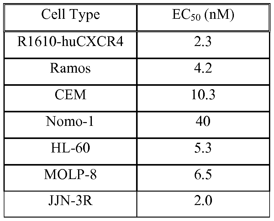


Claims
Priority Applications (21)
| Application Number | Priority Date | Filing Date | Title |
|---|---|---|---|
| KR1020147012396A KR102024957B1 (en) | 2011-11-09 | 2012-11-09 | Treatment of hematologic malignancies with an anti-cxcr4 antibody |
| SG11201401386XA SG11201401386XA (en) | 2011-11-09 | 2012-11-09 | Treatment of hematologic malignancies with an anti-cxcr4 antibody |
| DK12787343.8T DK2776032T3 (en) | 2011-11-09 | 2012-11-09 | TREATMENT OF HEMATOLOGICAL MALIGNITIES WITH ANTI-CXCR4 ANTIBODY |
| PL12787343T PL2776032T3 (en) | 2011-11-09 | 2012-11-09 | Treatment of hematologic malignancies with an anti-cxcr4 antibody |
| JP2014541315A JP6541350B2 (en) | 2011-11-09 | 2012-11-09 | Treatment of hematologic malignancies with anti-CXCR4 antibody |
| LTEP12787343.8T LT2776032T (en) | 2011-11-09 | 2012-11-09 | Treatment of hematologic malignancies with an anti-cxcr4 antibody |
| SI201231459T SI2776032T1 (en) | 2011-11-09 | 2012-11-09 | Treatment of hematologic malignancies with an anti-cxcr4 antibody |
| BR112014011144A BR112014011144A2 (en) | 2011-11-09 | 2012-11-09 | treatment of hematological malignancies with an anti-cxcr4 antibody |
| US14/356,996 US10428151B2 (en) | 2011-11-09 | 2012-11-09 | Treatment of hematologic malignancies with an anti-CXCR4 antibody |
| CN201280066551.7A CN104039320B (en) | 2011-11-09 | 2012-11-09 | The treatment of the anti-CXCR4 antibody of hematologic malignancies |
| MX2014005267A MX357100B (en) | 2011-11-09 | 2012-11-09 | Treatment of hematologic malignancies with an anti-cxcr4 antibody. |
| CA2855155A CA2855155A1 (en) | 2011-11-09 | 2012-11-09 | Treatment of hematologic malignancies with an anti-cxcr4 antibody |
| ES12787343T ES2705001T3 (en) | 2011-11-09 | 2012-11-09 | Treatment of hematological malignancies with an anti-CXCR4 antibody |
| EP12787343.8A EP2776032B1 (en) | 2011-11-09 | 2012-11-09 | Treatment of hematologic malignancies with an anti-cxcr4 antibody |
| EA201490957A EA026153B1 (en) | 2011-11-09 | 2012-11-09 | Treatment of hematologic malignancies with an anti-cxcr4 antibody |
| AU2012335553A AU2012335553B2 (en) | 2011-11-09 | 2012-11-09 | Treatment of hematologic malignancies with an anti-CXCR4 antibody |
| RS20181552A RS58102B1 (en) | 2011-11-09 | 2012-11-09 | Treatment of hematologic malignancies with an anti-cxcr4 antibody |
| IL232517A IL232517B (en) | 2011-11-09 | 2014-05-08 | Anti-cxcr4 antibody for treatment of hematologic malignancies |
| HRP20182183TT HRP20182183T1 (en) | 2011-11-09 | 2018-12-27 | Treatment of hematologic malignancies with an anti-cxcr4 antibody |
| CY20191100051T CY1121331T1 (en) | 2011-11-09 | 2019-01-15 | TREATMENT OF HEMATOLOGICAL MALIGRANTS WITH ANTI-CXCR4 |
| US16/590,152 US20200231683A1 (en) | 2011-11-09 | 2019-10-01 | Treatment of hematologic malignancies with an anti-cxcr4 antibody |
Applications Claiming Priority (4)
| Application Number | Priority Date | Filing Date | Title |
|---|---|---|---|
| US201161557815P | 2011-11-09 | 2011-11-09 | |
| US61/557,815 | 2011-11-09 | ||
| US201161569113P | 2011-12-09 | 2011-12-09 | |
| US61/569,113 | 2011-12-09 |
Related Child Applications (2)
| Application Number | Title | Priority Date | Filing Date |
|---|---|---|---|
| US14/356,996 A-371-Of-International US10428151B2 (en) | 2011-11-09 | 2012-11-09 | Treatment of hematologic malignancies with an anti-CXCR4 antibody |
| US16/590,152 Division US20200231683A1 (en) | 2011-11-09 | 2019-10-01 | Treatment of hematologic malignancies with an anti-cxcr4 antibody |
Publications (2)
| Publication Number | Publication Date |
|---|---|
| WO2013071068A2 true WO2013071068A2 (en) | 2013-05-16 |
| WO2013071068A3 WO2013071068A3 (en) | 2013-07-18 |
Family
ID=47179025
Family Applications (1)
| Application Number | Title | Priority Date | Filing Date |
|---|---|---|---|
| PCT/US2012/064395 WO2013071068A2 (en) | 2011-11-09 | 2012-11-09 | Treatment of hematologic malignancies with an anti-cxcr4 antibody |
Country Status (23)
| Country | Link |
|---|---|
| US (2) | US10428151B2 (en) |
| EP (1) | EP2776032B1 (en) |
| JP (1) | JP6541350B2 (en) |
| KR (1) | KR102024957B1 (en) |
| CN (1) | CN104039320B (en) |
| AU (1) | AU2012335553B2 (en) |
| BR (1) | BR112014011144A2 (en) |
| CA (1) | CA2855155A1 (en) |
| CY (1) | CY1121331T1 (en) |
| DK (1) | DK2776032T3 (en) |
| EA (1) | EA026153B1 (en) |
| ES (1) | ES2705001T3 (en) |
| HR (1) | HRP20182183T1 (en) |
| HU (1) | HUE043001T2 (en) |
| IL (1) | IL232517B (en) |
| LT (1) | LT2776032T (en) |
| MX (1) | MX357100B (en) |
| PL (1) | PL2776032T3 (en) |
| PT (1) | PT2776032T (en) |
| RS (1) | RS58102B1 (en) |
| SG (1) | SG11201401386XA (en) |
| SI (1) | SI2776032T1 (en) |
| WO (1) | WO2013071068A2 (en) |
Cited By (19)
| Publication number | Priority date | Publication date | Assignee | Title |
|---|---|---|---|---|
| WO2015038887A1 (en) * | 2013-09-12 | 2015-03-19 | Dana-Farber Cancer Institute Inc. | Methods for evaluating and treating waldenstrom's macroglobulinemia |
| WO2015069874A1 (en) | 2013-11-06 | 2015-05-14 | Bristol-Myers Squibb Company | Treatment of c1013g/cxcr4-associated waldenström's macroglobulinemia with an anti-cxcr4 antibody |
| WO2015093949A2 (en) | 2013-12-17 | 2015-06-25 | Aimm Therapeutics B.V. | Means and methods for counteracting myeloproliferative or lymphoproliferative disorders |
| US20150216843A1 (en) * | 2013-08-05 | 2015-08-06 | Cambridge Enterprise Limited | Inhibition of cxcr4 signaling in cancer immunotherapy |
| WO2015123818A1 (en) * | 2014-02-19 | 2015-08-27 | Microconstants China Inc | Chemokine receptor antagonist and its combinational therapy |
| WO2016201425A1 (en) | 2015-06-12 | 2016-12-15 | Bristol-Myers Squibb Company | Treatment of cancer by combined blockade of the pd-1 and cxcr4 signaling pathways |
| WO2017053170A1 (en) * | 2015-09-24 | 2017-03-30 | The University Of North Carolina At Chapel Hill | Methods and compositions for reducing metastases |
| WO2017216368A1 (en) * | 2016-06-16 | 2017-12-21 | Centre National De La Recherche Scientifique | Compounds useful for decreasing interferon level |
| WO2017220989A1 (en) | 2016-06-20 | 2017-12-28 | Kymab Limited | Anti-pd-l1 and il-2 cytokines |
| WO2019175802A1 (en) | 2018-03-13 | 2019-09-19 | Fundación Para La Investigación Biomédica Del Hospital Universitario La Paz | Anti-cxcr4 antibody combined with activated and expanded natural killer cells for cancer immunotherapy |
| WO2019191133A1 (en) | 2018-03-27 | 2019-10-03 | Bristol-Myers Squibb Company | Real-time monitoring of protein concentration using ultraviolet signal |
| US10465247B2 (en) | 2011-07-01 | 2019-11-05 | Dana-Farber Cancer Institute, Inc. | Discovery of a somatic mutation in MYD88 gene in lymphoplasmacytic lymphoma |
| US10525060B2 (en) | 2016-04-29 | 2020-01-07 | Dana-Farber Cancer Institute, Inc. | HCK as a therapeutic target in MYD88 mutated diseases |
| US10612095B2 (en) | 2013-12-06 | 2020-04-07 | Dana-Farber Cancer Institute, Inc. | Methods to distinguish Waldenström's Macroglobulinemia from IgM monoclonal gammopathy of undetermined significance |
| WO2020172658A1 (en) | 2019-02-24 | 2020-08-27 | Bristol-Myers Squibb Company | Methods of isolating a protein |
| EP3576787A4 (en) * | 2017-01-31 | 2020-09-23 | MSM Protein Technologies Inc. | Anti-cxcr4 antibodies |
| WO2020237221A1 (en) | 2019-05-23 | 2020-11-26 | Bristol-Myers Squibb Company | Methods of monitoring cell culture media |
| WO2022076318A1 (en) | 2020-10-05 | 2022-04-14 | Bristol-Myers Squibb Company | Methods for concentrating proteins |
| WO2023173011A1 (en) | 2022-03-09 | 2023-09-14 | Bristol-Myers Squibb Company | Transient expression of therapeutic proteins |
Families Citing this family (15)
| Publication number | Priority date | Publication date | Assignee | Title |
|---|---|---|---|---|
| PL2066351T3 (en) * | 2006-10-02 | 2016-02-29 | Squibb & Sons Llc | Human antibodies that bind cxcr4 and uses thereof |
| EP3102607B1 (en) | 2014-02-06 | 2018-08-22 | Yeda Research and Development Co. Ltd. | Anti cd84 antibodies, compositions comprising same and uses thereof |
| HRP20220860T1 (en) * | 2015-01-09 | 2022-10-14 | Adalta Limited | Cxcr4 binding molecules |
| EP3283107B1 (en) | 2015-04-17 | 2020-05-27 | Bristol-Myers Squibb Company | Compositions comprising a combination of ipilimumab and nivolumab |
| CN111375066B (en) | 2015-07-16 | 2023-04-25 | 百欧肯治疗有限公司 | Compositions and methods for treating cancer |
| US20180353472A1 (en) * | 2015-11-09 | 2018-12-13 | The General Hospital Corporation D.B.A Massachusetts General Hospital | Unit dose formulations for use as an anti-fugetactic agent |
| AU2017222495B2 (en) * | 2016-02-23 | 2019-08-08 | Biolinerx Ltd. | Methods of treating acute myeloid leukemia |
| SG11201809669RA (en) | 2016-05-20 | 2018-11-29 | Biohaven Pharm Holding Co Ltd | Use of glutamate modulating agents with immunotherapies to treat cancer |
| BR112019017375A2 (en) * | 2017-02-21 | 2020-03-31 | Kura Oncology, Inc. | METHOD FOR TREATING A CANCER EXPRESSING CXCL12 AND T-CELL ANGIOIMMUNOBLASTIC LYMPHOMA (AITL) IN AN INDIVIDUAL |
| KR20190133213A (en) | 2017-03-31 | 2019-12-02 | 브리스톨-마이어스 스큅 컴퍼니 | How to treat a tumor |
| US20200239577A1 (en) | 2017-10-15 | 2020-07-30 | Bristol-Myers Squibb Company | Methods of treating tumor |
| US20210162009A1 (en) * | 2017-12-08 | 2021-06-03 | The Medical College Of Wisconsin, Inc. | Chemokine variants as immune stimulants |
| TW202003565A (en) | 2018-03-23 | 2020-01-16 | 美商必治妥美雅史谷比公司 | Antibodies against MICA and/or MICB and uses thereof |
| WO2019191676A1 (en) | 2018-03-30 | 2019-10-03 | Bristol-Myers Squibb Company | Methods of treating tumor |
| EP4314068A1 (en) | 2021-04-02 | 2024-02-07 | The Regents Of The University Of California | Antibodies against cleaved cdcp1 and uses thereof |
Citations (11)
| Publication number | Priority date | Publication date | Assignee | Title |
|---|---|---|---|---|
| US4941880A (en) | 1987-06-19 | 1990-07-17 | Bioject, Inc. | Pre-filled ampule and non-invasive hypodermic injection device assembly |
| US5383851A (en) | 1992-07-24 | 1995-01-24 | Bioject Inc. | Needleless hypodermic injection device |
| US5756084A (en) | 1993-10-14 | 1998-05-26 | Ono Pharmaceutical Co., Ltd. | Human stromal derived factor 1α and 1β |
| WO2002043478A2 (en) | 2000-11-30 | 2002-06-06 | Medarex, Inc. | Transgenic transchromosomal rodents for making human antibodies |
| US6794132B2 (en) | 1999-10-02 | 2004-09-21 | Biosite, Inc. | Human antibodies |
| WO2008060367A2 (en) | 2006-10-02 | 2008-05-22 | Medarex, Inc. | Human antibodies that bind cxcr4 and uses thereof |
| WO2008142303A2 (en) | 2007-04-23 | 2008-11-27 | Pierre Fabre Medicament | Use of an anti-cxcr4 antibody for treating cancer |
| WO2009140124A1 (en) | 2008-05-14 | 2009-11-19 | Eli Lilly And Company | Anti-cxcr4 antibodies |
| US7674618B2 (en) | 2003-09-04 | 2010-03-09 | Medarex, Inc. | Expression vector |
| WO2010043650A2 (en) | 2008-10-14 | 2010-04-22 | Ablynx Nv | Amino acid sequences directed against cellular receptors for viruses and bacteria |
| US20120052097A1 (en) | 2010-08-20 | 2012-03-01 | Cerulean Pharma Inc. | Therapeutic peptide-polymer conjugates, particles, compositions, and related methods |
Family Cites Families (5)
| Publication number | Priority date | Publication date | Assignee | Title |
|---|---|---|---|---|
| WO2006089141A2 (en) | 2005-02-18 | 2006-08-24 | Dana-Farber Cancer Institute | Antibodies against cxcr4 and methods of use thereof |
| US8569280B2 (en) * | 2005-04-25 | 2013-10-29 | Immune Disease Institute | Methods for the treatment of multiple myeloma |
| EP2029155B1 (en) | 2006-02-28 | 2016-04-13 | Pharma Mar S.A. | Improved treatment of multiple myeloma |
| US8536161B2 (en) * | 2009-03-12 | 2013-09-17 | Genentech, Inc. | Combinations of phosphoinositide 3-kinase inhibitor compounds and chemotherapeutic agents for the treatment of hematopoietic malignancies |
| SG10201402346UA (en) * | 2009-05-19 | 2014-10-30 | Vivia Biotech Sl | Methods for providing personalized medicine tests ex vivo for hematological neoplasms |
-
2012
- 2012-11-09 HU HUE12787343A patent/HUE043001T2/en unknown
- 2012-11-09 MX MX2014005267A patent/MX357100B/en active IP Right Grant
- 2012-11-09 DK DK12787343.8T patent/DK2776032T3/en active
- 2012-11-09 PL PL12787343T patent/PL2776032T3/en unknown
- 2012-11-09 US US14/356,996 patent/US10428151B2/en active Active
- 2012-11-09 LT LTEP12787343.8T patent/LT2776032T/en unknown
- 2012-11-09 EP EP12787343.8A patent/EP2776032B1/en active Active
- 2012-11-09 WO PCT/US2012/064395 patent/WO2013071068A2/en active Application Filing
- 2012-11-09 PT PT12787343T patent/PT2776032T/en unknown
- 2012-11-09 CA CA2855155A patent/CA2855155A1/en not_active Abandoned
- 2012-11-09 SG SG11201401386XA patent/SG11201401386XA/en unknown
- 2012-11-09 CN CN201280066551.7A patent/CN104039320B/en active Active
- 2012-11-09 ES ES12787343T patent/ES2705001T3/en active Active
- 2012-11-09 RS RS20181552A patent/RS58102B1/en unknown
- 2012-11-09 BR BR112014011144A patent/BR112014011144A2/en not_active Application Discontinuation
- 2012-11-09 JP JP2014541315A patent/JP6541350B2/en not_active Expired - Fee Related
- 2012-11-09 KR KR1020147012396A patent/KR102024957B1/en active IP Right Grant
- 2012-11-09 EA EA201490957A patent/EA026153B1/en not_active IP Right Cessation
- 2012-11-09 SI SI201231459T patent/SI2776032T1/en unknown
- 2012-11-09 AU AU2012335553A patent/AU2012335553B2/en not_active Ceased
-
2014
- 2014-05-08 IL IL232517A patent/IL232517B/en active IP Right Grant
-
2018
- 2018-12-27 HR HRP20182183TT patent/HRP20182183T1/en unknown
-
2019
- 2019-01-15 CY CY20191100051T patent/CY1121331T1/en unknown
- 2019-10-01 US US16/590,152 patent/US20200231683A1/en not_active Abandoned
Patent Citations (13)
| Publication number | Priority date | Publication date | Assignee | Title |
|---|---|---|---|---|
| US4941880A (en) | 1987-06-19 | 1990-07-17 | Bioject, Inc. | Pre-filled ampule and non-invasive hypodermic injection device assembly |
| US5383851A (en) | 1992-07-24 | 1995-01-24 | Bioject Inc. | Needleless hypodermic injection device |
| US5399163A (en) | 1992-07-24 | 1995-03-21 | Bioject Inc. | Needleless hypodermic injection methods and device |
| US5756084A (en) | 1993-10-14 | 1998-05-26 | Ono Pharmaceutical Co., Ltd. | Human stromal derived factor 1α and 1β |
| US6794132B2 (en) | 1999-10-02 | 2004-09-21 | Biosite, Inc. | Human antibodies |
| US7041870B2 (en) | 2000-11-30 | 2006-05-09 | Medarex, Inc. | Transgenic transchromosomal rodents for making human antibodies |
| WO2002043478A2 (en) | 2000-11-30 | 2002-06-06 | Medarex, Inc. | Transgenic transchromosomal rodents for making human antibodies |
| US7674618B2 (en) | 2003-09-04 | 2010-03-09 | Medarex, Inc. | Expression vector |
| WO2008060367A2 (en) | 2006-10-02 | 2008-05-22 | Medarex, Inc. | Human antibodies that bind cxcr4 and uses thereof |
| WO2008142303A2 (en) | 2007-04-23 | 2008-11-27 | Pierre Fabre Medicament | Use of an anti-cxcr4 antibody for treating cancer |
| WO2009140124A1 (en) | 2008-05-14 | 2009-11-19 | Eli Lilly And Company | Anti-cxcr4 antibodies |
| WO2010043650A2 (en) | 2008-10-14 | 2010-04-22 | Ablynx Nv | Amino acid sequences directed against cellular receptors for viruses and bacteria |
| US20120052097A1 (en) | 2010-08-20 | 2012-03-01 | Cerulean Pharma Inc. | Therapeutic peptide-polymer conjugates, particles, compositions, and related methods |
Non-Patent Citations (92)
| Title |
|---|
| "Cancer Facts and Figures", 2008, AMERICAN CANCER SOCIETY |
| ALSAYED Y ET AL., BLOOD, vol. 109, no. 7, 2007, pages 2708 - 17 |
| AMADORI S ET AL., JCLIN ONCOL, vol. 9, no. 7, 1991, pages 1210 - 4 |
| ANGAL S ET AL., MOL IMMUNOL, vol. 30, no. 1, 1993, pages 105 - 8 |
| AZAB AK ET AL., BLOOD, vol. 113, no. 18, 2009, pages 4341 - 51 |
| BEIDER K ET AL., EXP HEMATOL, vol. 39, no. 3, 2011, pages 282 - 92 |
| BERNDT C ET AL., PROC NATL ACAD SCI USA, vol. 95, 1998, pages 12556 - 61 |
| BERTOLINI F ET AL., CANCER RES, vol. 62, no. 11, 2002, pages 3106 - 12 |
| BLEUL CC ET AL., NATURE, vol. 382, no. 6594, 1996, pages 829 - 33 |
| BREEMS DA ET AL., J CLIN ONCOL, vol. 23, no. 9, 2005, pages 1969 - 78 |
| BROXMEYER HE ET AL., J EXP MED, vol. 201, 2005, pages 1307 - 18 |
| BURGER JA ET AL., BLOOD, vol. 94, 1999, pages 3658 - 67 |
| CARNEC X ET AL., J VIROL, vol. 79, 2005, pages 1930 - 8 |
| CHOW KU ET AL., HAEMATOLOGICA, vol. 87, 2002, pages 33 - 43 |
| CORCIONE A ET AL., J NATL CANCER INST, vol. 92, 2000, pages 628 - 35 |
| CORVATA N ET AL., POSTER PRESENTED AT 102ND ANNUAL MEETING OF THE AMERICAN ASSOCIATION FOR CANCER RESEARCH, 2 April 2011 (2011-04-02) |
| CRAZZOLARA R ET AL., BR JHAEMATOL, vol. 115, 2001, pages 545 - 53 |
| DAR A ET AL., LEUKEMIA, vol. 25, no. 8, 2011, pages 1286 - 96 |
| DEVINE SM ET AL., J CLIN ONCOL, vol. 22, 2004, pages 1095 - 102 |
| DI SALVO, EUR JPHARMACOL, vol. 409, 2000, pages 143 - 154 |
| FENG Y ET AL., SCIENCE, vol. 272, 1996, pages 872 - 7 |
| FLOMENBERG N ET AL., BLOOD, vol. 106, 2005, pages 1867 - 74 |
| FRIEDBERG JW ET AL., J CLIN ONCOL, vol. 26, no. 2, 2008, pages 204 - 10 |
| GARG H ET AL., JLEUKOCYTE BIOL, vol. 79, 2006, pages 351 - 62 |
| GAZITT Y, JHEMATOTHER STEM CELL RES, vol. 10, 2001, pages 229 - 36 |
| GEMINDER ET AL., J IMMUNOL, vol. 167, 2001, pages 4747 - 57 |
| GISSELBRECHT C ET AL., J CLIN ONCOL, vol. 28, 2010, pages 4184 - 90 |
| GISSELBRECHT C ET AL.: "Novel Agents for Diffuse Large B-cell Lymphoma", 2011, ASCO EDUCATION BOOK 2011, pages: 321 |
| GONZALO JA ET AL., J IMMUNOL, vol. 165, 2000, pages 499 - 508 |
| GULENG B ET AL., CANCER RES, vol. 65, no. 13, 2005, pages 5864 - 71 |
| GUPTA SK ET AL., JBIOL CHEM, vol. 273, no. 7, 1998, pages 4282 - 7 |
| HESSELGESSER J ET AL., CURR BIOL, vol. 7, no. 2, 1997, pages 112 - 21 |
| HILLER DJ ET AL., SURGERY, vol. 150, no. 3, 2011, pages 459 - 65 |
| HOLLINGER ET AL., NATURE BIOTECH, vol. 23, no. 9, 2005, pages 1126 - 36 |
| HOSOKAWA Y ET AL., CLIN EXP IMMUNOL, vol. 141, 2005, pages 467 - 74 |
| HOU T ET AL., J IMMUNOL, vol. 160, 1998, pages 180 - 8 |
| HWANG JH ET AL., J CLIN ENDOCRINOL METAB, vol. 88, 2003, pages 408 - 16 |
| JEMAL A ET AL., CA CANCER J CLIN, vol. 55, 2005, pages 10 - 50 |
| JEMAL A ET AL., CA CANCER J CLIN, vol. 58, 2008, pages 71 - 96 |
| JEMAL A ET AL., CA CANCER J CLIN, vol. 59, 2009, pages 225 - 49 |
| JIANG YP ET AL., GYNECOL ONCOL, vol. 103, no. 1, 2006, pages 226 - 33 |
| JIN DK ET AL., NAT MED, vol. 12, 2006, pages 557 - 67 |
| JIN L ET AL., MOL CANCER THER, vol. 7, 2008, pages 48 - 58 |
| KABAT EA ET AL.: "Sequences of Proteins of Immunological Interest, Fifth Edition,", 1991, NIH PUBLICATION NO. 91-3242 |
| KASHYAP MK; KUMAR D; JONES H; MELO-CARDENAS J; KUHNE MR; SABBATINI P; COHEN LJ; CARDARELLI JM; KIPPS TJ; CASTRO JE, ABSTRACT FOR PRESENTATION AT AMERICAN SOCIETY OF HEMATOLOGY (ASH) 54TH ANNUAL MEETING, 8 December 2012 (2012-12-08) |
| KEWALRAMANI T ET AL., BLOOD, vol. 103, no. 10, 2004, pages 3684 - 8 |
| KOSHIBA T ET AL., CLIN CANCER RES, vol. 6, 2000, pages 3530 - 5 |
| LAVERDIERE C ET AL., CLIN CANCER RES, vol. 11, 2005, pages 2561 - 7 |
| LEE B ET AL., PROC NATL ACAD SCI USA, vol. 96, no. 9, 1999, pages 5215 - 20 |
| LIBURA J ET AL., BLOOD, vol. 100, 2002, pages 2597 - 606 |
| LIU Z ET AL., DIABETOLOGIA, vol. 54, no. 8, 2011, pages 2067 - 76 |
| LOETSCHER M ET AL., JBIOL CHEM, vol. 269, no. 1, 1994, pages 232 - 7 |
| LONBERG N ET AL., NATURE, vol. 368, no. 6474, 1994, pages 856 - 859 |
| MARECHAL R ET AL., BRIT J CANCER, vol. 100, no. 9, 2009, pages 1444 - 51 |
| MATTHYS P ET AL., J IMMUNOL, vol. 167, 2001, pages 4686 - 92 |
| MOHLE R ET AL., BLOOD, vol. 91, no. 12, 1998, pages 4523 - 30 |
| MOHLE R ET AL., LEUKEMIA, vol. 13, 1999, pages 1954 - 9 |
| MULLER A ET AL., NATURE, vol. 410, 2001, pages 50 - 6 |
| MURPHY PM, NEW ENGL J MED, vol. 345, no. 11, 2001, pages 833 - 5 |
| NERVI B ET AL., BLOOD, vol. 113, 2009, pages 6206 - 14 |
| OBERLIN E ET AL., NATURE, vol. 382, 1996, pages 833 - 5 |
| OLAFSEN ET AL., SEMIN NUCL MED, vol. 40, no. 3, 2010, pages 167 - 81 |
| OTTAIANO A ET AL., CLIN CANCER RES, vol. 12, no. 9, 2006, pages 2795 - 803 |
| PING YF ET AL., JPATHOL, vol. 224, no. 3, 2011, pages 344 - 54 |
| PORVASNIK S ET AL., THE PROSTATE, vol. 69, 2009, pages 1460 - 9 |
| RAAB MS ET AL., LANCET, vol. 374, no. 9686, 2009, pages 324 - 39 |
| REMPEL SA ET AL., CLIN CANCER RES, vol. 6, 2000, pages 102 - 11 |
| RICHERT M ET AL., ONCOL REP, vol. 21, 2009, pages 76 - 7 |
| RIGHI E ET AL., CANCER RES, vol. 71, no. 16, 2011, pages 5522 - 34 |
| ROBINSON JR: "Sustained and Controlled Release Drug Delivery Systems", 1978, MARCEL DEKKER, INC. |
| ROCCARO AM; SACCO A; KUHNE M; AZAB A; MAISO P; ZHANG Y; LIU Y; MICHAELA R; NGO HT; QUANG P, ABSTRACT FOR PRESENTATION AT AMERICAN SOCIETY OF HEMATOLOGY (ASH) 54TH ANNUAL MEETING, 2012 |
| ROMBOUTS EJC ET AL., BLOOD, vol. 104, 2004, pages 550 - 7 |
| RUMMEL MJ ET AL., SEMIN ONCOL, vol. 29, no. 13, 2002, pages 12 - 4 |
| SCALA S ET AL., CLIN CANCER RES, vol. 11, 2005, pages 183541 |
| SCHRADER AJ ET AL., BR J CANCER, vol. 86, 2002, pages 1250 - 6 |
| SCOTTON C ET AL., BR. J. CANCER, vol. 85, 2001, pages 891 - 7 |
| SIEGEL R ET AL., CA CANCER J CLIN, vol. 61, no. 4, 2011, pages 212 - 36 |
| SINGER CR; GOLDSTONE AH, CLIN HAEMATOL, vol. 15, 1986, pages 105 |
| SPANO JP ET AL., ANN ONCOL, vol. 15, 2004, pages 613 - 7 |
| SPOO AC ET AL., BLOOD, vol. 109, no. 2, 2007, pages 786 - 91 |
| STALLER PET, NATURE, vol. 425, 2003, pages 307 - 11 |
| TACHIBANA K ET AL., NATURE, vol. 393, no. 6685, 1998, pages 591 - 4 |
| TAICHMAN RS ET AL., CANCER RES, vol. 62, 2002, pages 1832 - 7 |
| TAVOR S ET AL., CANCER RES, vol. 64, no. 8, 2004, pages 2817 - 24 |
| TERADA R ET AL., LAB INVEST, vol. 83, 2003, pages 665 - 72 |
| VICARI AP ET AL., CYTOKINE GROWTH FACTOR REV, vol. 13, 2002, pages 143 - 154 |
| WANG N ET AL., J TRANSL MED, vol. 3, 2005, pages 26 - 33 |
| WENG AP ET AL., AM JCLIN PATHOL, vol. 119, 2003, pages 424 - 30 |
| ZEELENBERG IS ET AL., CANCER RES, vol. 63, 2003, pages 3833 - 9 |
| ZENG Z ET AL., BLOOD, vol. 113, no. 24, 2009, pages 6215 - 24 |
| ZHANG ET AL., JBIOL CHEM, vol. 277, no. 27, 2002, pages 24515 - 21 |
| ZOU Y ET AL., NATURE, vol. 393, no. 6685, 1998, pages 595 - 9 |
Cited By (34)
| Publication number | Priority date | Publication date | Assignee | Title |
|---|---|---|---|---|
| US10465247B2 (en) | 2011-07-01 | 2019-11-05 | Dana-Farber Cancer Institute, Inc. | Discovery of a somatic mutation in MYD88 gene in lymphoplasmacytic lymphoma |
| US11739385B2 (en) | 2011-07-01 | 2023-08-29 | Dana-Farber Cancer Institute, Inc. | Discovery of a somatic mutation in MYD88 gene in lymphoplasmacytic lymphoma |
| US20150216843A1 (en) * | 2013-08-05 | 2015-08-06 | Cambridge Enterprise Limited | Inhibition of cxcr4 signaling in cancer immunotherapy |
| WO2015038887A1 (en) * | 2013-09-12 | 2015-03-19 | Dana-Farber Cancer Institute Inc. | Methods for evaluating and treating waldenstrom's macroglobulinemia |
| US10526660B2 (en) | 2013-09-12 | 2020-01-07 | Dana-Farber Cancer Institute, Inc. | Methods for evaluating and treating Waldenstrom's macroglobulinemia |
| JP2020023499A (en) * | 2013-11-06 | 2020-02-13 | ブリストル−マイヤーズ スクイブ カンパニーBristol−Myers Squibb Company | Treatment of c1013g/cxcr4-associated waldenstrom's macroglobulinemia with anti-cxcr4 antibody |
| JP2017502923A (en) * | 2013-11-06 | 2017-01-26 | ブリストル−マイヤーズ スクイブ カンパニーBristol−Myers Squibb Company | Treatment of C1013G / CXCR4-related Waldenstrom's macroglobulinemia with anti-CXCR4 antibodies |
| CN105899536A (en) * | 2013-11-06 | 2016-08-24 | 百时美施贵宝公司 | Treatment of C1013g/cxcr4-associated waldenstr m's macroglobulinemia with an anti-cxcr4 antibody |
| US10233248B2 (en) | 2013-11-06 | 2019-03-19 | Bristol-Myers Squibb Company | Treatment of C1013G/CXCR4-associated waldenstroöm's macroglobulinemia with an anti-CXCR4 antibody |
| EP3599248A1 (en) | 2013-11-06 | 2020-01-29 | Bristol-Myers Squibb Company | Treatment of c1013g/cxcr4-associated waldenström's macroglobulinemia with an anti-cxcr4 antibody |
| US11220548B2 (en) | 2013-11-06 | 2022-01-11 | Bristol-Myers Squibb Company | Treatment of C1013G/CXCR4-associated Waldenstrom's macroglobulinemia with an anti-CXCR4 antibody |
| WO2015069874A1 (en) | 2013-11-06 | 2015-05-14 | Bristol-Myers Squibb Company | Treatment of c1013g/cxcr4-associated waldenström's macroglobulinemia with an anti-cxcr4 antibody |
| US10612095B2 (en) | 2013-12-06 | 2020-04-07 | Dana-Farber Cancer Institute, Inc. | Methods to distinguish Waldenström's Macroglobulinemia from IgM monoclonal gammopathy of undetermined significance |
| JP2017502664A (en) * | 2013-12-17 | 2017-01-26 | アイム・セラピューティクス・べー・フェー | Means and methods for combating myeloproliferative or lymphoproliferative disorders |
| EP3572425A1 (en) | 2013-12-17 | 2019-11-27 | AIMM Therapeutics B.V. | Means and methods for counteracting myeloproliferative or lymphoproliferative disorders |
| WO2015093949A2 (en) | 2013-12-17 | 2015-06-25 | Aimm Therapeutics B.V. | Means and methods for counteracting myeloproliferative or lymphoproliferative disorders |
| US11629200B2 (en) | 2013-12-17 | 2023-04-18 | Kling Biotherapeutics B.V. | Means and methods for counteracting myeloproliferative or lymphoproliferative disorders |
| WO2015123818A1 (en) * | 2014-02-19 | 2015-08-27 | Microconstants China Inc | Chemokine receptor antagonist and its combinational therapy |
| WO2016201425A1 (en) | 2015-06-12 | 2016-12-15 | Bristol-Myers Squibb Company | Treatment of cancer by combined blockade of the pd-1 and cxcr4 signaling pathways |
| US11219694B2 (en) | 2015-09-24 | 2022-01-11 | The University Of North Carolina At Chapel Hill | Methods and compositions for reducing metastases |
| WO2017053170A1 (en) * | 2015-09-24 | 2017-03-30 | The University Of North Carolina At Chapel Hill | Methods and compositions for reducing metastases |
| CN108289966A (en) * | 2015-09-24 | 2018-07-17 | 北卡罗来纳-查佩尔山大学 | Method and composition for reducing transfer |
| US10525060B2 (en) | 2016-04-29 | 2020-01-07 | Dana-Farber Cancer Institute, Inc. | HCK as a therapeutic target in MYD88 mutated diseases |
| WO2017216368A1 (en) * | 2016-06-16 | 2017-12-21 | Centre National De La Recherche Scientifique | Compounds useful for decreasing interferon level |
| WO2017220988A1 (en) | 2016-06-20 | 2017-12-28 | Kymab Limited | Multispecific antibodies for immuno-oncology |
| WO2017220989A1 (en) | 2016-06-20 | 2017-12-28 | Kymab Limited | Anti-pd-l1 and il-2 cytokines |
| WO2017220990A1 (en) | 2016-06-20 | 2017-12-28 | Kymab Limited | Anti-pd-l1 antibodies |
| EP3576787A4 (en) * | 2017-01-31 | 2020-09-23 | MSM Protein Technologies Inc. | Anti-cxcr4 antibodies |
| WO2019175802A1 (en) | 2018-03-13 | 2019-09-19 | Fundación Para La Investigación Biomédica Del Hospital Universitario La Paz | Anti-cxcr4 antibody combined with activated and expanded natural killer cells for cancer immunotherapy |
| WO2019191133A1 (en) | 2018-03-27 | 2019-10-03 | Bristol-Myers Squibb Company | Real-time monitoring of protein concentration using ultraviolet signal |
| WO2020172658A1 (en) | 2019-02-24 | 2020-08-27 | Bristol-Myers Squibb Company | Methods of isolating a protein |
| WO2020237221A1 (en) | 2019-05-23 | 2020-11-26 | Bristol-Myers Squibb Company | Methods of monitoring cell culture media |
| WO2022076318A1 (en) | 2020-10-05 | 2022-04-14 | Bristol-Myers Squibb Company | Methods for concentrating proteins |
| WO2023173011A1 (en) | 2022-03-09 | 2023-09-14 | Bristol-Myers Squibb Company | Transient expression of therapeutic proteins |
Also Published As
| Publication number | Publication date |
|---|---|
| PL2776032T3 (en) | 2019-03-29 |
| WO2013071068A3 (en) | 2013-07-18 |
| RS58102B1 (en) | 2019-02-28 |
| EP2776032A2 (en) | 2014-09-17 |
| KR102024957B1 (en) | 2019-09-24 |
| MX357100B (en) | 2018-06-26 |
| CA2855155A1 (en) | 2013-05-16 |
| KR20140093945A (en) | 2014-07-29 |
| IL232517B (en) | 2018-05-31 |
| CN104039320B (en) | 2019-10-15 |
| ES2705001T3 (en) | 2019-03-21 |
| PT2776032T (en) | 2019-01-21 |
| CY1121331T1 (en) | 2020-05-29 |
| BR112014011144A2 (en) | 2017-05-16 |
| MX2014005267A (en) | 2014-07-28 |
| JP2014533279A (en) | 2014-12-11 |
| US20200231683A1 (en) | 2020-07-23 |
| EP2776032B1 (en) | 2018-10-17 |
| DK2776032T3 (en) | 2019-02-04 |
| AU2012335553B2 (en) | 2017-09-21 |
| HRP20182183T1 (en) | 2019-02-22 |
| HUE043001T2 (en) | 2019-07-29 |
| JP6541350B2 (en) | 2019-07-10 |
| AU2012335553A1 (en) | 2014-07-03 |
| CN104039320A (en) | 2014-09-10 |
| IL232517A0 (en) | 2014-06-30 |
| LT2776032T (en) | 2018-12-10 |
| EA026153B1 (en) | 2017-03-31 |
| EA201490957A1 (en) | 2014-08-29 |
| US10428151B2 (en) | 2019-10-01 |
| SI2776032T1 (en) | 2018-12-31 |
| US20140322208A1 (en) | 2014-10-30 |
| SG11201401386XA (en) | 2014-10-30 |
Similar Documents
| Publication | Publication Date | Title |
|---|---|---|
| US20200231683A1 (en) | Treatment of hematologic malignancies with an anti-cxcr4 antibody | |
| US11103579B2 (en) | Combination of DR5 agonist and anti-PD-1 antagonist and methods of use | |
| US20200179511A1 (en) | Bcma-targeting agent, and combination therapy with a gamma secretase inhibitor | |
| US11220548B2 (en) | Treatment of C1013G/CXCR4-associated Waldenstrom's macroglobulinemia with an anti-CXCR4 antibody | |
| US20180179282A1 (en) | Treatment of cancer by combined blockade of the pd-1 and cxcr4 signaling pathways | |
| JP2019516712A (en) | Combination of glutamate modulator and immunotherapy to treat cancer | |
| JP2018523652A (en) | Combination of PD-1 antagonist and EGFR inhibitor | |
| JP2015083613A (en) | Cd37 immunotherapeutic and combination with bifunctional chemotherapeutic thereof | |
| EP3142697A1 (en) | Treatment of lung cancer using a combination of an anti-pd-1 antibody and another anti-cancer agent | |
| JP6184952B2 (en) | Combination therapy with anti-CD19 antibody and nitrogen mustard | |
| WO2015116729A2 (en) | Anti-cd37 antibody and anti-cd20 antibody combination therapy for treatment of b-cell malignancies and disorders | |
| JP2014525926A5 (en) | ||
| WO2020033283A1 (en) | Compositions and methods for treating cancer with a combination of programmed death receptor (pd-1) antibodies and vicriviroc | |
| WO2019067332A1 (en) | Compositions and methods for treating cancer with a combination of programmed death receptor (pd-1) antibodies and a cxcr2 antagonist | |
| TW202222833A (en) | Cd40 agonistic antibody and method of use | |
| JP2022553649A (en) | Novel therapeutic combinations containing derivatives of oxyazaphosphorine for the treatment of cancer |
Legal Events
| Date | Code | Title | Description |
|---|---|---|---|
| 121 | Ep: the epo has been informed by wipo that ep was designated in this application |
Ref document number: 12787343 Country of ref document: EP Kind code of ref document: A2 |
|
| WWE | Wipo information: entry into national phase |
Ref document number: MX/A/2014/005267 Country of ref document: MX |
|
| ENP | Entry into the national phase |
Ref document number: 2855155 Country of ref document: CA Ref document number: 2014541315 Country of ref document: JP Kind code of ref document: A Ref document number: 20147012396 Country of ref document: KR Kind code of ref document: A |
|
| WWE | Wipo information: entry into national phase |
Ref document number: 14356996 Country of ref document: US Ref document number: 232517 Country of ref document: IL |
|
| WWE | Wipo information: entry into national phase |
Ref document number: 2012787343 Country of ref document: EP |
|
| WWE | Wipo information: entry into national phase |
Ref document number: 201490957 Country of ref document: EA |
|
| ENP | Entry into the national phase |
Ref document number: 2012335553 Country of ref document: AU Date of ref document: 20121109 Kind code of ref document: A |
|
| REG | Reference to national code |
Ref country code: BR Ref legal event code: B01A Ref document number: 112014011144 Country of ref document: BR |
|
| ENP | Entry into the national phase |
Ref document number: 112014011144 Country of ref document: BR Kind code of ref document: A2 Effective date: 20140508 |