WO2015019120A2 - Method for the analysis of immunoreactivity, and a device suitable for carrying out the method - Google Patents
Method for the analysis of immunoreactivity, and a device suitable for carrying out the method Download PDFInfo
- Publication number
- WO2015019120A2 WO2015019120A2 PCT/HU2014/000068 HU2014000068W WO2015019120A2 WO 2015019120 A2 WO2015019120 A2 WO 2015019120A2 HU 2014000068 W HU2014000068 W HU 2014000068W WO 2015019120 A2 WO2015019120 A2 WO 2015019120A2
- Authority
- WO
- WIPO (PCT)
- Prior art keywords
- cells
- antigen
- sample
- capillary channel
- capillary
- Prior art date
Links
- 238000000034 method Methods 0.000 title claims abstract description 134
- 238000004458 analytical method Methods 0.000 title claims abstract description 27
- 102000036639 antigens Human genes 0.000 claims abstract description 415
- 108091007433 antigens Proteins 0.000 claims abstract description 415
- 239000000427 antigen Substances 0.000 claims abstract description 414
- 239000007788 liquid Substances 0.000 claims abstract description 106
- 239000013642 negative control Substances 0.000 claims abstract description 97
- 239000013641 positive control Substances 0.000 claims abstract description 64
- 238000000576 coating method Methods 0.000 claims abstract description 53
- 239000011248 coating agent Substances 0.000 claims abstract description 48
- 239000013558 reference substance Substances 0.000 claims abstract description 24
- 239000000126 substance Substances 0.000 claims abstract description 20
- 230000000007 visual effect Effects 0.000 claims abstract description 6
- 210000004027 cell Anatomy 0.000 claims description 322
- 239000000523 sample Substances 0.000 claims description 205
- 238000001514 detection method Methods 0.000 claims description 109
- 239000008280 blood Substances 0.000 claims description 71
- 210000004369 blood Anatomy 0.000 claims description 67
- 238000005259 measurement Methods 0.000 claims description 60
- 238000002493 microarray Methods 0.000 claims description 60
- 239000000463 material Substances 0.000 claims description 46
- 238000012360 testing method Methods 0.000 claims description 42
- 108090000623 proteins and genes Proteins 0.000 claims description 36
- 230000021164 cell adhesion Effects 0.000 claims description 35
- 102000004169 proteins and genes Human genes 0.000 claims description 35
- 210000000440 neutrophil Anatomy 0.000 claims description 28
- 230000004913 activation Effects 0.000 claims description 26
- 210000001616 monocyte Anatomy 0.000 claims description 25
- 230000000295 complement effect Effects 0.000 claims description 24
- 210000002966 serum Anatomy 0.000 claims description 22
- 208000037265 diseases, disorders, signs and symptoms Diseases 0.000 claims description 21
- 230000001413 cellular effect Effects 0.000 claims description 14
- 238000003745 diagnosis Methods 0.000 claims description 13
- 239000013060 biological fluid Substances 0.000 claims description 12
- 230000020411 cell activation Effects 0.000 claims description 12
- 239000012472 biological sample Substances 0.000 claims description 11
- 239000000203 mixture Substances 0.000 claims description 11
- 201000000596 systemic lupus erythematosus Diseases 0.000 claims description 10
- 230000036755 cellular response Effects 0.000 claims description 9
- 210000002381 plasma Anatomy 0.000 claims description 8
- 108090000765 processed proteins & peptides Proteins 0.000 claims description 8
- 230000001464 adherent effect Effects 0.000 claims description 7
- 210000002808 connective tissue Anatomy 0.000 claims description 7
- 208000035475 disorder Diseases 0.000 claims description 7
- 230000036039 immunity Effects 0.000 claims description 7
- 230000003287 optical effect Effects 0.000 claims description 6
- 102000004196 processed proteins & peptides Human genes 0.000 claims description 6
- 231100000617 superantigen Toxicity 0.000 claims description 6
- 239000013566 allergen Substances 0.000 claims description 5
- 238000011493 immune profiling Methods 0.000 claims description 5
- 241000700605 Viruses Species 0.000 claims description 4
- 230000003834 intracellular effect Effects 0.000 claims description 4
- 210000002540 macrophage Anatomy 0.000 claims description 4
- 238000004519 manufacturing process Methods 0.000 claims description 4
- 238000004451 qualitative analysis Methods 0.000 claims description 4
- 238000012764 semi-quantitative analysis Methods 0.000 claims description 4
- 102000029797 Prion Human genes 0.000 claims description 3
- 108091000054 Prion Proteins 0.000 claims description 3
- 239000011324 bead Substances 0.000 claims description 3
- 150000001720 carbohydrates Chemical class 0.000 claims description 3
- 239000013592 cell lysate Substances 0.000 claims description 3
- 210000004443 dendritic cell Anatomy 0.000 claims description 3
- 208000002672 hepatitis B Diseases 0.000 claims description 3
- 150000002632 lipids Chemical class 0.000 claims description 3
- 108020004707 nucleic acids Proteins 0.000 claims description 3
- 102000039446 nucleic acids Human genes 0.000 claims description 3
- 150000007523 nucleic acids Chemical class 0.000 claims description 3
- 238000009589 serological test Methods 0.000 claims description 3
- 230000011664 signaling Effects 0.000 claims description 3
- 239000006228 supernatant Substances 0.000 claims description 3
- 238000009007 Diagnostic Kit Methods 0.000 claims description 2
- 229930186217 Glycolipid Natural products 0.000 claims description 2
- 102000003886 Glycoproteins Human genes 0.000 claims description 2
- 108090000288 Glycoproteins Proteins 0.000 claims description 2
- 101000686985 Mouse mammary tumor virus (strain C3H) Protein PR73 Proteins 0.000 claims description 2
- 230000004075 alteration Effects 0.000 claims description 2
- 235000014633 carbohydrates Nutrition 0.000 claims description 2
- 239000003795 chemical substances by application Substances 0.000 claims description 2
- 210000004748 cultured cell Anatomy 0.000 claims description 2
- 244000045947 parasite Species 0.000 claims description 2
- 230000026731 phosphorylation Effects 0.000 claims description 2
- 238000006366 phosphorylation reaction Methods 0.000 claims description 2
- 150000003384 small molecules Chemical class 0.000 claims description 2
- 102000004895 Lipoproteins Human genes 0.000 claims 1
- 108090001030 Lipoproteins Proteins 0.000 claims 1
- 108091006146 Channels Proteins 0.000 description 417
- 230000027455 binding Effects 0.000 description 49
- 101100217502 Caenorhabditis elegans lgg-3 gene Proteins 0.000 description 34
- 235000018102 proteins Nutrition 0.000 description 33
- 239000000243 solution Substances 0.000 description 30
- 238000003491 array Methods 0.000 description 29
- 102000005962 receptors Human genes 0.000 description 29
- 108020003175 receptors Proteins 0.000 description 29
- 108091003079 Bovine Serum Albumin Proteins 0.000 description 27
- 239000002953 phosphate buffered saline Substances 0.000 description 24
- 229940098773 bovine serum albumin Drugs 0.000 description 23
- 230000003993 interaction Effects 0.000 description 23
- 238000002474 experimental method Methods 0.000 description 22
- 230000009257 reactivity Effects 0.000 description 21
- 239000007787 solid Substances 0.000 description 17
- 238000005406 washing Methods 0.000 description 17
- 244000052769 pathogen Species 0.000 description 16
- 239000004205 dimethyl polysiloxane Substances 0.000 description 14
- 235000013870 dimethyl polysiloxane Nutrition 0.000 description 14
- CXQXSVUQTKDNFP-UHFFFAOYSA-N octamethyltrisiloxane Chemical compound C[Si](C)(C)O[Si](C)(C)O[Si](C)(C)C CXQXSVUQTKDNFP-UHFFFAOYSA-N 0.000 description 14
- 238000004987 plasma desorption mass spectroscopy Methods 0.000 description 14
- 229920000435 poly(dimethylsiloxane) Polymers 0.000 description 14
- 201000010099 disease Diseases 0.000 description 13
- 101100454808 Caenorhabditis elegans lgg-2 gene Proteins 0.000 description 12
- 102000009109 Fc receptors Human genes 0.000 description 12
- 108010087819 Fc receptors Proteins 0.000 description 12
- 239000000020 Nitrocellulose Substances 0.000 description 12
- 238000006243 chemical reaction Methods 0.000 description 12
- 230000004154 complement system Effects 0.000 description 12
- 238000011534 incubation Methods 0.000 description 12
- 229920001220 nitrocellulos Polymers 0.000 description 12
- 230000001363 autoimmune Effects 0.000 description 11
- 239000012634 fragment Substances 0.000 description 11
- 239000012895 dilution Substances 0.000 description 10
- 238000010790 dilution Methods 0.000 description 10
- 238000001035 drying Methods 0.000 description 10
- 239000011521 glass Substances 0.000 description 10
- 230000017531 blood circulation Effects 0.000 description 9
- 238000005516 engineering process Methods 0.000 description 9
- 239000003446 ligand Substances 0.000 description 9
- 230000001717 pathogenic effect Effects 0.000 description 9
- 230000035945 sensitivity Effects 0.000 description 9
- 108060003951 Immunoglobulin Proteins 0.000 description 8
- 238000012512 characterization method Methods 0.000 description 8
- 230000000694 effects Effects 0.000 description 8
- 230000006870 function Effects 0.000 description 8
- 102000018358 immunoglobulin Human genes 0.000 description 8
- 108010088751 Albumins Proteins 0.000 description 7
- 102000009027 Albumins Human genes 0.000 description 7
- KCXVZYZYPLLWCC-UHFFFAOYSA-N EDTA Chemical compound OC(=O)CN(CC(O)=O)CCN(CC(O)=O)CC(O)=O KCXVZYZYPLLWCC-UHFFFAOYSA-N 0.000 description 7
- 239000000872 buffer Substances 0.000 description 7
- 230000001965 increasing effect Effects 0.000 description 7
- 239000012528 membrane Substances 0.000 description 7
- XLYOFNOQVPJJNP-UHFFFAOYSA-N water Substances O XLYOFNOQVPJJNP-UHFFFAOYSA-N 0.000 description 7
- 101100454807 Caenorhabditis elegans lgg-1 gene Proteins 0.000 description 6
- RTZKZFJDLAIYFH-UHFFFAOYSA-N Diethyl ether Chemical compound CCOCC RTZKZFJDLAIYFH-UHFFFAOYSA-N 0.000 description 6
- LFQSCWFLJHTTHZ-UHFFFAOYSA-N Ethanol Chemical compound CCO LFQSCWFLJHTTHZ-UHFFFAOYSA-N 0.000 description 6
- 230000004071 biological effect Effects 0.000 description 6
- 230000000903 blocking effect Effects 0.000 description 6
- 230000008859 change Effects 0.000 description 6
- 239000003153 chemical reaction reagent Substances 0.000 description 6
- 238000002405 diagnostic procedure Methods 0.000 description 6
- 239000000975 dye Substances 0.000 description 6
- 238000011156 evaluation Methods 0.000 description 6
- 230000028993 immune response Effects 0.000 description 6
- 208000015181 infectious disease Diseases 0.000 description 6
- 238000002360 preparation method Methods 0.000 description 6
- 230000008569 process Effects 0.000 description 6
- 238000011282 treatment Methods 0.000 description 6
- 238000002255 vaccination Methods 0.000 description 6
- 108010028780 Complement C3 Proteins 0.000 description 5
- 102000016918 Complement C3 Human genes 0.000 description 5
- 241001465754 Metazoa Species 0.000 description 5
- 206010028980 Neoplasm Diseases 0.000 description 5
- 241000191940 Staphylococcus Species 0.000 description 5
- 238000003556 assay Methods 0.000 description 5
- 210000001124 body fluid Anatomy 0.000 description 5
- 239000012530 fluid Substances 0.000 description 5
- 210000002865 immune cell Anatomy 0.000 description 5
- 210000000987 immune system Anatomy 0.000 description 5
- 238000002372 labelling Methods 0.000 description 5
- 239000004033 plastic Substances 0.000 description 5
- 229920003023 plastic Polymers 0.000 description 5
- 239000000047 product Substances 0.000 description 5
- 238000012124 rapid diagnostic test Methods 0.000 description 5
- IPJDHSYCSQAODE-UHFFFAOYSA-N 5-chloromethylfluorescein diacetate Chemical compound O1C(=O)C2=CC(CCl)=CC=C2C21C1=CC=C(OC(C)=O)C=C1OC1=CC(OC(=O)C)=CC=C21 IPJDHSYCSQAODE-UHFFFAOYSA-N 0.000 description 4
- CSCPPACGZOOCGX-UHFFFAOYSA-N Acetone Chemical compound CC(C)=O CSCPPACGZOOCGX-UHFFFAOYSA-N 0.000 description 4
- 206010020751 Hypersensitivity Diseases 0.000 description 4
- 102000009112 Mannose-Binding Lectin Human genes 0.000 description 4
- 108010087870 Mannose-Binding Lectin Proteins 0.000 description 4
- 241000186359 Mycobacterium Species 0.000 description 4
- BLRPTPMANUNPDV-UHFFFAOYSA-N Silane Chemical compound [SiH4] BLRPTPMANUNPDV-UHFFFAOYSA-N 0.000 description 4
- 208000026935 allergic disease Diseases 0.000 description 4
- 230000007815 allergy Effects 0.000 description 4
- 239000003146 anticoagulant agent Substances 0.000 description 4
- 229940127219 anticoagulant drug Drugs 0.000 description 4
- 230000001580 bacterial effect Effects 0.000 description 4
- 230000008901 benefit Effects 0.000 description 4
- 230000008827 biological function Effects 0.000 description 4
- 230000015572 biosynthetic process Effects 0.000 description 4
- 210000001772 blood platelet Anatomy 0.000 description 4
- 230000001900 immune effect Effects 0.000 description 4
- 230000008073 immune recognition Effects 0.000 description 4
- 229940072221 immunoglobulins Drugs 0.000 description 4
- 229920000642 polymer Polymers 0.000 description 4
- 239000012925 reference material Substances 0.000 description 4
- 230000001105 regulatory effect Effects 0.000 description 4
- 229910000077 silane Inorganic materials 0.000 description 4
- -1 sols Substances 0.000 description 4
- 229960000814 tetanus toxoid Drugs 0.000 description 4
- 241000712461 unidentified influenza virus Species 0.000 description 4
- 241000222120 Candida <Saccharomycetales> Species 0.000 description 3
- 102100022133 Complement C3 Human genes 0.000 description 3
- 108010069112 Complement System Proteins Proteins 0.000 description 3
- 102000000989 Complement System Proteins Human genes 0.000 description 3
- 108020004414 DNA Proteins 0.000 description 3
- 241000588652 Neisseria gonorrhoeae Species 0.000 description 3
- 239000012980 RPMI-1640 medium Substances 0.000 description 3
- 239000007983 Tris buffer Substances 0.000 description 3
- 241000251539 Vertebrata <Metazoa> Species 0.000 description 3
- 230000003213 activating effect Effects 0.000 description 3
- 230000003321 amplification Effects 0.000 description 3
- 210000003050 axon Anatomy 0.000 description 3
- 230000024203 complement activation Effects 0.000 description 3
- 102000006834 complement receptors Human genes 0.000 description 3
- 108010047295 complement receptors Proteins 0.000 description 3
- 230000001419 dependent effect Effects 0.000 description 3
- 239000012636 effector Substances 0.000 description 3
- 230000002255 enzymatic effect Effects 0.000 description 3
- 238000001943 fluorescence-activated cell sorting Methods 0.000 description 3
- 238000012239 gene modification Methods 0.000 description 3
- 230000005017 genetic modification Effects 0.000 description 3
- 235000013617 genetically modified food Nutrition 0.000 description 3
- 239000001963 growth medium Substances 0.000 description 3
- 239000000017 hydrogel Substances 0.000 description 3
- 230000001939 inductive effect Effects 0.000 description 3
- 230000004054 inflammatory process Effects 0.000 description 3
- 210000000265 leukocyte Anatomy 0.000 description 3
- 230000001404 mediated effect Effects 0.000 description 3
- 239000002609 medium Substances 0.000 description 3
- 230000004048 modification Effects 0.000 description 3
- 238000012986 modification Methods 0.000 description 3
- 238000012544 monitoring process Methods 0.000 description 3
- 238000003199 nucleic acid amplification method Methods 0.000 description 3
- 230000037361 pathway Effects 0.000 description 3
- 210000001539 phagocyte Anatomy 0.000 description 3
- 229920000136 polysorbate Polymers 0.000 description 3
- 238000003498 protein array Methods 0.000 description 3
- 230000000405 serological effect Effects 0.000 description 3
- 241000894007 species Species 0.000 description 3
- 230000009870 specific binding Effects 0.000 description 3
- 208000011580 syndromic disease Diseases 0.000 description 3
- LENZDBCJOHFCAS-UHFFFAOYSA-N tris Chemical compound OCC(N)(CO)CO LENZDBCJOHFCAS-UHFFFAOYSA-N 0.000 description 3
- 241000701161 unidentified adenovirus Species 0.000 description 3
- FTOAOBMCPZCFFF-UHFFFAOYSA-N 5,5-diethylbarbituric acid Chemical compound CCC1(CC)C(=O)NC(=O)NC1=O FTOAOBMCPZCFFF-UHFFFAOYSA-N 0.000 description 2
- 241000186046 Actinomyces Species 0.000 description 2
- 229920000936 Agarose Polymers 0.000 description 2
- 241000894006 Bacteria Species 0.000 description 2
- 241000589969 Borreliella burgdorferi Species 0.000 description 2
- 241000589562 Brucella Species 0.000 description 2
- 101150077194 CAP1 gene Proteins 0.000 description 2
- 101150014715 CAP2 gene Proteins 0.000 description 2
- 241000711573 Coronaviridae Species 0.000 description 2
- 241000606768 Haemophilus influenzae Species 0.000 description 2
- 241000282412 Homo Species 0.000 description 2
- 102000008394 Immunoglobulin Fragments Human genes 0.000 description 2
- 206010061218 Inflammation Diseases 0.000 description 2
- 102000052508 Lipopolysaccharide-binding protein Human genes 0.000 description 2
- 108010053632 Lipopolysaccharide-binding protein Proteins 0.000 description 2
- 241000124008 Mammalia Species 0.000 description 2
- 201000009906 Meningitis Diseases 0.000 description 2
- 239000012901 Milli-Q water Substances 0.000 description 2
- 241001529936 Murinae Species 0.000 description 2
- 101100245221 Mus musculus Prss8 gene Proteins 0.000 description 2
- 101100260872 Mus musculus Tmprss4 gene Proteins 0.000 description 2
- 241000204048 Mycoplasma hominis Species 0.000 description 2
- 229920001213 Polysorbate 20 Polymers 0.000 description 2
- 108010071390 Serum Albumin Proteins 0.000 description 2
- 102000007562 Serum Albumin Human genes 0.000 description 2
- XUIMIQQOPSSXEZ-UHFFFAOYSA-N Silicon Chemical compound [Si] XUIMIQQOPSSXEZ-UHFFFAOYSA-N 0.000 description 2
- 241000589884 Treponema pallidum Species 0.000 description 2
- 108010031318 Vitronectin Proteins 0.000 description 2
- 102100035140 Vitronectin Human genes 0.000 description 2
- 230000009471 action Effects 0.000 description 2
- 230000033228 biological regulation Effects 0.000 description 2
- 239000010839 body fluid Substances 0.000 description 2
- 210000001185 bone marrow Anatomy 0.000 description 2
- 150000001768 cations Chemical class 0.000 description 2
- 239000006285 cell suspension Substances 0.000 description 2
- 239000002738 chelating agent Substances 0.000 description 2
- 238000007385 chemical modification Methods 0.000 description 2
- 238000003776 cleavage reaction Methods 0.000 description 2
- 230000015271 coagulation Effects 0.000 description 2
- 238000005345 coagulation Methods 0.000 description 2
- 229920001436 collagen Polymers 0.000 description 2
- 230000009918 complex formation Effects 0.000 description 2
- 230000006378 damage Effects 0.000 description 2
- 238000000708 deep reactive-ion etching Methods 0.000 description 2
- 238000013461 design Methods 0.000 description 2
- 238000011161 development Methods 0.000 description 2
- 229960004132 diethyl ether Drugs 0.000 description 2
- 238000009826 distribution Methods 0.000 description 2
- 210000002969 egg yolk Anatomy 0.000 description 2
- 210000003743 erythrocyte Anatomy 0.000 description 2
- 239000007850 fluorescent dye Substances 0.000 description 2
- 239000007789 gas Substances 0.000 description 2
- 230000013595 glycosylation Effects 0.000 description 2
- 238000006206 glycosylation reaction Methods 0.000 description 2
- 229940047650 haemophilus influenzae Drugs 0.000 description 2
- 208000006454 hepatitis Diseases 0.000 description 2
- 231100000283 hepatitis Toxicity 0.000 description 2
- 238000007654 immersion Methods 0.000 description 2
- NOESYZHRGYRDHS-UHFFFAOYSA-N insulin Chemical compound N1C(=O)C(NC(=O)C(CCC(N)=O)NC(=O)C(CCC(O)=O)NC(=O)C(C(C)C)NC(=O)C(NC(=O)CN)C(C)CC)CSSCC(C(NC(CO)C(=O)NC(CC(C)C)C(=O)NC(CC=2C=CC(O)=CC=2)C(=O)NC(CCC(N)=O)C(=O)NC(CC(C)C)C(=O)NC(CCC(O)=O)C(=O)NC(CC(N)=O)C(=O)NC(CC=2C=CC(O)=CC=2)C(=O)NC(CSSCC(NC(=O)C(C(C)C)NC(=O)C(CC(C)C)NC(=O)C(CC=2C=CC(O)=CC=2)NC(=O)C(CC(C)C)NC(=O)C(C)NC(=O)C(CCC(O)=O)NC(=O)C(C(C)C)NC(=O)C(CC(C)C)NC(=O)C(CC=2NC=NC=2)NC(=O)C(CO)NC(=O)CNC2=O)C(=O)NCC(=O)NC(CCC(O)=O)C(=O)NC(CCCNC(N)=N)C(=O)NCC(=O)NC(CC=3C=CC=CC=3)C(=O)NC(CC=3C=CC=CC=3)C(=O)NC(CC=3C=CC(O)=CC=3)C(=O)NC(C(C)O)C(=O)N3C(CCC3)C(=O)NC(CCCCN)C(=O)NC(C)C(O)=O)C(=O)NC(CC(N)=O)C(O)=O)=O)NC(=O)C(C(C)CC)NC(=O)C(CO)NC(=O)C(C(C)O)NC(=O)C1CSSCC2NC(=O)C(CC(C)C)NC(=O)C(NC(=O)C(CCC(N)=O)NC(=O)C(CC(N)=O)NC(=O)C(NC(=O)C(N)CC=1C=CC=CC=1)C(C)C)CC1=CN=CN1 NOESYZHRGYRDHS-UHFFFAOYSA-N 0.000 description 2
- 102000006495 integrins Human genes 0.000 description 2
- 108010044426 integrins Proteins 0.000 description 2
- 150000002500 ions Chemical class 0.000 description 2
- 230000001788 irregular Effects 0.000 description 2
- 238000003368 label free method Methods 0.000 description 2
- 208000032839 leukemia Diseases 0.000 description 2
- 238000009630 liquid culture Methods 0.000 description 2
- 229920002521 macromolecule Polymers 0.000 description 2
- 210000000066 myeloid cell Anatomy 0.000 description 2
- 210000004940 nucleus Anatomy 0.000 description 2
- 210000005259 peripheral blood Anatomy 0.000 description 2
- 239000011886 peripheral blood Substances 0.000 description 2
- 239000008363 phosphate buffer Substances 0.000 description 2
- 229920000729 poly(L-lysine) polymer Polymers 0.000 description 2
- 239000000256 polyoxyethylene sorbitan monolaurate Substances 0.000 description 2
- 235000010486 polyoxyethylene sorbitan monolaurate Nutrition 0.000 description 2
- 239000011148 porous material Substances 0.000 description 2
- 238000011002 quantification Methods 0.000 description 2
- 230000004044 response Effects 0.000 description 2
- 150000003839 salts Chemical class 0.000 description 2
- 230000007017 scission Effects 0.000 description 2
- 238000012216 screening Methods 0.000 description 2
- 229910052710 silicon Inorganic materials 0.000 description 2
- 239000010703 silicon Substances 0.000 description 2
- 239000001509 sodium citrate Substances 0.000 description 2
- NLJMYIDDQXHKNR-UHFFFAOYSA-K sodium citrate Chemical compound O.O.[Na+].[Na+].[Na+].[O-]C(=O)CC(O)(CC([O-])=O)C([O-])=O NLJMYIDDQXHKNR-UHFFFAOYSA-K 0.000 description 2
- 229960001790 sodium citrate Drugs 0.000 description 2
- 235000011083 sodium citrates Nutrition 0.000 description 2
- 239000011343 solid material Substances 0.000 description 2
- 239000000725 suspension Substances 0.000 description 2
- 238000009966 trimming Methods 0.000 description 2
- 238000001262 western blot Methods 0.000 description 2
- 108091032973 (ribonucleotides)n+m Proteins 0.000 description 1
- ACNUVXZPCIABEX-UHFFFAOYSA-N 3',6'-diaminospiro[2-benzofuran-3,9'-xanthene]-1-one Chemical compound O1C(=O)C2=CC=CC=C2C21C1=CC=C(N)C=C1OC1=CC(N)=CC=C21 ACNUVXZPCIABEX-UHFFFAOYSA-N 0.000 description 1
- XGWFJBFNAQHLEF-UHFFFAOYSA-N 9-anthroic acid Chemical compound C1=CC=C2C(C(=O)O)=C(C=CC=C3)C3=CC2=C1 XGWFJBFNAQHLEF-UHFFFAOYSA-N 0.000 description 1
- 102000007469 Actins Human genes 0.000 description 1
- 108010085238 Actins Proteins 0.000 description 1
- 241001552669 Adonis annua Species 0.000 description 1
- 108700042778 Antimicrobial Peptides Proteins 0.000 description 1
- 102000044503 Antimicrobial Peptides Human genes 0.000 description 1
- 102000010637 Aquaporins Human genes 0.000 description 1
- 108010063290 Aquaporins Proteins 0.000 description 1
- 206010060968 Arthritis infective Diseases 0.000 description 1
- 241000228212 Aspergillus Species 0.000 description 1
- 201000001320 Atherosclerosis Diseases 0.000 description 1
- 208000023275 Autoimmune disease Diseases 0.000 description 1
- 101000585552 Bacillus anthracis Protective antigen Proteins 0.000 description 1
- 208000035143 Bacterial infection Diseases 0.000 description 1
- 241000588832 Bordetella pertussis Species 0.000 description 1
- 241000589968 Borrelia Species 0.000 description 1
- 241000283707 Capra Species 0.000 description 1
- 101710132601 Capsid protein Proteins 0.000 description 1
- 208000031229 Cardiomyopathies Diseases 0.000 description 1
- 102000014914 Carrier Proteins Human genes 0.000 description 1
- 102000000844 Cell Surface Receptors Human genes 0.000 description 1
- 108010001857 Cell Surface Receptors Proteins 0.000 description 1
- 108010076305 Centromere Protein B Proteins 0.000 description 1
- 102000011683 Centromere Protein B Human genes 0.000 description 1
- 241000606161 Chlamydia Species 0.000 description 1
- 241001647372 Chlamydia pneumoniae Species 0.000 description 1
- 108010077544 Chromatin Proteins 0.000 description 1
- 101900068981 Clostridium tetani Tetanus toxin Proteins 0.000 description 1
- 241000223203 Coccidioides Species 0.000 description 1
- 241000223205 Coccidioides immitis Species 0.000 description 1
- 208000015943 Coeliac disease Diseases 0.000 description 1
- 108010035532 Collagen Proteins 0.000 description 1
- 102000008186 Collagen Human genes 0.000 description 1
- 208000035473 Communicable disease Diseases 0.000 description 1
- 102000014447 Complement C1q Human genes 0.000 description 1
- 108010078043 Complement C1q Proteins 0.000 description 1
- 108010078015 Complement C3b Proteins 0.000 description 1
- 108010028778 Complement C4 Proteins 0.000 description 1
- 201000007336 Cryptococcosis Diseases 0.000 description 1
- 241000221204 Cryptococcus neoformans Species 0.000 description 1
- 102000053602 DNA Human genes 0.000 description 1
- 241000725619 Dengue virus Species 0.000 description 1
- 102000011799 Desmoglein Human genes 0.000 description 1
- 108050002238 Desmoglein Proteins 0.000 description 1
- 238000002965 ELISA Methods 0.000 description 1
- 241000588878 Eikenella corrodens Species 0.000 description 1
- 241000588914 Enterobacter Species 0.000 description 1
- 241000194033 Enterococcus Species 0.000 description 1
- 241000709661 Enterovirus Species 0.000 description 1
- 241000991587 Enterovirus C Species 0.000 description 1
- 101710126503 Envelope glycoprotein G Proteins 0.000 description 1
- 101710091045 Envelope protein Proteins 0.000 description 1
- 102000004190 Enzymes Human genes 0.000 description 1
- 108090000790 Enzymes Proteins 0.000 description 1
- 241000588724 Escherichia coli Species 0.000 description 1
- 108010037362 Extracellular Matrix Proteins Proteins 0.000 description 1
- 102000010834 Extracellular Matrix Proteins Human genes 0.000 description 1
- 102000002090 Fibronectin type III Human genes 0.000 description 1
- 108050009401 Fibronectin type III Proteins 0.000 description 1
- 108010067306 Fibronectins Proteins 0.000 description 1
- 102000016359 Fibronectins Human genes 0.000 description 1
- 206010016654 Fibrosis Diseases 0.000 description 1
- 229920001917 Ficoll Polymers 0.000 description 1
- 208000004262 Food Hypersensitivity Diseases 0.000 description 1
- 206010017533 Fungal infection Diseases 0.000 description 1
- 241000207201 Gardnerella vaginalis Species 0.000 description 1
- 108010010803 Gelatin Proteins 0.000 description 1
- 206010048461 Genital infection Diseases 0.000 description 1
- SXRSQZLOMIGNAQ-UHFFFAOYSA-N Glutaraldehyde Chemical compound O=CCCCC=O SXRSQZLOMIGNAQ-UHFFFAOYSA-N 0.000 description 1
- 108010068370 Glutens Proteins 0.000 description 1
- HTTJABKRGRZYRN-UHFFFAOYSA-N Heparin Chemical compound OC1C(NC(=O)C)C(O)OC(COS(O)(=O)=O)C1OC1C(OS(O)(=O)=O)C(O)C(OC2C(C(OS(O)(=O)=O)C(OC3C(C(O)C(O)C(O3)C(O)=O)OS(O)(=O)=O)C(CO)O2)NS(O)(=O)=O)C(C(O)=O)O1 HTTJABKRGRZYRN-UHFFFAOYSA-N 0.000 description 1
- 241000700721 Hepatitis B virus Species 0.000 description 1
- 102000007625 Hirudins Human genes 0.000 description 1
- 108010007267 Hirudins Proteins 0.000 description 1
- 241000228402 Histoplasma Species 0.000 description 1
- 241000228404 Histoplasma capsulatum Species 0.000 description 1
- 101001074035 Homo sapiens Zinc finger protein GLI2 Proteins 0.000 description 1
- 241000700588 Human alphaherpesvirus 1 Species 0.000 description 1
- 241000701044 Human gammaherpesvirus 4 Species 0.000 description 1
- 108010003272 Hyaluronate lyase Proteins 0.000 description 1
- 102000001974 Hyaluronidases Human genes 0.000 description 1
- 108010058683 Immobilized Proteins Proteins 0.000 description 1
- 108010021625 Immunoglobulin Fragments Proteins 0.000 description 1
- 102000004877 Insulin Human genes 0.000 description 1
- 108090001061 Insulin Proteins 0.000 description 1
- 102100034349 Integrase Human genes 0.000 description 1
- 241000588748 Klebsiella Species 0.000 description 1
- 241001534216 Klebsiella granulomatis Species 0.000 description 1
- 102000004856 Lectins Human genes 0.000 description 1
- 108090001090 Lectins Proteins 0.000 description 1
- 208000016604 Lyme disease Diseases 0.000 description 1
- 241000588621 Moraxella Species 0.000 description 1
- 208000005647 Mumps Diseases 0.000 description 1
- 206010028372 Muscular weakness Diseases 0.000 description 1
- 208000001572 Mycoplasma Pneumonia Diseases 0.000 description 1
- 201000008235 Mycoplasma pneumoniae pneumonia Diseases 0.000 description 1
- 208000031888 Mycoses Diseases 0.000 description 1
- 102000006386 Myelin Proteins Human genes 0.000 description 1
- 108010083674 Myelin Proteins Proteins 0.000 description 1
- 102000012064 NLR Proteins Human genes 0.000 description 1
- 108091005686 NOD-like receptors Proteins 0.000 description 1
- 241000588653 Neisseria Species 0.000 description 1
- 241000187654 Nocardia Species 0.000 description 1
- 238000000636 Northern blotting Methods 0.000 description 1
- 239000004677 Nylon Substances 0.000 description 1
- 239000002033 PVDF binder Substances 0.000 description 1
- 241001631646 Papillomaviridae Species 0.000 description 1
- 208000030852 Parasitic disease Diseases 0.000 description 1
- 241000606856 Pasteurella multocida Species 0.000 description 1
- 241000721454 Pemphigus Species 0.000 description 1
- 206010057249 Phagocytosis Diseases 0.000 description 1
- 241000224016 Plasmodium Species 0.000 description 1
- 206010035664 Pneumonia Diseases 0.000 description 1
- 239000004793 Polystyrene Substances 0.000 description 1
- 101710188315 Protein X Proteins 0.000 description 1
- 241000588770 Proteus mirabilis Species 0.000 description 1
- 102000007056 Recombinant Fusion Proteins Human genes 0.000 description 1
- 108010008281 Recombinant Fusion Proteins Proteins 0.000 description 1
- 241000702670 Rotavirus Species 0.000 description 1
- 229920001486 SU-8 photoresist Polymers 0.000 description 1
- 241000293871 Salmonella enterica subsp. enterica serovar Typhi Species 0.000 description 1
- 241000013732 Salvia moniliformis Species 0.000 description 1
- 206010039710 Scleroderma Diseases 0.000 description 1
- 208000034189 Sclerosis Diseases 0.000 description 1
- 108010003723 Single-Domain Antibodies Proteins 0.000 description 1
- 208000021386 Sjogren Syndrome Diseases 0.000 description 1
- 238000002105 Southern blotting Methods 0.000 description 1
- 241001149962 Sporothrix Species 0.000 description 1
- 241001149963 Sporothrix schenckii Species 0.000 description 1
- 241000191967 Staphylococcus aureus Species 0.000 description 1
- 241000194017 Streptococcus Species 0.000 description 1
- 101000874347 Streptococcus agalactiae IgA FC receptor Proteins 0.000 description 1
- 241000193998 Streptococcus pneumoniae Species 0.000 description 1
- 241000193996 Streptococcus pyogenes Species 0.000 description 1
- 241001312524 Streptococcus viridans Species 0.000 description 1
- 208000018359 Systemic autoimmune disease Diseases 0.000 description 1
- 230000024932 T cell mediated immunity Effects 0.000 description 1
- 210000001744 T-lymphocyte Anatomy 0.000 description 1
- 108010034949 Thyroglobulin Proteins 0.000 description 1
- 102000009843 Thyroglobulin Human genes 0.000 description 1
- 102000002689 Toll-like receptor Human genes 0.000 description 1
- 108020000411 Toll-like receptor Proteins 0.000 description 1
- 101710183280 Topoisomerase Proteins 0.000 description 1
- 101710120037 Toxin CcdB Proteins 0.000 description 1
- 108091023040 Transcription factor Proteins 0.000 description 1
- 102000040945 Transcription factor Human genes 0.000 description 1
- 101800001690 Transmembrane protein gp41 Proteins 0.000 description 1
- 241000202921 Ureaplasma urealyticum Species 0.000 description 1
- 102000013127 Vimentin Human genes 0.000 description 1
- 108010065472 Vimentin Proteins 0.000 description 1
- 108010067390 Viral Proteins Proteins 0.000 description 1
- 208000036142 Viral infection Diseases 0.000 description 1
- 102100035558 Zinc finger protein GLI2 Human genes 0.000 description 1
- 238000010521 absorption reaction Methods 0.000 description 1
- 102000034337 acetylcholine receptors Human genes 0.000 description 1
- 108020000715 acetylcholine receptors Proteins 0.000 description 1
- 239000013543 active substance Substances 0.000 description 1
- 102000019997 adhesion receptor Human genes 0.000 description 1
- 108010013985 adhesion receptor Proteins 0.000 description 1
- 239000002390 adhesive tape Substances 0.000 description 1
- 230000002411 adverse Effects 0.000 description 1
- 238000001261 affinity purification Methods 0.000 description 1
- 230000004520 agglutination Effects 0.000 description 1
- 238000007818 agglutination assay Methods 0.000 description 1
- 230000002009 allergenic effect Effects 0.000 description 1
- 239000012491 analyte Substances 0.000 description 1
- 208000007502 anemia Diseases 0.000 description 1
- 230000002096 anti-tetanic effect Effects 0.000 description 1
- 230000009830 antibody antigen interaction Effects 0.000 description 1
- 230000008350 antigen-specific antibody response Effects 0.000 description 1
- 230000000890 antigenic effect Effects 0.000 description 1
- 238000013459 approach Methods 0.000 description 1
- 206010003246 arthritis Diseases 0.000 description 1
- 230000000712 assembly Effects 0.000 description 1
- 238000000429 assembly Methods 0.000 description 1
- 239000012298 atmosphere Substances 0.000 description 1
- 230000005784 autoimmunity Effects 0.000 description 1
- 210000003719 b-lymphocyte Anatomy 0.000 description 1
- 208000022362 bacterial infectious disease Diseases 0.000 description 1
- 229960002319 barbital Drugs 0.000 description 1
- 102000016967 beta-1 Adrenergic Receptors Human genes 0.000 description 1
- 108010014494 beta-1 Adrenergic Receptors Proteins 0.000 description 1
- 108091008324 binding proteins Proteins 0.000 description 1
- 239000000090 biomarker Substances 0.000 description 1
- 206010006451 bronchitis Diseases 0.000 description 1
- 201000011510 cancer Diseases 0.000 description 1
- 230000004956 cell adhesive effect Effects 0.000 description 1
- 238000004113 cell culture Methods 0.000 description 1
- 230000003915 cell function Effects 0.000 description 1
- 210000000170 cell membrane Anatomy 0.000 description 1
- 230000017455 cell-cell adhesion Effects 0.000 description 1
- 238000005119 centrifugation Methods 0.000 description 1
- 239000000919 ceramic Substances 0.000 description 1
- 210000001175 cerebrospinal fluid Anatomy 0.000 description 1
- 210000003483 chromatin Anatomy 0.000 description 1
- 230000001684 chronic effect Effects 0.000 description 1
- 230000004087 circulation Effects 0.000 description 1
- 230000007882 cirrhosis Effects 0.000 description 1
- 208000019425 cirrhosis of liver Diseases 0.000 description 1
- 239000003086 colorant Substances 0.000 description 1
- 238000004040 coloring Methods 0.000 description 1
- 230000002301 combined effect Effects 0.000 description 1
- 238000010668 complexation reaction Methods 0.000 description 1
- 230000002153 concerted effect Effects 0.000 description 1
- 239000000470 constituent Substances 0.000 description 1
- 230000002517 constrictor effect Effects 0.000 description 1
- 210000000805 cytoplasm Anatomy 0.000 description 1
- 230000003111 delayed effect Effects 0.000 description 1
- 238000004925 denaturation Methods 0.000 description 1
- 230000036425 denaturation Effects 0.000 description 1
- 238000000432 density-gradient centrifugation Methods 0.000 description 1
- 108010004029 deoxyribonuclease B Proteins 0.000 description 1
- 230000008021 deposition Effects 0.000 description 1
- 201000001981 dermatomyositis Diseases 0.000 description 1
- 206010012601 diabetes mellitus Diseases 0.000 description 1
- 238000010586 diagram Methods 0.000 description 1
- 230000004069 differentiation Effects 0.000 description 1
- 238000009792 diffusion process Methods 0.000 description 1
- 238000007865 diluting Methods 0.000 description 1
- LOKCTEFSRHRXRJ-UHFFFAOYSA-I dipotassium trisodium dihydrogen phosphate hydrogen phosphate dichloride Chemical compound P(=O)(O)(O)[O-].[K+].P(=O)(O)([O-])[O-].[Na+].[Na+].[Cl-].[K+].[Cl-].[Na+] LOKCTEFSRHRXRJ-UHFFFAOYSA-I 0.000 description 1
- 239000003814 drug Substances 0.000 description 1
- 238000004043 dyeing Methods 0.000 description 1
- 229920001971 elastomer Polymers 0.000 description 1
- 239000000806 elastomer Substances 0.000 description 1
- 230000007613 environmental effect Effects 0.000 description 1
- 229940088598 enzyme Drugs 0.000 description 1
- 238000003114 enzyme-linked immunosorbent spot assay Methods 0.000 description 1
- 210000002744 extracellular matrix Anatomy 0.000 description 1
- 238000009313 farming Methods 0.000 description 1
- 229950003499 fibrin Drugs 0.000 description 1
- 238000011049 filling Methods 0.000 description 1
- 238000005189 flocculation Methods 0.000 description 1
- 230000016615 flocculation Effects 0.000 description 1
- 238000001215 fluorescent labelling Methods 0.000 description 1
- 102000034287 fluorescent proteins Human genes 0.000 description 1
- 108091006047 fluorescent proteins Proteins 0.000 description 1
- 235000013305 food Nutrition 0.000 description 1
- 235000020932 food allergy Nutrition 0.000 description 1
- 239000000989 food dye Substances 0.000 description 1
- 238000005194 fractionation Methods 0.000 description 1
- 230000008014 freezing Effects 0.000 description 1
- 238000007710 freezing Methods 0.000 description 1
- 230000002538 fungal effect Effects 0.000 description 1
- 230000004927 fusion Effects 0.000 description 1
- 102000037865 fusion proteins Human genes 0.000 description 1
- 108020001507 fusion proteins Proteins 0.000 description 1
- 108010074605 gamma-Globulins Proteins 0.000 description 1
- 239000000499 gel Substances 0.000 description 1
- 239000008273 gelatin Substances 0.000 description 1
- 229920000159 gelatin Polymers 0.000 description 1
- 235000019322 gelatine Nutrition 0.000 description 1
- 235000011852 gelatine desserts Nutrition 0.000 description 1
- 235000021312 gluten Nutrition 0.000 description 1
- 150000004676 glycans Chemical class 0.000 description 1
- 210000003714 granulocyte Anatomy 0.000 description 1
- 208000007475 hemolytic anemia Diseases 0.000 description 1
- 229960002897 heparin Drugs 0.000 description 1
- 229920000669 heparin Polymers 0.000 description 1
- 210000003494 hepatocyte Anatomy 0.000 description 1
- 229940006607 hirudin Drugs 0.000 description 1
- WQPDUTSPKFMPDP-OUMQNGNKSA-N hirudin Chemical compound C([C@@H](C(=O)N[C@@H](CCC(O)=O)C(=O)N[C@@H](CCC(O)=O)C(=O)N[C@@H]([C@@H](C)CC)C(=O)N1[C@@H](CCC1)C(=O)N[C@@H](CCC(O)=O)C(=O)N[C@@H](CCC(O)=O)C(=O)N[C@@H](CC=1C=CC(OS(O)(=O)=O)=CC=1)C(=O)N[C@@H](CC(C)C)C(=O)N[C@@H](CCC(N)=O)C(O)=O)NC(=O)[C@H](CC(O)=O)NC(=O)CNC(=O)[C@H](CC(O)=O)NC(=O)[C@H](CC(N)=O)NC(=O)[C@H](CC=1NC=NC=1)NC(=O)[C@H](CO)NC(=O)[C@H](CCC(N)=O)NC(=O)[C@H]1N(CCC1)C(=O)[C@H](CCCCN)NC(=O)[C@H]1N(CCC1)C(=O)[C@@H](NC(=O)CNC(=O)[C@H](CCC(O)=O)NC(=O)CNC(=O)[C@@H](NC(=O)[C@@H](NC(=O)[C@H]1NC(=O)[C@H](CCC(N)=O)NC(=O)[C@H](CC(N)=O)NC(=O)[C@H](CCCCN)NC(=O)[C@H](CCC(O)=O)NC(=O)CNC(=O)[C@H](CC(O)=O)NC(=O)[C@H](CO)NC(=O)CNC(=O)[C@H](CC(C)C)NC(=O)[C@H]([C@@H](C)CC)NC(=O)[C@@H]2CSSC[C@@H](C(=O)N[C@@H](CCC(O)=O)C(=O)NCC(=O)N[C@@H](CO)C(=O)N[C@@H](CC(N)=O)C(=O)N[C@H](C(=O)N[C@H](C(NCC(=O)N[C@@H](CCC(N)=O)C(=O)NCC(=O)N[C@@H](CC(N)=O)C(=O)N[C@@H](CCCCN)C(=O)N2)=O)CSSC1)C(C)C)NC(=O)[C@H](CC(C)C)NC(=O)[C@H]1NC(=O)[C@H](CC(C)C)NC(=O)[C@H](CC(N)=O)NC(=O)[C@H](CCC(N)=O)NC(=O)CNC(=O)[C@H](CO)NC(=O)[C@H](CCC(O)=O)NC(=O)[C@H]([C@@H](C)O)NC(=O)[C@@H](NC(=O)[C@H](CC(O)=O)NC(=O)[C@@H](NC(=O)[C@H](CC=2C=CC(O)=CC=2)NC(=O)[C@@H](NC(=O)[C@@H](N)C(C)C)C(C)C)[C@@H](C)O)CSSC1)C(C)C)[C@@H](C)O)[C@@H](C)O)C1=CC=CC=C1 WQPDUTSPKFMPDP-OUMQNGNKSA-N 0.000 description 1
- 229960002773 hyaluronidase Drugs 0.000 description 1
- 239000001257 hydrogen Substances 0.000 description 1
- 229910052739 hydrogen Inorganic materials 0.000 description 1
- 230000005660 hydrophilic surface Effects 0.000 description 1
- 230000002209 hydrophobic effect Effects 0.000 description 1
- 230000003100 immobilizing effect Effects 0.000 description 1
- 238000003365 immunocytochemistry Methods 0.000 description 1
- 238000010166 immunofluorescence Methods 0.000 description 1
- 230000016784 immunoglobulin production Effects 0.000 description 1
- 238000003364 immunohistochemistry Methods 0.000 description 1
- 238000013394 immunophenotyping Methods 0.000 description 1
- 238000001114 immunoprecipitation Methods 0.000 description 1
- 238000007901 in situ hybridization Methods 0.000 description 1
- 238000001727 in vivo Methods 0.000 description 1
- 238000007373 indentation Methods 0.000 description 1
- 230000006698 induction Effects 0.000 description 1
- 208000000509 infertility Diseases 0.000 description 1
- 230000036512 infertility Effects 0.000 description 1
- 231100000535 infertility Toxicity 0.000 description 1
- 230000002401 inhibitory effect Effects 0.000 description 1
- 230000000977 initiatory effect Effects 0.000 description 1
- 230000015788 innate immune response Effects 0.000 description 1
- 229940125396 insulin Drugs 0.000 description 1
- 238000010329 laser etching Methods 0.000 description 1
- 238000001001 laser micro-dissection Methods 0.000 description 1
- 239000002523 lectin Substances 0.000 description 1
- 238000011068 loading method Methods 0.000 description 1
- 230000033001 locomotion Effects 0.000 description 1
- 206010025135 lupus erythematosus Diseases 0.000 description 1
- 230000002934 lysing effect Effects 0.000 description 1
- 239000003550 marker Substances 0.000 description 1
- 102000027540 membrane-bound PRRs Human genes 0.000 description 1
- 108091008872 membrane-bound PRRs Proteins 0.000 description 1
- 230000005499 meniscus Effects 0.000 description 1
- 230000002503 metabolic effect Effects 0.000 description 1
- 229910052751 metal Inorganic materials 0.000 description 1
- 239000002184 metal Substances 0.000 description 1
- 150000002739 metals Chemical class 0.000 description 1
- 239000011325 microbead Substances 0.000 description 1
- 238000005459 micromachining Methods 0.000 description 1
- 238000000386 microscopy Methods 0.000 description 1
- 210000005087 mononuclear cell Anatomy 0.000 description 1
- 208000010805 mumps infectious disease Diseases 0.000 description 1
- 230000036473 myasthenia Effects 0.000 description 1
- 210000005012 myelin Anatomy 0.000 description 1
- AEMBWNDIEFEPTH-UHFFFAOYSA-N n-tert-butyl-n-ethylnitrous amide Chemical compound CCN(N=O)C(C)(C)C AEMBWNDIEFEPTH-UHFFFAOYSA-N 0.000 description 1
- 201000001119 neuropathy Diseases 0.000 description 1
- 230000007823 neuropathy Effects 0.000 description 1
- 229920001778 nylon Polymers 0.000 description 1
- QYSGYZVSCZSLHT-UHFFFAOYSA-N octafluoropropane Chemical compound FC(F)(F)C(F)(F)C(F)(F)F QYSGYZVSCZSLHT-UHFFFAOYSA-N 0.000 description 1
- 230000001151 other effect Effects 0.000 description 1
- 108010071584 oxidized low density lipoprotein Proteins 0.000 description 1
- 239000001301 oxygen Substances 0.000 description 1
- 229910052760 oxygen Inorganic materials 0.000 description 1
- 238000004091 panning Methods 0.000 description 1
- 230000001575 pathological effect Effects 0.000 description 1
- 239000008188 pellet Substances 0.000 description 1
- 210000003819 peripheral blood mononuclear cell Anatomy 0.000 description 1
- 208000033808 peripheral neuropathy Diseases 0.000 description 1
- 230000008782 phagocytosis Effects 0.000 description 1
- 238000000206 photolithography Methods 0.000 description 1
- 238000012123 point-of-care testing Methods 0.000 description 1
- 229920001184 polypeptide Polymers 0.000 description 1
- 229920002223 polystyrene Polymers 0.000 description 1
- 229920002981 polyvinylidene fluoride Polymers 0.000 description 1
- 230000016412 positive regulation of cytokine production Effects 0.000 description 1
- 238000001556 precipitation Methods 0.000 description 1
- 238000000327 preparative centrifugation Methods 0.000 description 1
- 238000002203 pretreatment Methods 0.000 description 1
- 201000007094 prostatitis Diseases 0.000 description 1
- 235000004252 protein component Nutrition 0.000 description 1
- 238000000746 purification Methods 0.000 description 1
- 238000012797 qualification Methods 0.000 description 1
- 238000011158 quantitative evaluation Methods 0.000 description 1
- 230000002285 radioactive effect Effects 0.000 description 1
- 230000002829 reductive effect Effects 0.000 description 1
- 238000009877 rendering Methods 0.000 description 1
- 238000011160 research Methods 0.000 description 1
- 210000002345 respiratory system Anatomy 0.000 description 1
- 102000014452 scavenger receptors Human genes 0.000 description 1
- 108010078070 scavenger receptors Proteins 0.000 description 1
- 238000013207 serial dilution Methods 0.000 description 1
- 238000010008 shearing Methods 0.000 description 1
- 229920002379 silicone rubber Polymers 0.000 description 1
- 201000009890 sinusitis Diseases 0.000 description 1
- 238000002174 soft lithography Methods 0.000 description 1
- 238000001179 sorption measurement Methods 0.000 description 1
- 238000011895 specific detection Methods 0.000 description 1
- 230000007480 spreading Effects 0.000 description 1
- 238000003892 spreading Methods 0.000 description 1
- 238000000528 statistical test Methods 0.000 description 1
- 239000000758 substrate Substances 0.000 description 1
- 230000008093 supporting effect Effects 0.000 description 1
- 230000009897 systematic effect Effects 0.000 description 1
- 230000009885 systemic effect Effects 0.000 description 1
- 229960002175 thyroglobulin Drugs 0.000 description 1
- 206010043778 thyroiditis Diseases 0.000 description 1
- 238000012546 transfer Methods 0.000 description 1
- 230000001960 triggered effect Effects 0.000 description 1
- 201000008827 tuberculosis Diseases 0.000 description 1
- 238000009827 uniform distribution Methods 0.000 description 1
- 238000010200 validation analysis Methods 0.000 description 1
- 230000035899 viability Effects 0.000 description 1
- 210000005048 vimentin Anatomy 0.000 description 1
- 230000003612 virological effect Effects 0.000 description 1
- 238000009736 wetting Methods 0.000 description 1
Classifications
-
- G—PHYSICS
- G01—MEASURING; TESTING
- G01N—INVESTIGATING OR ANALYSING MATERIALS BY DETERMINING THEIR CHEMICAL OR PHYSICAL PROPERTIES
- G01N33/00—Investigating or analysing materials by specific methods not covered by groups G01N1/00 - G01N31/00
- G01N33/48—Biological material, e.g. blood, urine; Haemocytometers
- G01N33/50—Chemical analysis of biological material, e.g. blood, urine; Testing involving biospecific ligand binding methods; Immunological testing
- G01N33/53—Immunoassay; Biospecific binding assay; Materials therefor
- G01N33/543—Immunoassay; Biospecific binding assay; Materials therefor with an insoluble carrier for immobilising immunochemicals
- G01N33/54366—Apparatus specially adapted for solid-phase testing
- G01N33/54386—Analytical elements
-
- B—PERFORMING OPERATIONS; TRANSPORTING
- B01—PHYSICAL OR CHEMICAL PROCESSES OR APPARATUS IN GENERAL
- B01L—CHEMICAL OR PHYSICAL LABORATORY APPARATUS FOR GENERAL USE
- B01L3/00—Containers or dishes for laboratory use, e.g. laboratory glassware; Droppers
- B01L3/50—Containers for the purpose of retaining a material to be analysed, e.g. test tubes
- B01L3/502—Containers for the purpose of retaining a material to be analysed, e.g. test tubes with fluid transport, e.g. in multi-compartment structures
- B01L3/5027—Containers for the purpose of retaining a material to be analysed, e.g. test tubes with fluid transport, e.g. in multi-compartment structures by integrated microfluidic structures, i.e. dimensions of channels and chambers are such that surface tension forces are important, e.g. lab-on-a-chip
- B01L3/502761—Containers for the purpose of retaining a material to be analysed, e.g. test tubes with fluid transport, e.g. in multi-compartment structures by integrated microfluidic structures, i.e. dimensions of channels and chambers are such that surface tension forces are important, e.g. lab-on-a-chip specially adapted for handling suspended solids or molecules independently from the bulk fluid flow, e.g. for trapping or sorting beads, for physically stretching molecules
-
- G—PHYSICS
- G01—MEASURING; TESTING
- G01N—INVESTIGATING OR ANALYSING MATERIALS BY DETERMINING THEIR CHEMICAL OR PHYSICAL PROPERTIES
- G01N33/00—Investigating or analysing materials by specific methods not covered by groups G01N1/00 - G01N31/00
- G01N33/48—Biological material, e.g. blood, urine; Haemocytometers
- G01N33/50—Chemical analysis of biological material, e.g. blood, urine; Testing involving biospecific ligand binding methods; Immunological testing
- G01N33/5005—Chemical analysis of biological material, e.g. blood, urine; Testing involving biospecific ligand binding methods; Immunological testing involving human or animal cells
-
- B—PERFORMING OPERATIONS; TRANSPORTING
- B01—PHYSICAL OR CHEMICAL PROCESSES OR APPARATUS IN GENERAL
- B01L—CHEMICAL OR PHYSICAL LABORATORY APPARATUS FOR GENERAL USE
- B01L2200/00—Solutions for specific problems relating to chemical or physical laboratory apparatus
- B01L2200/06—Fluid handling related problems
- B01L2200/0647—Handling flowable solids, e.g. microscopic beads, cells, particles
- B01L2200/0668—Trapping microscopic beads
-
- B—PERFORMING OPERATIONS; TRANSPORTING
- B01—PHYSICAL OR CHEMICAL PROCESSES OR APPARATUS IN GENERAL
- B01L—CHEMICAL OR PHYSICAL LABORATORY APPARATUS FOR GENERAL USE
- B01L2200/00—Solutions for specific problems relating to chemical or physical laboratory apparatus
- B01L2200/10—Integrating sample preparation and analysis in single entity, e.g. lab-on-a-chip concept
-
- B—PERFORMING OPERATIONS; TRANSPORTING
- B01—PHYSICAL OR CHEMICAL PROCESSES OR APPARATUS IN GENERAL
- B01L—CHEMICAL OR PHYSICAL LABORATORY APPARATUS FOR GENERAL USE
- B01L2300/00—Additional constructional details
- B01L2300/06—Auxiliary integrated devices, integrated components
- B01L2300/0627—Sensor or part of a sensor is integrated
- B01L2300/0636—Integrated biosensor, microarrays
-
- B—PERFORMING OPERATIONS; TRANSPORTING
- B01—PHYSICAL OR CHEMICAL PROCESSES OR APPARATUS IN GENERAL
- B01L—CHEMICAL OR PHYSICAL LABORATORY APPARATUS FOR GENERAL USE
- B01L2300/00—Additional constructional details
- B01L2300/06—Auxiliary integrated devices, integrated components
- B01L2300/0627—Sensor or part of a sensor is integrated
- B01L2300/0663—Whole sensors
-
- B—PERFORMING OPERATIONS; TRANSPORTING
- B01—PHYSICAL OR CHEMICAL PROCESSES OR APPARATUS IN GENERAL
- B01L—CHEMICAL OR PHYSICAL LABORATORY APPARATUS FOR GENERAL USE
- B01L2300/00—Additional constructional details
- B01L2300/08—Geometry, shape and general structure
- B01L2300/0809—Geometry, shape and general structure rectangular shaped
- B01L2300/0819—Microarrays; Biochips
-
- B—PERFORMING OPERATIONS; TRANSPORTING
- B01—PHYSICAL OR CHEMICAL PROCESSES OR APPARATUS IN GENERAL
- B01L—CHEMICAL OR PHYSICAL LABORATORY APPARATUS FOR GENERAL USE
- B01L2300/00—Additional constructional details
- B01L2300/08—Geometry, shape and general structure
- B01L2300/0848—Specific forms of parts of containers
- B01L2300/0858—Side walls
-
- B—PERFORMING OPERATIONS; TRANSPORTING
- B01—PHYSICAL OR CHEMICAL PROCESSES OR APPARATUS IN GENERAL
- B01L—CHEMICAL OR PHYSICAL LABORATORY APPARATUS FOR GENERAL USE
- B01L2300/00—Additional constructional details
- B01L2300/08—Geometry, shape and general structure
- B01L2300/0861—Configuration of multiple channels and/or chambers in a single devices
- B01L2300/0864—Configuration of multiple channels and/or chambers in a single devices comprising only one inlet and multiple receiving wells, e.g. for separation, splitting
-
- B—PERFORMING OPERATIONS; TRANSPORTING
- B01—PHYSICAL OR CHEMICAL PROCESSES OR APPARATUS IN GENERAL
- B01L—CHEMICAL OR PHYSICAL LABORATORY APPARATUS FOR GENERAL USE
- B01L2300/00—Additional constructional details
- B01L2300/08—Geometry, shape and general structure
- B01L2300/0861—Configuration of multiple channels and/or chambers in a single devices
- B01L2300/0867—Multiple inlets and one sample wells, e.g. mixing, dilution
-
- B—PERFORMING OPERATIONS; TRANSPORTING
- B01—PHYSICAL OR CHEMICAL PROCESSES OR APPARATUS IN GENERAL
- B01L—CHEMICAL OR PHYSICAL LABORATORY APPARATUS FOR GENERAL USE
- B01L2400/00—Moving or stopping fluids
- B01L2400/08—Regulating or influencing the flow resistance
- B01L2400/084—Passive control of flow resistance
- B01L2400/086—Passive control of flow resistance using baffles or other fixed flow obstructions
-
- B—PERFORMING OPERATIONS; TRANSPORTING
- B01—PHYSICAL OR CHEMICAL PROCESSES OR APPARATUS IN GENERAL
- B01L—CHEMICAL OR PHYSICAL LABORATORY APPARATUS FOR GENERAL USE
- B01L3/00—Containers or dishes for laboratory use, e.g. laboratory glassware; Droppers
- B01L3/50—Containers for the purpose of retaining a material to be analysed, e.g. test tubes
- B01L3/502—Containers for the purpose of retaining a material to be analysed, e.g. test tubes with fluid transport, e.g. in multi-compartment structures
- B01L3/5027—Containers for the purpose of retaining a material to be analysed, e.g. test tubes with fluid transport, e.g. in multi-compartment structures by integrated microfluidic structures, i.e. dimensions of channels and chambers are such that surface tension forces are important, e.g. lab-on-a-chip
- B01L3/502746—Containers for the purpose of retaining a material to be analysed, e.g. test tubes with fluid transport, e.g. in multi-compartment structures by integrated microfluidic structures, i.e. dimensions of channels and chambers are such that surface tension forces are important, e.g. lab-on-a-chip characterised by the means for controlling flow resistance, e.g. flow controllers, baffles
Definitions
- the present invention generally relates to antigen arrays, more particularly to systems and methods using live cells for the detection of interactions between antigens on an array, preferably on a microarray, and components of the sample, which is preferably a biological sample, e.g. a biological fluid contacted with the array.
- Proteomics is the systematic study of the properties of the numerous protein components present in cells and organisms.
- the purpose of functional immunomics is to understand the highly diverse interactions between the molecular and cellular components of the immune system and integrate this knowledge into systems biological perspective of immunity.
- Protein arrays are tools for the study of the various interactions between macromolecules, especially proteins, and are used in proteomics and functional immunomics.
- antigen arrays form a subset of protein microarrays.
- the characterization of the specificity of antibodies present in the blood is carried out by serological measurements. Diagnostic measurements (testing) carried out at or by the place of medical attendance are called 'point-of-care (POC)' testing or 'bed-side testing'. Within POC testing measurements those that can be implemented rapidly, without instruments or by simple devices, are called rapid diagnostic tests (RDT).
- an antigen microarray When antigens are bound to a support thereby forming an antigen array, optionally an antigen microarray, and the antigen array, preferably the antigen microarray, is contacted with a blood sample or other biological fluid, and the antibodies of the sample specifically binding to such antigens are detected, a binding pattern referred to as an antibody profile, which is characteristic of the tested sample, is generated (J.Prechl, K.Papp, and A.Erdei. 2010. Antigen microarrays: descriptive chemistry or functional immunomics? Trends Immunol. 31 :133-137.).
- this binding pattern is revealed by detection (detector) antibodies labelled suitably to allow qualitative and quantitative detection of the immune complexes generated.
- the binding pattern indicates the antigens (i.e., the spots) of the antigen array where immune complexes are generated and the quantities thereof.
- a disadvantage of the detection method using detection antibodies is that in living organisms, effector functions elicited by immune complexes are controlled by all the constituents of the complex, including not one but all the antibody classes, complement components and potentially all other recognition molecules. Therefore, detection antibodies only provide indirect information regarding the biological functions of immune complexes, i.e., whether they are actually functional in the living biological environment. Detection antibodies recognise the tested antibodies (i.e.
- antigen-specific antibodies on the basis of the antigenicity of said tested antibodies in the host animals used to generate such detection antibodies.
- This antigenicity is unrelated to the effector functions of the primary antibodies, which are exerted through the receptors of immune cells.
- detection antibodies recognise certain components of the immune complexes, only, e.g. a certain subclass of antibodies forming immune complexes, therefore, binding patterns, antibody profiles generated using detection antibodies indicate a segment of the entire immunoreactivity, only.
- monocytes and their progeny, and neutrophil granulocytes are responsible for recognising, analysing and removing immune complexes.
- Monocytes, macrophages and dendritic cells are constantly monitoring the body and alerting other cells when potentially dangerous molecules are found.
- Neutrophil granulocytes migrating to the area of inflammation participate in the recognition and destruction of pathogens. These cells are therefore ideal as cellular sensors for probing antibody-antigen interactions.
- most mature cells bear some kind of receptor(s) for immune complexes, and are therefore also potentially suitable for this use.
- T lymphocytes bind to MHC-peptide complexes printed as arrays (Soen,Y., D.S.Chen, D.L.Kraft, M.M.Davis, and P.O.Brown, 2003, Detection and characterization of cellular immune responses using peptide-MHC microarrays.
- PLoS Biol 1 :E65 leukaemia cells are captured by surface marker-specific capture antibodies (Belov.L, l.de, V, C.G.dos Remedios, S. P.
- hepatocytes adhere to glycans (Nimrichter,L, A.Gargir, M.Gortler, R.T.AItstock, A.Shtevi, O.Weisshaus, E.Fire, N.Dotan, and R.L.Schnaar, 2004, Intact cell adhesion to glycan microarrays. Glycobiology 14:197-203), just to mention a few types of binding.
- Monocytes carry a number of receptors for antibodies (IgG, IgA, IgM and IgE Fc receptors) and for complement components.
- Serological rapid diagnostic tests are based on detecting changes which are due to specific binding of antigen(s) to antibody(ies) and which are easy to recognise and/or to measure. Such changes include e.g. agglutination, precipitation, colour reaction with labelled reagents. Serological rapid diagnostic tests currently used operate in cell-free manner, the ability of cells to recognise immune complexes is not utilized.
- a further object of the invention is to provide a novel method and a novel device useful in diagnostics, which, when compared to known technical solutions, better reflect the biological effects of the detected antibodies on cells, and provide unambiguous, semi-quantitative, multiplex results of measurement.
- a fundamental observation providing basis for the present invention is that the above object can be achieved by using cells, preferably adherent cells, e.g. immune cells, e.g. monocytes, macrophages, dendritic cells, neutrophil granulocytes.
- adherent cells e.g. immune cells, e.g. monocytes, macrophages, dendritic cells, neutrophil granulocytes.
- Cells act as biosensors when detecting the immune complex patterns which are generated on the antigen array, preferably on antigen microarray, and which reflect the reactivity of the humoral immune system.
- the cells able to adhere (adherent cells) and having specific receptors to the components of immune complexes adhere to the surface of the antigen array, which is preferably a microarray, in a manner ensuring that the number and distribution of the adhered cells depend on the quantity and quality (immunoglobulin isotypes and complement product types) of the immune complexes generated on that surface and characterise them.
- the antigen array which is preferably a microarray
- t difference between said areas concerning the extent of cell activation and cell adhesion is observable or measurable and can be used to generate patterns.
- immune cells preferably bone marrow-derived phagocyte cells, most preferably monocytes, macrophages and neutrophil granulocytes, wherein the immune cells are optionally labelled
- detection antibodies preferably monocytes, macrophages and neutrophil granulocytes, wherein the immune cells are optionally labelled
- cells integrate all biologically relevant information, are superior in reflecting biological functions of antigen array-bound antibodies and represent a relatively simple but robust alternative for profiling immune responses, e.g. for profiling interactions between proteins responsible for humoral immunoreactivity and antigens.
- the adhesion of cells depends on the quantity of IgG present in the immune complex and on the relative IgG subclass composition of the antibodies captured. It has been found that cells, preferably monocyte cell lines and/or neutrophil granulocytes prefer certain subclasses to others when adhering, giving the following pattern: lgG3>lgG1>lgG4>lgG2. This finding confirms that IgG subclasses responsible for immunological effector functions can be detected more efficiently, that is, the binding pattern carries biologically relevant information.
- cellular detection can be used to determine not only the presence of antigen-specific antibodies but also a function thereof, namely, that the immune complexes formed by them bind to the IgG Fc receptors of the cell.
- IgG Fc receptors play a key role in the regulation of immune cell functions including e.g. phagocyte activation, stimulation of cytokine production and initiation of inflammatory processes.
- cellular detection indicates that the body can produce such responses against the antigen in question.
- the adherence initiated by the IgG Fc receptors of the cell is strong enough to resist the liquid (i.e. fluid) flow shearing forces e.g. generated during washing steps or by capillary forces and/or by external action.
- the flow slows down measurably.
- immobilising antigens on the surface of capillaries of proper diameter, and then flowing blood or diluted blood comprising antigen-specific antibodies, binding of antibodies takes place and then adherence of cells, thereby causing a measurably slowing down of the flow of the blood or diluted blood.
- the novel devices according to the invention include capillary channels and capillary channel systems (microfluidic systems).
- the term 'capillary channel' includes any channel in which capillary forces act.
- the microfluidic devices, capillary channels and capillary channel systems according to the invention are particularly suitable for carrying out the methods of the invention.
- the invention provides methods, uses and devices, e.g. those useful in diagnostics, as defined in the main claims.
- the dependent claims describe certain preferred embodiments according to the invention.
- the invention provides a method for the analysis of immunoreactivity on an antigen array, the method comprises contacting with a sample an antigen array comprising one or more antigens and optionally one or more reference substances; and detecting by cells the complexes formed by the one or more antigens and by one or more components of the sample which may be captured by the antigens and, wherein for the detecting by cells the sample serves as a source of the cells, or the cells are from a source other than the sample.
- the step of detecting by cells comprises contacting the array with cells.
- the step of detecting by cells comprises one or more of the following steps i) to v), thereby obtaining data for the areas of the individual features of the array:
- the analysis of immunoreactivity is aimed at the diagnosis of a condition or disorder accompanied by the presence of one or more specific antibodies
- the array comprises at least one antigen capable to bind to said one or more specific antibodies
- the method further comprises comparing the data observed or measured for the areas of the individual antigens and for the areas of the individual reference substances.
- the invention further provides a method for the analysis of immunoreactivity on an antigen array, the method comprises a) contacting with a sample an antigen array comprising one or more antigens and optionally one or more reference substances,
- the invention also encompasses a method for the qualitative and/or semiquantitative analysis of the composition of one or more immune complexes on an antigen array, the method comprises contacting with cells the antigen array comprising the one or more immune complexes, wherein for the contacting with cells either the sample serves as a source of the cells, or the cells are from a source other than the sample, and detecting the cells adhered to the one or more immune complexes, determining the amount of cells adhered, measuring a quantity proportional to the amount of cells adhered, measuring cell adhesion on the one or more immune complexes and/or measuring cell activation of the cells adhered.
- the invention also provides a capillary channel, which comprises a trap (also called as trap section) having a coating, preferably an antigen coating, and further comprises a collector section (also called as collector or collector container or collector container section) having a volume which is at least as large as that of the trap section, and a reader section (also called as reader) adjacent to the collector section, wherein the reader section optionally comprises one or more physical and/or chemical sensors adapted for measuring a flow rate and/or one or more markings for visual reading of a liquid front (i.e. fluid front) position, and wherein the collector section is located in the part between the trap section and the reader section of the capillary channel.
- a trap also called as trap section
- collector section also called as collector or collector container or collector container section
- the reader section optionally comprises one or more physical and/or chemical sensors adapted for measuring a flow rate and/or one or more markings for visual reading of a liquid front (i.e. fluid front) position
- the collector section is located in the part between the trap
- the invention also provides a capillary channel system, which comprises at least one measuring channel, which measuring channel is a capillary channel according to the invention, wherein the coating comprises an antigen, and the system comprises at least one negative control channel and optionally at least one positive control channel, and optionally at least one sample distributing part which communicates with the channels.
- the invention also provides a microfluidic device comprising a capillary channel according to the invention or a capillary channel system according to the invention, wherein the capillary channel or the capillary channel system is formed in the material or materials constituting the device.
- the invention provides also a method for the analysis of immunoreactivity, comprising introducing a sample into a capillary channel system according to the invention, and after the liquid front of the sample in the negative control channel having reached the reader section, and, preferably, after both the liquid front of the sample in the antigen-containing capillary channel(s) and the liquid front of the sample in the negative control channel having reached the reader section, observing or measuring on the reader sections of the individual channels a feature of the flow of the sample, preferably the flow rate of the sample or a quantity proportional thereto.
- the analysis of immunoreactivity is aimed at the diagnosis of a condition or disorder accompanied by the presence of one or more specific antibodies
- the trap section of at least one measuring channel comprises an antigen capable to bind to at least one of said specific antibodies
- the method optionally further comprises comparing the flow features observed or measured in the individual channels, wherein the flow features are preferably the flow rates.
- kits comprising a support suitable for creating antigen arrays, preferably antigen microarrays, one or more antigens and, optionally, one or more reference substances, all preferably arranged as array features on the solid support; the kits preferably comprise cells, preferably monocytes, optionally a washing liquid; optionally a buffer for diluting the sample; optionally a substance suitable for cell labelling or labelled cells.
- the support suitable for creating the antigen array, preferably antigen microarray is preferably a capillary; for two-dimensional antigen arrays, preferably antigen microarrays, the support is preferably a plate.
- the kit may comprise written and/or pictorial instructions for the use of the kits.
- the methods, capillary channel systems, microfluidic devices and kits according to the invention are preferably used in the diagnosis of a condition or a disorder accompanied by the presence of one or more specific antibodies, preferably in the diagnosis of systemic lupus erythematosus, hepatitis B infection or immunity.
- kits of the present invention are used in the methods of the present invention and/or for the detection of a disease or condition related to immune profile changes.
- the capillary channel, capillary channel system or microfluidic device according to the invention is used as a rapid diagnostic device for point-of-care serological tests.
- Antigens applied to a solid support in the form of antigen arrays are used for identifying targets of samples, preferably biological samples, e.g. biological fluids such as serum antibodies, and for generating binding profiles.
- biological samples e.g. biological fluids such as serum antibodies
- the antigen array is a capillary channel system according to the invention.
- the invention also encompasses use of cells for the analysis of immunoreactivity on an antigen array, for the detection of immune complexes on an antigen array, or for the qualitative and/or semi-quantitative analysis of the composition of immune complexes on an antigen array.
- Figure 1 shows the result of the detection of different antibody subclasses by detection antibodies to human IgG, complement C3 fixing ability and adhesion of U937 monocytoid cells.
- Figure 2 shows the detection of the binding patterns of serum antibodies by detection antibodies to human IgG and by U937 monocytoid cells, as well as by IgG subclass-specific detection antibodies.
- Figure 3 outlines the principle of the technical solution of the prior art (A) and that of the invention (B).
- Figure 4 demonstrates that cell adhesion can be inhibited by blocking (IgGBL) the interaction between IgGs and their receptors.
- Figure 5 shows the use of monocytes purified from human blood as detector cells.
- Figure 6 shows the use of an antigen array created in the form of a capillary.
- Figure 7 shows the identifiers of the antigens present on the SLE chip according to Example 6, in accordance with the arrangement of the array.
- the microarray carrying autoantigens and reference substances was reacted with the serum of a patient with systemic lupus erythematosus (SLE), then fluorescently labelled anti-SLE
- Figure 8 shows the SLE chip as detected by anti-lgG detection antibody.
- Figure 9 shows the SLE chip as detected by U937 monocytoid cells.
- Figure 10 is the graphical representation of the data from the SLE chip as detected by anti-lgG detection antibody.
- Figure 11 is the graphical representation of the data from the SLE chip as detected by U937 monocytoid cells.
- Figure 12 is a schematic cross-sectional view of a preferred embodiment of the capillary channel according to the invention.
- Figure 13 illustrates the binding of antibodies and cell adhesion in a trap used according to the invention.
- Figure 14 shows that in a capillary treated with a material (lgG3) which facilitates cell adhesion, the liquid flow slows down as compared to the liquid flow observed in a capillary treated with materials (albumin or, lgG2) that do not support cell adhesion.
- a material lgG3 which facilitates cell adhesion
- Figure 15 illustrates an array of the capillary channels according to the invention.
- Figure 16 illustrates the working of an array of capillary channels.
- Figure 17 shows the result of the experiment carried out as described in Example
- Figure 18 illustrates the structure of the HBsAg test as described in Example 8.
- Figure 19 is the chart of the results of the experiment performed as described in Example 8.
- Figure 20 shows a part of the result of the experiment carried out as described in Example 8.
- Figure 21 illustrates cross-sectional views of traps of preferred configuration.
- Figure 22 illustrates cross-sectional views of two further traps of preferred configuration.
- Figure 23A illustrates the test results of a sample with ++ positivity, in a reader section having a fixed negative control endpoint.
- Figure 23B illustrates the test results of a sample with +++ positivity, in a reader section having a fixed measuring channel endpoint.
- Figure 24A illustrates the test results of a negative sample in a reader section having a fixed negative control endpoint.
- Figure 24B illustrates an invalid (not evaluable) test result in a reader section having a fixed measuring channel endpoint.
- Figure 25 illustrates a negative test result
- Figure 26 shows the application of detection cells: by the application of human blood or washed neutrophil granulocytes from blood.
- Figure 27 illustrates in a top view an embodiment of the capillary channel system according to the invention, the direction of flow is also indicated.
- Figure 28 illustrates the configuration of the trap according to the embodiment according to Figure 27 in enlarged drawings.
- Figure 29A illustrates a configuration of the trap in a further embodiment.
- Figure 29B illustrates a configuration of the traversable cross sections in the trap according to the embodiment according to Figure 29A.
- Figure 30A illustrates the treated chambers, according to Example 11.
- Figures 30B and 30C show the result of the experiment carried out according to Example 11.
- Figure 30D shows a Nexterion plate used in the experiments according to Examples 12 and 13. The place of the application of lgG3 and BSA solutions are indicated by arrows.
- Figure 30E shows the result of the experiment carried out according to Example 12.
- Figures 30F and 30G show the result of the experiment carried out according to Example 3.
- Figure 30H illustrates a PDMS cast and a Nexterion plate fitted to each other and used in the experiments carried out according to Examples 12 and 13.
- immune complexes are allowed to form by the one or more antigens and by the one or more components of the sample which can be captured by said antigens.
- Antigens are printed in an array format thereby forming an antigen array, preferably an antigen microarray, and are reacted with a sample, e.g. with a serum sample.
- the antigens are arranged, immobilized on a solid support, formed by the walls of the capillary channels.
- Immunoglobulins e.g. IgG
- complement fragments e.g. C3b
- detection antibodies are used for detecting bound serum components.
- cells e.g. monocytes
- said cells act as sensor cells and are able to integrate multiple signals from all receptor-ligand interactions originating from the immune complex on the antigen array.
- Cell adhesion or activation can be utilised for read-out.
- Immunoreactivity is the ability of the immune system to recognise, bind and influence a target by its soluble molecules or cells.
- the targets are typically antigens.
- the immunoreactivity of an individual is determined through testing a sample from said individual.
- the individual may be a human or an animal, e.g. a vertebrate animal such as a mammal or a bird.
- an immune profile is determined.
- Immune profile refers to the pattern obtainable through the determination of the reactivity against more than one antigens, i.e. a set of reactivities.
- the method of the present invention may form a part of a diagnostic method.
- the immune profile or immunoreactivity of a patient can be determined and may be compared to the immune profile or immunoreactivity of a healthy subject.
- Antigens are selected in a manner to generate an immune profile characteristic for the condition, e.g. disease to be diagnosed or excluded.
- the term 'diagnostic methods' includes also those methods which are directed e.g. to the analysis of immunity.
- the methods of the invention are intended to be used in diagnostics, it is preferable to measure samples already determined by other methods to show negative or positive reaction against one or more relevant antigens. That is, on the basis of measurement results for known positive samples and/or negative samples the method can be calibrated, i.e. calibration data can be obtained.
- the method of the invention may comprise the step of comparing the data obtained for the areas of the individual antigen-comprising array features with said calibration data. ' Antigens - Antigen arrays - Antibodies - Immune complexes
- An 'antigen' is a substance that in the organism of a vertebrate is capable of eliciting an immune response.
- antigens may be, but are not limited to, parasites, microbes, viruses, prions, components thereof, peptides, polypeptides, glycoproteins, proteins, lipids, glycolipids, carbohydrates, nucleic acids, DNA, RNA, small molecules, saccharides, allergens and autoantigens, combinations thereof, fractions thereof, or mimetics thereof.
- An 'autoantigen' is a self antigen, which is a molecular component of the organism that responds to this particular molecule with antibody production.
- the one or more antigens are selected from those listed in Table 1 below.
- the methods of the invention are used for the detection of a disease or condition listed in Table 1 and the respective one or more antigens are selected from those listed in Table 1 below.
- the term 'antigen array' refers to an ordered arrangement of antigens and, optionally, of reference substances, wherein the antigens and reference substances are on a solid support.
- the antigen array is preferably an antigen microarray.
- the features are arranged in a manner to ensure that the array preferably has at least one, more preferably at least two or more different array features, more preferably at least 10 array features, and most preferably at least 100 array features, and even more preferably at least 1 ,000, on a surface of a few cm 2 .
- an antigen array is used, wherein antigens and reference substances are printed side-by-side, preferably as spots of a small diameter on the support, and preferably at high spatial density.
- the antigen array preferably comprises at least one antigen which is able to bind specifically to an antibody which is specific for a condition or disease to be tested for.
- the antigen array or antigen microarray may have identical features.
- the antigen array comprises a mixture of distinguishable beads.
- a one dimensional antigen microarray is used as antigen array, which is of linear arrangement, e.g. an ordered series of antigens is formed on a support.
- the support is preferably a capillary (tube).
- the inner surface of e.g. a capillary tube, e.g. glass tube is coated with antigens in separated lanes.
- the cells binding (adhering) in the lanes indicate the quantity and quality of immune complexes formed in the individual lanes.
- the sample in which immunoreactivity is analysed comprise cells, which optionally have been made detectable beforehand.
- the sample, preferably blood, and the detection cell are from the same organism, wherein the detection cell is preferably neutrophil granulocyte and monocyte present in the blood used as sample.
- a series of capillaries in other words an array of capillaries, i.e. a capillary channel system is used as an antigen array
- these embodiments will be described in details further below.
- antigens are used which themselves do not cause cell adhesion, neither directly, nor through binding ligands recognised by the adhesion receptors of the detection cells from the sample, thereby ensuring that the detection cells indeed detect the antigen- specific antibodies and the immune complexes comprising same, respectively.
- Receptors responsible for adhesion of detection cells include e.g. integrins having various ligands such as connective tissue proteins.
- Antigen arrays, antigen microarrays can be purchased from commercial sources or can be prepared in the desired form. Protein collections, autoantigens, pathogen-derived antigens, and recombinant proteins are commercially available in solution or bound to a solid support in the form of an antigen array, which is preferably an antigen microarray. Antigen arrays, antigen microarrays can be prepared manually, by mechanical equipment, or by robots, according to the prior art.
- 'solid support' refers to a well-known solid material to which various components used according to the invention can be physically attached, i.e. on which the components used according to the present invention can be immobilized.
- solid supports include, but are not limited to, membranes, sheets, plates, plastics, gels, sols, glass, ceramics and metals.
- suitable supports see Shalon et al. [Genome Research 6: 639-645 (1996)], LeGendre [BioTechniques 9: 788-805 (1990)], U.S. Pat. No. 6,197,599 and U.S. Pat. No. 6,140,045, each of which is incorporated herein by reference.
- the array is formed with a membrane made from polymeric, elastomeric or other suitable membrane material.
- an inner surface of the capillary channels (capillaries) or the constricting elements optionally present in the capillary channels are preferably covered by a coating or a reagent (e.g. PVDF, nitrocellulose, nylon, poly-L-lysine, silane, agarose or modified variants thereof) that increase the antigen binding capacity of the material of the capillary channel (e.g. glass or plastic).
- a coating or a reagent e.g. PVDF, nitrocellulose, nylon, poly-L-lysine, silane, agarose or modified variants thereof
- the present invention contemplates the use of any such material as known to those skilled in the art for use in Northern, Southern or Western blotting.
- the solid support may display at most minimal cell- binding and cell-activation properties on its own, only, thereby the detection cells will be bound to and activated by the complexes formed by the antigen and the components of the fluid tested on the features of the antigen array (which is preferably an antigen microarray), only, and that background signals have to be minimal.
- the antigen is applied to the solid support in a way to ensure that its antigenicity is preserved and that the components (typically antibodies) of the sample, e.g. of a biological fluid, which are able to recognise the native form of the antigen also recognise it in the solid support-bound form.
- Antigens can be identified by their spatial location as features of a one- or two-dimensional array.
- antigen array which is preferably an antigen microarray
- useful techniques include, but are not limited to, contact or liquid jet printers, liquid dispensing, piezoelectric devices and photolithography.
- Antigens may be bound through diffusion, adsorption, absorption, covalent chemical binding or affinity binding.
- attachment to the solid surface is preferably performed under controlled temperature and moisture content. The arrangement and dimensions of the antigen arrays and the diameter of the antigen spots may be varied depending on the use and according to the needs.
- the antigens and reference materials (e.g. controls) forming the antigen array are called the features of the antigen array, wherein the surface and area necessary for the structural forming of the array features are inclusive.
- One or more antigens' used throughout the description and claims refers to one sort of or more sorts of (i.e. different) antigens.
- One or more reference substances' used throughout the description and claims refers to one sort of or more sorts of reference substances.
- Each feature of the array comprises an antigen or a reference substance, which characterizes the particular feature of the array.
- a given feature of the array comprises one sort of antigen or one sort of reference substance.
- the 'antibody' may be a polyclonal or monoclonal antibody. Typical examples of antibodies are the immunoglobulins. Further, the term 'antibody' encompasses intact immunoglobulin molecules, chimeric immunoglobulin molecules, or Fab or F(ab') 2 or scFv or single-domain antibody fragments. Such antibodies and antibody fragments can be produced by techniques well known by a skilled person, such as those described in Harlow and Lane [Antibodies: A Laboratory Manual, Cold Spring Harbor, N.Y. (1989)] and Kohler et al. [Nature 256: 495-97 (1975)] and U.S. Pat. Nos. 5,545,806, 5,569,825 and 5,625,126.
- the antibodies of the present invention may be of any isotype (of classes IgG, IgA, IgD, IgE and IgM and of subclasses lgG1 , lgG2, lgG3, lgG4, lgA1 , lgA2 in humans).
- the antibodies of the present invention may be of any glycoform. Both the isotype and glycosylation status of antibodies are known to affect the recognition of antibodies by cells.
- Antibodies produced in vertebrates, their isotypes and glycoforms are known by those skilled in the art, and are all included in the definition of the antibody according to the present invention.
- the complement system is a network of dissolved and membrane-bound proteins.
- the activation of the complement system can occur through any pathways known in the field (classical, lectin and alternative pathway), and this results in the activation of successive elements of the system, accompanied by enzymatic cleavage steps.
- certain elements C4b, C3b
- C4b, C3b covalently bind to macromolecules in the environment; they form molecular complexes with enzymatic activity (C3bBb, C4b2b, C3bBbC3b, C4b2b3b) or with pore forming capacity (C5b-9).
- the native forms of complement proteins do not have receptors; however, activation products and cleavage fragments have various receptors. These complement fragments facilitate the attachment of phagocytes to antigens and to antigen-carrying pathogens.
- the complement system only works in the presence of bivalent cations.
- the functioning (i.e. working) of the complement system may be inhibited through binding the bivalent cations by chelating agents. Therefore, when treating the antigen array, which is preferably an antigen microarray, with a sample, chelators may be used to inhibit the functioning of the complement system while the binding of antibodies to antigens is maintained.
- 'Immune profiling' refers to the determination of the immunoreactivity of an individual using an antigen array comprising more than one antigen.
- the term 'immunoreactivity', as defined herein, does not only include the reactivity of antibodies but also the reactivity of other recognition molecules and the activation of the complement system initiated by the antigens. It follows that immune profiling includes antibody profiling.
- the functioning of the complement system may be inhibited according to the above, thereby reducing the immune profile if necessary while antibodies are still able to bind to their targets, i.e. a specific antigen.
- the immune profiling performed by cells may be restricted to antibody profiling, if desired.
- the determination of the immunoreactivity performed under these conditions provides information about the capacity of antibodies to recognise antigens and to activate cells.
- Conditions whereby an antigen/antibody complex can form as well as assays for the detection of the formation of an antigen/antibody complex and quantification of the detected protein are standard in the art.
- assays include, but are not limited to, Western blotting, immunoprecipitation, immunofluorescence, immunocytochemistry, immunohistochemistry, fluorescence-activated cell sorting (FACS), fluorescence in situ hybridization (FISH), immunomagnetic assays, ELISA, ELISPOT (Coligan, J. E., et al., eds. 1995. Current Protocols in Immunology.
- the term 'immune complex' refers to a combination consisting of at least one antigen molecule and one antibody molecule kept together by non- covalent bonds.
- the term 'immune complexes' also includes more complex combinations of many antibodies binding to a single antigen, as well as structures comprising other immune recognition molecules [mannose-binding lectin (MBL), complement C1q, pentraxins] and various products of complement activation (C3b, iC3b, C3d, C4b, C4c, C4d, C5b-9).
- the term 'qualitative analysis of the composition of the immune complex' refers to determining in the immune complexes the presence of the components binding to the antigens and reflecting immunoreactivity, that are preferably antibodies and complement components, preferably in terms of their biological activity. It is known to those skilled in the art that in a given organism, antibodies with different isotypes and glycosylation status may bind to a given antigen. Similarly, antigens are known to be bound by various components of the complement system partly via the above antibodies, depending on the pathway and progression status of the activation. The presence of biologically more active antibodies and complement components in the immune complexes formed on the antigen array results in the adhesion of higher number of cells.
- composition of the immune complex' refers to the determination of the amount (i.e. quantity) of the components binding to the antigens and reflecting immunoreactivity, that are preferably antibodies and complement components, preferably in terms of their biological activity. It is known to those skilled in the art that in the case of a receptor-ligand interaction of a given affinity lower quantities of the ligand are less able to activate a cell through its receptor.
- An increase in the quantity of ligands, preferably the antibodies and/or complement fragments taking part in the formation of the immune complexes, will be accompanied by an increase in avidity and thereby stronger interactions between the antigen array and the cells, and will result in the adhesion of a higher number of cells.
- binding' includes the well-known antigen/antibody binding as well as other non-random associations between an antigen and an antibody.
- 'Specifically binding' or 'specific binding' describes the behaviour of an antibody or molecule by which it does not substantially cross-react with any antigen other than the antigen, or antigens, specified. For instance, 'specific binding' of an antibody to a class of antigens having an epitope in common is included in the term.
- sample' includes biological samples of various types and/or origins, such as blood and other liquid (i.e. fluid) samples of biological origin.
- biological sample' also includes any sample of biological origin.
- the sample contains at least one component which may be captured by the antigens arranged on the antigen array, such components include e.g. antibodies and complement components.
- sample' may also refer to a clinical sample, as well as to whole blood, cell supernatants, cell lysates, serum, plasma, cerebrospinal fluid, cultured cells and other biological fluids, and any purified or enriched sample obtained therefrom.
- a sample may be derived from animals, e.g. mammals such as humans. These samples can be prepared by methods known in the art such as lysing, fractionation, purification, e.g. affinity purification, FACS, laser microdissection or preparative centrifugation.
- the term 'whole blood' refers to blood which may contain an anticoagulant or may be anti-coagulated blood. Concerning its source, the blood may be e.g. venous blood or, preferably, capillary blood taken from fingertip.
- Anticoagulants commonly used EDTA, sodium citrate, heparin
- EDTA sodium citrate, heparin
- the coagulation system can be inhibited independently of the complement system.
- such an anti-coagulant is used which does not influence the ability of the cells to adhere, or the influence is exerted in a planned manner.
- 'capture protein' refers to an immobilized protein which binds, or forms a complex with, one or more analytes of interest in the sample to be tested. Capture proteins are able to bind to their targets with high affinity, thereby capturing them from the tested sample, e.g. a biological fluid. Capture proteins may be used as features of the antigen microarray to provide controls for the measurement, to facilitate the evaluation of the results as reference substances. Antibodies are examples of capture proteins.
- 'analyte' refers to any molecule that contributes to the immunoreactivity of tested sample and binds specifically to a feature, or to multiple features, of the antigen array, which is preferably an antigen microarray.
- Analytes are preferably antibodies, most preferably antibodies of the IgG class, as well as complement components, preferably complement C3 and C4 fragments.
- the antigens themselves also act as capture proteins and bind at least one component, preferably an antibody, of the sample tested, thereby forming an immune complex.
- reference substances refers to the features of the antigen array which are used in the evaluation of the measurement rather than for the characterisation of the immunoreactivity of the sample.
- reference substances include positive controls, negative controls and standards.
- Positive controls used for the cellular detection carried out on the antigen array guarantee cell adhesion in the analysis (testing) of any sample.
- Useful positive controls include proteins derived from connective tissues, preferably collagen; antibodies, preferably IgG molecules; capture proteins, preferably bacterial superantigens, as well as anti-human IgG antibodies; complement proteins, preferably vitronectin.
- the positive controls are selected from the group consisting of superantigens, connective tissue proteins and materials other than antigens (non-antigenic materials), preferably antibodies and complement proteins.
- a capture protein printed on the support of the antigen array which is preferably an antigen microarray, as reference substance is an antibody specific for a component of the biological fluid to be tested.
- a human IgG specific antibody is used as a capture protein, whereby the captured human IgG serves as a positive control for the adhesion and activation of the detection cells.
- the adhesion of the cells is guaranteed by connective tissue proteins or suitable peptide segments thereof as positive controls.
- Negative controls used in the cellular detection carried out on the antigen array do not enable cell adhesion.
- E.g. serum albumin may be used as negative control.
- the negative controls are selected from the group consisting of albumins and materials other than antigens.
- 'Standards' as used herein, allow for the quantitative evaluation of the tests and for the compensation of the technical variability of the technology.
- Serial dilutions, i.e. solutions of appropriate dilution, of positive controls may be used as standards.
- Cells' refer to the basic units of life, in the biological sense. As the simplest biological units, cells are equipped with a machinery to decipher stimuli from the outside world and to respond to those stimuli. Current scientific knowledge and technology allow the modification of cells, these modifications range from the semi-synthetic forms of cells to genetic modifications, to metabolic modifications and to chemical modifications. All of such modified cells or cell forms are included in the scope of the present invention.
- Cells of the immune system e.g. leukocytes or white blood cells, are particularly sensitive to complexes comprising immune recognition molecules, especially antibodies and complement components.
- Detection cells refers to cells which will be bound by the one or more immune complexes formed by the sample and the antigens, or which bind to, or form a complex with the immune complexes.
- Detection cells are preferably bone marrow-derived cells, e.g. monocytes, most preferably neutrophil granulocytes.
- Detection cells may carry a detectable, measurable label. Examples of measurable labels include, but are not limited to, nucleic acid labels, chemically reactive labels, fluorescence labels, enzymatic labels and radioactive labels.
- Detection cells can be used also in unlabelled form, if the adhesion itself and/or its physical consequences are measurable.
- the adhesion of detection cells may have optical or electric effects or effects exerted on the flow of liquid (i.e. flow of fluid).
- a combination of different sorts of cells and/or of cell fragments may be also used as detection cells, as an example the combination of monocytes, neutrophil granulocytes and thrombocytes in the blood can be mentioned.
- the term 'cells' encompasses also those cells and cell fragments of the sample itself, which are suitable for implementing the method of the invention. As whole cells neutrophil granulocytes, as cell fragments platelets can be given as examples.
- cell lines with appropriate receptors are used for detection.
- Human immortal monocytoid cells expressing various immunoglobulin Fc and complement receptors are well known to those skilled in the art (for example, U937, THP1 , MonoMac).
- immortal cells are transfected with receptors specific for immunoglobulins and complement components, thereby rendering them suitable for detecting immune complexes on antigen arrays, which are preferably microarrays.
- primary human monocytes are used as detection cells. The array-based detection provides information about the function of the monocytes and this reflects the immune status of the blood donor individual.
- a 'receptor' refers to a protein which is embedded in the plasma membrane or in the cytoplasm of a cell and is able to bind a 'ligand', i.e. a specific molecule that fits into the binding pocket of the receptor.
- a 'ligand' i.e. a specific molecule that fits into the binding pocket of the receptor.
- Each cell typically has many receptors of different kinds, with different ligand-binding abilities. Binding of the ligand induces a change in the state of the receptor regarding its conformation or oligomerisation, and also perturbs the signaling molecules attached to the receptor.
- Cells will integrate various signals from the different receptors and certain cellular responses will follow.
- genetically engineered cell lines are used for detection in a method of the present invention or in the kits according to the invention.
- detection cells are fluorescently labelled to allow the measurement using fluorescence microarray scanners. Methods for obtaining fluorescent signals in cells are known to those skilled in the art. Both constitutive and inducible fluorescence can be achieved by chemical and/or genetic modification of the cells.
- CFSE cell permeable dyes
- Rhodamin 123 dyes sensitive to oxygen radical species
- GFP fluorescent fusion partners
- intracellular components, surface markers or cell products may also be labelled posteriorly, that is, when cells had adhered to, been activated by or responded to the complexes on the antigen array, which is preferably an antigen microarray.
- Detection cells may derive directly from the donor of the biological sample, that is, in this embodiment both the biological sample and the detection cells are derived from the same subject, e.g. individual.
- An outstanding advantage of such a system is that direct information can be obtained as to what happens when the antigen is contacted with the serum of the donor (e.g. a patient) and with the donor's own cells, thereby approximating the processes actually occurring in the body ('personalized medicine').
- neutrophil granulocyte In human blood the white blood cell of highest number is neutrophil granulocyte, in healthy adults the number of neutrophil granulocytes is 1 500 - 8 000 per microliters. These cells are responsible for destroying pathogens, however, in autoimmune processes they may attack also the self organism. In these processes the interactions between the Fc receptors and the target-bound IgG antibodies play an important role. On the basis of the above, neutrophil granulocytes present in whole human blood are suitable as detection cells on the antigen array. In an embodiment of the invention, it is utilised that the activation of neutrophil granulocytes may lead to the discharge of nuclear content and antimicrobial peptides.
- NETosis deriving from the English term 'Neutrophil Extracellular Traps' (Brinkmann, V. et al. 2004. Neutrophil extracellular traps kill bacteria. Science. 303:1532-1535.).
- the further activation of the already activated and immunocomplex-bound neutrophil granulocytes may lead to the induction of NETosis.
- the discharge of the nucleus content leads to the formation of chromatine network (NET): the chromatin present in the nucleus in a compact form occupies a much larger space outside the cell in an unfolded form.
- the extracellular chromatine can be detected through DNA dyeing, or, without labelling, through slowing down of the liquid flow.
- whole blood is used on antigen arrays formed of capillaries.
- blood obtainable from fingertip is used, which has the advantages that no medical qualification is needed for taking the sample and that the blood obtained from fingertip contains a higher number of neutrophil granulocytes compared to the venous blood.
- detection is carried out without labelling, on a capillary array device suitable for rapid diagnostic method. So, in the methods of the invention, the cells act as detection cells (sensors) that bind to complexes formed by selected antigens and the components (typically molecules) of the bodily fluid or solution. Contacting the antigen array with bodily fluid or solution comprising said molecules allows the binding of certain immune recognition molecules, preferably antibodies, to the antigens.
- the detection indicates the presence of molecules responsible for immunoreactivity in the bodily fluid or solution, which molecules act as recognition molecules for certain antigens, and also indicates the fact that cells recognise these antigen-bound molecules by their receptors, the receptors promote the adhesion and activation of these cells.
- such components are antibodies and/or complement fragments.
- the antigens are autoantigens and/or pathogen-derived antigens.
- the formation of complexes between the antigens and antigen recognition molecules can be achieved under a variety of conditions.
- physiological pH, salt concentrations and temperatures are used.
- components can be deliberately controlled to bind or not to bind. Dilution of the sample favours the binding of high affinity agents.
- Conditions that favour the activation of the complement system will result in the deposition of complement components that will have a serious impact on the cellular recognition of the complexes thus formed.
- the present invention incorporates all kinds of conditions as long as they do not inhibit cell binding (adherence) to components on the antigen array.
- physiological pH, salt concentrations and temperatures are used.
- Label-dependent methods use affinity reagents, e.g. antibodies, labelled appropriately to allow their binding to particular surfaces of the protein array.
- Label-free methods sense and quantify physical events induced by macromolecular interactions. Both approaches interpret macromolecular interactions as biophysical-biochemical events and the experimenter extrapolates to potential biological events that could occur under in vivo conditions.
- the inventors have applied cells to the arrays with the purpose of probing the printed array content.
- macromolecular interactions take place between a printed array feature and a cellular component.
- the present invention takes one further step by using cells for the analysis of interactions between a printed microarray feature, or features, and a component, or components, of a tested sample, that is, to assess whether any interaction takes place, and if so, with what intensity, assuming that complexes formed by the macromolecular t
- interactions further interact with the components of the detection cell in a way that characterizes the former complex.
- those components of the tested sample will interact with the antigens which are antibodies and complement components, and the receptors of the detection cells will bind to said sample components.
- Antibodies bind to those features of the antigen microarray towards which the tested sample shows immunoreactivity.
- the quantity and quality of bound antibodies characterise the immunoreactivity of the organism providing the sample.
- the quantity and quality of bound antibodies determine the quality and intensity of complement activation.
- the components of the sample which may be captured by the antigens, typically antibodies and complement components, bind to the features of the antigen array, which is preferably an antigen microarray, and form immune complexes on the surface of the solid support.
- the receptors of the cells used for the detection interact with them with an intensity depending on the composition of the immune complexes, mediate signals into the cells and facilitate their adhesion to the immune complexes on the surface of the antigen array, which is preferably an antigen microarray.
- the adhesion and activation of the detection cells will characterise the quality of the immune complexes, and the efficiency of the adhesion may be measured by monitoring the number and activation status of the adhered cells.
- Antibodies efficiently recognising a given feature of the antigen array, which is preferably an antigen microarray, promote an increase in the number of cells adhering to the area of a given feature of the array.
- Immune recognition molecules are also present in the serum in soluble forms, and these include, for example, pentraxins, MBL (mannose-binding lectin), LBP (lipopolysaccharide-binding protein), etc.
- MBL mannose-binding lectin
- LBP lipopolysaccharide-binding protein
- antibodies are also soluble receptors of the B lymphocytes. It is also commonly known that the binding of soluble recognition molecules to antigens will enhance the recognition of that antigen by the concerted action of cell surface receptors.
- the analysis of the cells adhered on the antigen arrays or of the immune complexes may be quantitative, semi-quantitative or qualitative.
- 'Detection by cells' or 'detecting by cells' refers to determining which tested antigens formed complexes, and, optionally, also the amounts (i.e. quantities) of the complexes formed, and/or to identifying those tested antigens that are not involved in complex formation on the antigen array (i.e. ascertaining lack of complexes), which array is preferably an antigen microarray.
- 'Lack' of binding or complex formation, or 'lack of detection of product', as used herein means insignificant levels.
- the cells are detectably labelled cells and the step of detecting by cells comprises measuring the amount of the cells that adhered to the immune complexes formed by the components of the sample and the antigens of the antigen array, or measuring a physico- chemical attribute proportional to said amount.
- the detecting by cells, the step of revealing the adhered cells, the determining the number of cells adhered and/or the measurement of the activation of adhered cells comprises measuring cellular response, wherein the cellular response preferably is selected from: production of reactive intermediers, increase in intracellular ion concentrations, phosphorylation of signaling molecules, and altered expression of surface markers, all induced by the contact between detection cells and immune complexes formed by components of the sample and antigens of the antigen array.
- the detecting by cells, the step of revealing the adhered cells, the determination of the number of cells adhered and/or the measurement of the activation of adhered cells comprises measuring cellular responses using genetically engineered cells, chemically modified cells, primary cells isolated from a donor or immortalised cell lines.
- the step of detecting by cells, the step of revealing the adhered cells, the determining the number of cells adhered and/or the measurement of the activation of adhered cells comprises a labelling-free measurement of cell adhesion or cell activation through exploiting physical, optical, electrical, mass or magnetic alterations induced by the adhesion or activation of the detection cell.
- the contacting time may range from 1 second to 1 day, but typical contacting times are 10 to 60 minutes.
- the temperature used for the contacting step ranges between the freezing point of water and the upper temperature value which does not cause damage to cells, and is typically about 37°C.
- Buffers used in the method have a close-to-neutral pH (6 to 9), physiological ion strength and osmolarity. The physiological functioning and viability of the cells are ensured by the liquid media used in the method.
- At least 1 cell per antigen preferably a number of cells able to cover entire antigen spots (the entire surface of the antigen feature), for example, at least 2,000 cells per antigen if antigen spots of a diameter of 500 micrometers are used;
- the antigen array which is preferably an antigen microarray, at least 1 cell for each feature of the antigen array (preferably an antigen microarray), preferably a number of cells able to completely cover each feature of the antigen array (preferably an antigen microarray), more preferably a number of cells able to cover the entire surface of the array, for example, 400,000 cells per 0.3 cm 2 .
- the antigen array is not shaken.
- the sample may be used in a concentrated form or in a diluted form, e.g. in a 5- fold dilution, in order to approximate natural conditions as much as possible.
- the 'contacting' according to the present invention comprises also incubation.
- a washing step is performed after contacting the antigen array with the biological sample and/or cells not able to interact with the complexes formed on the array, which is preferably an antigen microarray, are removed by washing.
- Various solutions for example, phosphate-buffered saline
- array slides may be archived by various methods, such as fixation or drying. For archiving such a method has to be selected wherein the signals localized to the array features are preserved.
- array slides carrying bound detection cells are analysed with a fluorescence array scanner. Fluorescence laser scanners commonly have an optical resolution that allows the revealing and quantification of individual detection cells. Other scanning methods for non-fluorescent labels and those for label-free measurements of cells and cellular responses may also be used in the present invention.
- the reaction can be considered as positive, if higher number of cells adhere on the area of the antigen tested (e.g. antigen spot) than on the area of the negative control antigen/substance, or, on the test surface not covered by antigen (background adhesion), respectively. If the number of cells adhering on the area of the antigen tested or the number of cells activated thereby is not higher than the numbers for the area of the negative control, and the positive control indeed worked, the reaction is considered as negative. Positive control can be considered as working if on its area the number of adhered cells or the number of activated cells is higher than on the area of the negative control substance. The reaction cannot be considered as negative if the number of cells adhered to the positive control is not higher than either that on the negative control antigen/substance, or on the test surface not covered by antigen (background adhesion).
- capillary channels capillary channels, capillary channel systems, microfluidic devices, and methods implemented through their application
- an array of capillary channels i.e. a capillary channel system is used as an antigen array.
- the invention encompasses a method for analysing immunological reactivity, comprising introducing a sample into a capillary channel system according to the invention, and observing or measuring in the reader section in each channel a flow characteristic, preferably the flow rate or a proportional quantity once a liquid front of the sample has reached the reader in the negative control channel, preferably once a liquid front of the sample has reached the reader in the negative control channel as well as in the measuring channel.
- the sample is preferably blood.
- the description above also applies to the implementation of the methods according to the invention in the capillary channel systems.
- the invention also relates to a capillary channel and a capillary channel system, preferably a microfluidic system, these are especially suitable for implementing the methods according to the invention.
- the capillary channel according to the invention comprises a trap (also called 'a trap section') having a coating, preferably an antigen coating, further comprises a collector section (also called 'a collector' or 'a collector container section') having a volume which is at least as large as that of the trap section, and a reader section (also called 'a reader') adjacent to the collector section, which reader section optionally comprises one or more physical and/or chemical sensors adapted for measuring a liquid flow rate and/or it comprises one or more markings for visual reading of a liquid front position, and wherein the collector section is located in the capillary channel between the trap section and the reader section. Hence, the reader section is adjacent to the collector section.
- the adjacency of the two sections may also mean according to the invention that the reader section is inside the collector section, in such a part thereof, which is farther from the trap at least with a distance corresponding to the volume of the trap section.
- the reader section preferably has a transparent wall. Liquid can be introduced into the capillary channel through its inlet aperture.
- the capillary channel system according to the invention comprises at least one measuring channel, which measuring channel is a capillary channel according to the invention, in which the coating comprises an antigen, and the system comprises at least one negative control channel, and furthermore optionally at least one positive control channel, and optionally at least one sample distributing part, which communicates with the channels.
- the negative control channel is preferably such a capillary channel which has a structural configuration identical with that of at least one measuring channel, and it is provided with a coating other than that of the measuring channel or it is formed without coating.
- the positive control channel is preferably such a capillary channel which has a structural configuration identical with that of at least one measuring channel, and it is provided with a coating other than that of the measuring channel and negative control channel of identical structural configurations.
- the one or more measuring channels may have identical or different structural configurations.
- the coatings in the trap section of the different measuring channels may be identical or different.
- separate control channels positive and optionally negative are provided for the measuring channels of different structural configuration. If there are measuring channels of different structural configuration in the capillary channel system, it is preferred to use at least one negative control channel corresponding to each structural configuration, and optionally at least one positive control channel corresponding to each structural configuration in the system.
- the control channel of a configuration identical with that of a given measuring channel is also called a measuring channel associated control.
- the capillary channel system according to the invention may be a microfluidic device/apparatus or a part thereof.
- Phenomena and rules governing liquid flow in the micrometer range are known to a person skilled in the art, and they are described by the science of microfluidics.
- the sections represent a communicating channel system, and the dimensions and inner volumes of the sections may change subject to the type of use of the capillary channel.
- Fig. 12 shows an embodiment of the capillary channel, which in the present embodiment comprises a trap section 1 , a collector section 2, and a reader section 3.
- the arrow shows the flow direction of the sample in the capillary channel, if the capillary channel is used in the method according to the invention.
- the capillary channel is a capillary tube, and the collector is located in the same capillary tube with the trap and the reader (the collector and the reader are only separated functionally and not physically).
- the reader can be defined as a capillary tube section downstream from the collector having the same volume as the trap. If the collector and the reader are not separated physically, the starting point of the reader may be shown by a marking on the capillary channel.
- a capillary channel may comprise more than three sections.
- a liquid may flow subject to its interactions with the capillary wall. If the liquid wets and adheres to the capillary wall, then the liquid is moved along, i.e. it is 'taken into' the capillary channel by the capillary forces. Accordingly, if desired, a liquid e.g. a biological sample is introduced into the capillary channel, e.g. into a capillary tube in accordance with the principle of capillarity. This means that if a capillary channel, e.g. a capillary tube is e.g. immersed into liquid, the sample enters into the capillary channel in accordance with the principle of capillarity.
- a capillary channel e.g. a capillary tube is e.g. immersed into liquid
- the capillary channel is not sufficiently hydrophilic to ensure that the sample flows in the channel on the basis of the capillarity principle, external intervention (e.g. centrifuging of the device after the introduction of the sample or moving the sample in the device by a suction or pressure force) may be required for forcing the sample flow through the device (capillary channel, capillary channel system).
- external intervention e.g. centrifuging of the device after the introduction of the sample or moving the sample in the device by a suction or pressure force
- the wall of the capillary channel is made of glass and/or polymer, and the inner hydrophilic surface assists capillary forces in moving aqueous liquid.
- the walls of the various sections of the capillary channel may also be made from different materials.
- the material of the capillary channel is preferably a material or several materials that do not enter into biochemical interaction with the sample to be analysed.
- the capillary channel may be, for example, a simple capillary tube, any cross- section of which is of the same size, or may be a complex capillary tube which consists of several simple capillary tubes, in which any cross-section of each simple capillary tube is of the same size, or may be a simple or complex capillary tube, the trap of which comprises a constriction as a result of applying one or more constricting elements or using a trap wall designed to have a constricting effect.
- the trap section is formed with at least one route (a passage which ensures passing between the beginning and end of the trap section) which has a traversable cross-section having a characteristic size of 1 to 50 micrometer.
- the material of the trap wall determines the extent of liquid capillary forces, and the options of coating the inner surface and linking the biological molecules.
- the coating of the trap is typically applied to a ground layer.
- the most important biological and biochemical characteristics of the trap are the material of the ground layer, and the quality of the coating, e.g. antigen coating (i.e. the applied antigen), and antigen density on the surface ground layer.
- antigen coating i.e. the applied antigen
- antigen density on the surface ground layer.
- the inner surface of the capillary channel wall (or in certain embodiments that of a route which ensures transfer between the beginning and end of the trap section) and/or the outer surface of one or more constricting elements located in the trap carry the coating, i.e. is (are) coated with a material, e.g. antigen (cf. the embodiment in Fig. 18).
- the trap section comprises at least one constricting element, and the traversable cross-section of at least one route is formed with at least one constricting element.
- the constricting element may have a varying size and/or shape along the length of the trap.
- the at least one passage in the trap section has a traversable cross-section, which - as shown in Figs. 21 and 22 - may even have an irregular shape, especially in case the passage is formed with constricting elements in the measuring channel.
- a characteristic size is assigned to the traversable cross- section, which specifies the linear size in which the section of maximum length can be inserted into the traversable cross-section. Since the shape of the objects going through the passage is close to spherical in many cases, the characteristic size of the traversable cross-section may also be defined by the diameter of the largest circle that can be inserted into the traversable cross-section. Such a definition of the characteristic size is illustrated in Figs. 21 and 22.
- the characteristic size of the traversable cross-section specifies the approximate characteristic size of the object which can still pass through the traversable cross-section.
- both the traversable cross-section and the objects passing through them have irregular shapes in many cases.
- the coating of the trap section of the measuring channel comprises an antigen which measures a given reactivity.
- a trap comprising an antigen enables the adhesion of cells of a sample, preferably neutrophil granulocytes present in the blood, maybe monocytes and thrombocytes, in this section to the immune complexes formed by the antigen present on the inner surface (optionally on ground layer) of the capillary channel - in certain embodiments the passage - wall or on the outer surface of one or more constricting elements in the trap (optionally on ground layer) and by the antibodies present in the sample flowing through, in such a way that the adhesion leads to a slow-down of the liquid flow as a result of reducing the traversable cross-section.
- the efficiency of cell adhesion, the resulting changes in the liquid flow and hence the sensitivity of measurement are regulated. Resulting from the dimensions of the trap, the processes taking place in the trap section are determined by the rules of microfluidics known to persons skilled in the art.
- the adhesion of cells may be facilitated e.g. by extending the length of the trap, forming the shape and the cross-sectional view so as to enhance turbulence, and reducing the traversable cross-sectional area.
- the adherent cells in the blood have a diameter smaller than 20 micrometers, and, accordingly, when creating a traversable cross-sectional area, preferably inner distances shorter than 20 micrometer, and even more preferably shorter than 10 micrometers have to be formed.
- the neutrophil granulocytes in the blood are able to change their shape substantially, and they are even able to pass through pores of 1 micrometer diameter, therefore the smallest diameter in the cross-sectional area may even be as small as 1 micrometer, if necessary. Excessively facilitating cell adhesion may not only lead to increasing the sensitivity, but also to extending the measuring time.
- the characteristic size of the traversable cross-section in an embodiment of the capillary channel according to the invention is between 1 and
- the lower limit of the characteristic size is determined by the fact that preferably most of the objects frequently emerging in the sample, especially those significant from the aspect of the invention, must be able to pass through.
- the characteristic size shall be at least 1 micrometer, because a narrower passage would not function.
- the characteristic size can be advisably larger so that it most probably does not represent an obstacle to any component of the sample to be analysed.
- the characteristic size may be up to 50 micrometers, i.e. it is 50 micrometers or less, because in the case of using as broad passage as possible, it may happen with an increasing probability that adhesions to the channel wall are unable to have an effect that changes the flow characteristics of the sample going through the passage.
- the characteristic size is preferably less than 50 micrometers so that a change in the flow characteristics of the sample may be observed already in the case of a relatively smaller adhesion rate.
- the trap section with a passage having a characteristic size of 5 to 20 micrometers.
- the impacts experienced optionally in the vicinity of the lower or upper boundary of the characteristic size may be preferably avoided, and furthermore in the case of applying these characteristic sizes, the capillary channel according to the invention works appropriately within a broad range of samples.
- Figs. 21 , 22 and 28 several passages may be located in the trap section. It is necessary to apply the characteristic sizes specified above to each of these passages to ensure that the capillary channel according to the invention works appropriately. The working of the capillary channel according to the invention would be detrimentally influenced, if even one of the characteristic sizes assignable to several passages were larger than 50 micrometers.
- the characteristic sizes of the passages located in the trap section are preferably identical. It is advisable to apply identical characteristic sizes, because in this case the flowing sample will be distributed evenly in the passages, and, therefore, due to the adhesion the flow characteristics will change roughly equally in each passage.
- the traversable cross-sectional area i.e. the characteristic size of the cross section of the route (i.e. that part of the cross-section which is accessible to a liquid flow) at any place may be up to preferably 20 micrometers, and especially preferably 10 micrometers or less, and it is permeable to regular spherical beads.
- a sphere 36 is illustrated in Fig. 21 on its own and in trap sections of various cross-sections.
- the inner diameter (ID) of the capillary is preferably 20 micrometers or less, and especially preferably 10 micrometers or less.
- the distance between the closer opposite walls or the inner minor axis of the capillary is preferably 20 micrometers or less, and especially preferably 10 micrometers or less.
- the largest diameter of a circle that can be inserted into the traversable cross-sectional areas is preferably 20 micrometers.
- Such further trap sections 42, 44, 46, 47 and 48 are illustrated in Figs. 21 and 22.
- the wall of the trap section 42 surrounds a triangular cross-section inner space, in which an inner constricting element of circular cross-section is arranged.
- the trap section 44 is formed in a way that in the inner space of the cylindrical capillary, a constricting element of square cross-section is positioned, and the largest distance between the constricting element and the capillary channel wall is preferably 20 micrometer, and especially preferably 10 micrometers.
- the trap section 46 has a cross section of circular outer periphery, and a protrusion extends into its inner space in a way shown in the figure, so that - preferably along a certain length of the capillary - the largest size of the traversable cross-section is reduced preferably to 20 micrometers and especially preferably to 10 micrometers.
- Fig. 22 illustrates two further capillary cross-sections.
- the protrusions extending towards the centre of the capillary are arranged like spokes in a way that the largest size of the traversable cross-section is preferably 20 micrometers and especially preferably 10 micrometers.
- the trap section 48 has an inner space of rectangular cross-section, in which inner constricting elements of circular cross-section are arranged in a way that in the inner space of the capillary channel, the largest size of the traversable cross-section is preferably 20 micrometers, especially preferably 10 micrometers.
- the trap is a section of 0.1 to 20 mm length.
- the solid support determines which coating, e.g. antigens, can be bound to the surface and how.
- coating e.g. antigens
- the materials, reagents and techniques adapted for applying (binding) various materials, e.g. antigens, are known to a person skilled in the art.
- poly-L-lysine, silane, nitrocellulose and agarose are suitable for binding protein materials, e.g. protein antigens.
- the ground layer may comprise a material which improves cell adhesion triggered by antibodies. Such materials are for example fibronectin and vitronectin.
- ground layer forming (surface coating) materials of higher capacity and antigens of higher concentration By using ground layer forming (surface coating) materials of higher capacity and antigens of higher concentration, the sensitivity of the measurement can be increased.
- the adhesion of cells, through prior binding of antibodies from the sample may be achieved preferably by the use of antigens of 0.1 to 10 mg/ml application concentration.
- the antigen density achieved can be determined in the trap by techniques known to a person skilled in the art, for example through the use of enzymatically labelled antigen specific antibodies, by measuring the bound enzyme activity.
- the biological-biochemical characteristics of the trap also regulate the sensitivity of the measurement.
- the role of the collector in the measuring channel comprising the antigen is to ensure the receiving of a sample in such a volume, which, at the time of entering the capillary channel, comprises an antibody quantity sufficient to trigger cell adhesion after the binding of the antibodies to the antigens of the trap.
- the collector plays a passive role during the methods according to the invention, but by selecting its volume, the sensitivity can be regulated: by increasing the volume, a larger quantity of sample can flow through the trap, and antibodies of lower concentration may also be bound there in a sufficient quantity, thereby increasing the sensitivity of the measurement.
- the rate of a liquid flow can be visually monitored and/or measured.
- the wall of the reader is transparent, which enables the tracking and observation of the liquid flow.
- the reader comprises one or more physical and/or chemical sensors adapted for the quantitative measurement of the rate of a liquid flow, and this sensor or these sensors provide(s) electronic signals in case an instrument is connected.
- the reader section preferably comprises one or more physical and/or chemical sensors and/or one or more markings which enable the visual reading of the position of the liquid front.
- traps with the same coating e.g. antigen-containing coating
- the volume of the collector is at least as large as the combined volume of the traps.
- each capillary channel comprises one type of characteristic antigen or reference material.
- the capillary channel system comprises at least one negative control capillary (also called as 'a negative control channel') and optionally also at least one positive control capillary (also called as 'a positive control channel').
- the positive control channel is a capillary channel according to the invention, the coating of the trap section of which comprises a positive control material.
- the negative control channel may be a capillary channel according to the invention, the trap section of which is coated, and optionally it comprises a negative control material, or it may be a capillary channel having preferably the same structural configuration as that of the measuring channel, but having no coating.
- Two channels have the same structural configuration, if the structures of the individual channels (the length, width and cross-section of the corresponding sections, the constricting elements optionally arranged in the trap section) are identical, or symmetrical projections of each other, i.e. the channels of identical structural configuration only differ in the coating applied in their trap sections.
- a preferred capillary channel system comprises a measuring channel and a negative control channel, which have identical structural configurations.
- the capillary channel system comprises such a negative control channel which - when an identical sample is flown through the measuring channel and the negative control channel - provides a flow rate identical with or higher than that in the measuring channel in the part downstream from the trap section.
- the associated channels of preferably identical structural configuration are different in their coatings or the negative control channel has no coating, and when identical sample is flown in both the measuring channel and the negative control channel, and the corresponding points of the part downstream from the trap section of the measuring channel and the part downstream from the trap section of the negative control channel are compared, the flow rate of the liquid front of the sample in the negative control channel will be identical with or higher than in the measuring channel, because the trap section of the negative control capillary has no coating or its coating typically does not trigger cell adhesion in any sample to be tested.
- the sample comprises at least one component which can be captured by the antigen present in the trap section of the measuring channel, which said at least one component is selected from immunologically reactive components, preferably antibodies and complementary components
- the negative control channel in the part downstream from the trap section the liquid flow will be faster than in the corresponding part of the measuring channel, provided that the measuring channel and the negative control channel are of identical structural configuration.
- a preferred capillary channel system comprises a measuring channel and a positive control channel, which have identical structural configurations.
- the capillary channel system comprises a positive control channel which - when an identical sample is flown through the measuring channel and the positive control channel - provides a flow rate identical with or lower than that in the measuring channel in the part downstream from the trap section.
- the associated channels of preferably identical structural configuration are different in their coatings, and when the same sample is flown in both the measuring channel and the positive control channel, and the corresponding points of the part downstream from the trap section of the measuring channel and the part downstream from the trap section of the positive control channel are compared, the flow rate of the liquid front of the sample in the positive control channel will be identical with or lower than in the measuring channel, because the trap section of the positive control capillary has a coating which is expected to lead to a strong reaction, i.e. cell adhesion in the case of all samples to be tested.
- a flow rate slower than that in the measuring channel i.e. a lower rate, encompasses also a meaning that the sample does not even reach the relevant point in the positive control channel, i.e. its rate is zero at the given point.
- the flow in the section of the positive control channel downstream from the trap section is preferably slower and the flow in the section of the negative control channel downstream from the trap section is preferably faster than in the measuring channel associated with them.
- Fig. 24B Such an example is shown in Fig. 24B. ru i /nu ⁇ -U 1 4/ U U U U U b 8
- the adhesion of the detection cells influences the rate of the liquid flow in the capillary.
- An optimal structure can be achieved by the appropriate selection of the internal diameter of the capillary, in addition to changing and regulating same in a direction perpendicular to the liquid flow.
- a flow characteristic of the sample e.g. its flow rate or a quantity proportional thereto is observed and/or measured.
- Observing the flow rate typically comprises that a reading is taken of the distance covered by the liquid front of the sample in a given time period.
- Measuring the flow rate comprises for example the measuring of conductivity or optical density.
- Cell adhesion from the sample causes a change in the flow rate of the sample.
- observing and/or measuring a flow characteristic comprises that the stopping of the flow of the sample is detected.
- the capillary channel system comprises more than one capillary channel comprising a specific antigen or reference material, which enables performing parallel (simultaneous) measurements; i.e. in the capillary channel system one given type of antigen or reference material is comprised optionally in one or more capillary channels.
- Fig. 27 shows a sample distributing part that can be applied in the capillary channel system according to the invention.
- This sample distributing structural member is also called a sample gate.
- the sample distributing part may be, for example, a chamber or a container or a suitably formed part of the surface, e.g. a surface indentation.
- the shape of the sample gate must ensure the uniform distribution of the liquid into the channels and also the simultaneous introduction of the sample into the channels.
- the sample is preferably blood, the colour of which in front of an appropriate background enables easy visual tracking.
- the liquid flow by stopping the liquid flow at an appropriate time, constant, unchanged and measurable liquid endpoints are formed within the reader section.
- the phenomenon occurring in the trap is illustrated schematically in Fig. 13, in the case when an antigen-containing capillary channel according to the invention is used in a method according to the invention.
- the antibodies bind from the flowing sample to the antigen applied (bound) to the inner surface of the capillary channel or to the outer surface of the constricting element (optionally to ground layer), (2) and then the cells adhere to the antibodies and to the generated immune complexes, respectively, after the binding of an appropriate quantity and quality of antibody; (3) cell adhesion slows down the rate of the liquid flow.
- Fig. 14 shows the slowed down liquid flow: three capillaries are used, wherein the trap of each capillary comprises a different material (albumin, lgG2 or lgG3), and the blood flowing in the capillary channels is tracked by naked eye. After 10 minutes, the liquid has covered a shorter distance in the case of lgG3 ensuring cell adhesion.
- Capillary channel systems can be formed also by using rules, materials, methods and apparatuses known in the science of microfluidics. Routes characterised by widths and/or depths varying in the micrometer range can be formed in solid materials, for example in silicon panels or glass sheets, by various micromachining procedures, for example by means of laser or DRIE (deep-reactive ion etching) technology.
- the material (substrate) of the microfluidic device may have one or more components.
- the capillary channel system may be formed e.g. by coating a glass surface with antigens and reference (control) materials, and then coating the glass surface by a polymer comprising pre-formed channels, preferably a silicone elastomer (PDMS), thereby making routes.
- PDMS silicone elastomer
- a chamber communicating with the channel system formed in the polymer layer serves as a sample gate.
- constricting elements with a coating preferably an antigen coating, may be fitted therein, hence creating a trap.
- a microfluidic device adapted to implement the methods according to the invention may also be made by using plastics (polymers).
- the sample gate is suitable for receiving blood drawn from the fingertip in the form of a blood drop, and accordingly for receiving a sample of 1 to 100 microliter, preferably a sample of 2 to 50 microliter volume.
- the sample gate takes up a sufficient volume of the sample to be tested so that the sample flowing into the channels during the measurement is replenished on an ongoing basis until the moment of reading.
- the sample gate is suitable at least for receiving a sample volume corresponding to the combined volume of traps plus collectors plus readers.
- the measuring channels and the associated negative and positive control channels have a common, or at least interconnected sample gate, because the measurement is based on comparing the flow of the sample in the various channels.
- Fig. 15 is a schematic illustration of a capillary channel system according to the invention, in other words a capillary channel array (i.e.
- capillary channels which comprises a negative control channel 10, a positive control capillary channel 16, and antigen-containing measuring channels 12, 14.
- the so created capillary channel array (capillary array) is a multi-feature antigen array and it is adapted for implementing the methods according to the invention.
- an array comprising one or more antigen-containing capillary channels (e.g. the measuring channels 12, 14) and one or more control channels (e.g. in the form of capillary tubes; the negative control capillary channel 10 and the positive control capillary channel 16) is used, and the capillary channel comprising the negative control ('negative control channel') allows free flow in the trap section, thereby enabling the fastest liquid flow - identical with or faster than the liquid flow in the measuring channel.
- the capillary channel comprising the positive control provides very efficient cell adhesion, thereby allowing a slow liquid flow - identical with or slower than the liquid flow in the measuring channel.
- the measuring channel is a capillary channel, which comprises the relevant antigen, i.e. the one at which the analysis is aimed; the capillary channel array may comprise one or more measuring channels.
- such an antigen is applied as an antigen, which is able to adhere specifically to such an antibody which is specific to a certain condition or disorder.
- the analysis is aimed at detecting antibodies of biomarker value, i.e.
- Fig. 15 shows a trap section 22, a collector section 20 and a reader section 18 of the identical channels 10, 12, 14, 16.
- the control material applied in the coating of the trap of the positive control channel is selected from the group consisting of superantigens, connective tissue proteins and materials other than antigens.
- the control material used as a coating of the trap is a material which does not function as an antigen, and it is typically an albumin, preferably BSA (bovine serum albumin).
- the control material applied in the coating of the trap of the negative control channel is selected from the group consisting of albumins and materials other than antigens.
- the configuration of the positive and negative control channels, e.g. the geometry (constrictions and enlargings, etc.) is identical with those of the associated measuring channels.
- the reader of the negative control channel is defined in a way that the reader is positioned in the negative control channel in the same place within the channel as the reader in the measuring channel.
- the flows occurring in several capillary channels can be tracked in the reader sections simultaneously.
- the various measuring capillary channels may differ in the configuration of their traps and collectors in order to ensure that a valuable liquid-flow pattern is established in the reader sections, from the aspect of the measurement.
- an individual control channel or individual channels is/are associated with each differently formed measuring channel.
- Fig. 16 is a schematic illustration of a capillary channel system (capillary array) according to a further embodiment of the invention.
- capillary channels 24 represent a negative control
- capillary channels 26 represent the measuring channels
- capillary channels 28 represent the positive control.
- each group (group of negative controls, group of measuring channels, group of positive controls) comprises five identical capillary channels; the results of five channels in each category can be already subjected to a statistical test.
- the capillary channel system according to the invention, several measuring channels of an identical structural configuration may be applied, with their antigen coatings being different from each other.
- a common negative control channel and/or positive control channel may be associated with the several measuring channels. Accordingly, in this embodiment, the number of negative control channels and/or positive control channels is lower than the number of measuring channels. In other embodiments, however, the numbers of measuring channels, positive control channels and negative control channels are identical.
- the whole structure i.e. the device comprising the capillary channel system is of a few centimeters lateral size, but the length of the capillary channels may be longer than the length of the whole structure in an appropriately formed microfluidic system.
- the invention extends to all capillary channels and capillary channel systems, which are made with a material, shape and structure that ensure as a result of the processes described above that the antibodies of given specificities of a biological sample can be measured by means of changes in the liquid flow resulting from the interaction between the antibodies bound to the antigen and cells present in the sample.
- the reading of the result of the measurement comprises reading the distance covered by the sample, preferably blood, over a certain period of time, and preferably comprises reading the distance covered by the sample, preferably blood, over a certain period of time in the negative control channel and in the one or more measuring channels and the comparison of these readings.
- the determination of the flow characteristic is carried out for example by observing/reading in the reader section where the liquid front in the measuring channel is currently located as compared to the liquid front (the very first part of the blood flow) in the negative control capillary channel.
- the determination of the flow rate comprises measuring the distances covered by the liquid front of the sample in the various channels per unit time.
- the obtained data are compared.
- the determination of the flow characteristic typically comprises measuring the distance covered in one unit of time by the liquid front of the sample in each channel.
- the liquid front of the sample in the measuring channel From the distance covered by the liquid front of the sample in the measuring channel (optionally per unit time), a conclusion is drawn about the reactivity of the sample to the antigen located in the measuring channel. If the liquid front of the sample covers a shorter distance in the measuring channel (optionally per unit time) than in the negative control channel, the sample exhibits reactivity to the analysed antigen present in the measuring channel. The comparison may be made e.g. at a time falling into a time period starting from the moment when the sample both in the negative control channel and in the measuring capillary channel has already reached the reader section, and ending with the moment when the liquid front of neither sample in these channels has left this section yet.
- Figs. 23A and 23B illustrate two preferred reading methods of the result.
- a liquid front 53 of the sample proceeds further in a negative control channel 52 than a liquid front 55 of the sample in a measuring channel 54.
- the liquid front 53 of the sample flowing in the negative control channel 52 reaches a predetermined (fixed) point in the reader section. This fixed point is marked with a vertical line in the figure; the measurement ends when the liquid front 53 of the sample reaches this point.
- Fig. 23A depicts a relatively strong (++) positive result.
- FIG. 23B Another reading method is illustrated in Fig. 23B.
- a liquid front 65 of the sample flowing in a measuring channel 64 of the capillary channel system according to a further embodiment and arranged on a slide 60 reaches a predetermined (fixed) point of the reader section; the measurement ends when the liquid front 65 reaches this point.
- the present embodiment of the capillary channel system is calibrated in a way that the flow of the sample slows down exactly to such an extent in the measuring channel 64 that when reaching the fixed point, exactly the appropriate result appears in a negative control channel 62, i.e. a liquid front 63 reaches exactly the appropriate range. Then, in the negative control channel 62, the measurement falls into the section marked by the markings +++, i.e. the result of the measurement shown in Fig. 23B is very strongly positive.
- liquid front of the sample fails to appear either in a reader of a positive control channel 56 or in a reader of a positive control channel 66, which means in the present embodiment that the measurement is valid (if the sample appeared in the positive control channel, the measurement would be invalid, as shown by the 'invalid' caption in Figs. 23A and 23B).
- the signals are typically positioned depending on the type of antigen(s) in the measuring channel.
- the capillary channel system is formed in a way that the flow stops when the blood flowing in the negative control channel or in the measuring channel reaches the end of the reader, and thereby the results are registered in the readers, with the option of taking the reading later on.
- a capillary channel system is shown in Fig. 25, * where on a slide 90 there are two windows showing a negative control channel 92 and a positive control channel 94.
- the capillary channel system provides a result, if the sample reaches the fixed point of the measuring channel - which is concealed (has no window) in the present embodiment - and thereby triggers automatic stopping of the flow in the negative control channel 92, i.e. the present embodiment is similar to the arrangement shown in Fig. 23B.
- the measuring result shown in Fig. 25 is negative, because a liquid front 96 of the sample only reaches the (-) zone associated with a negative result. The measurement is valid, because the sample does not appear in the reader of the positive control channel 94.
- the result of the analysis can be considered to be positive, i.e. immune reactivity can be ascertained (found) regarding the tested antigen, if
- Fig. 24B depicts a measurement performed on a slide 80. The measurement shown in Fig. 24B comes to an end when reaching a fixed point located in a measuring channel 84. Fig. 24B shows the status similarly to Figs. 23A-25 when the measurement is already completed.
- a liquid front 83 of the sample in a negative control channel 82, a liquid front 83 of the sample, in the measuring channel 84 a liquid front 85 of the sample, and in a positive control channel 86 a liquid front 87 of the sample practically cover distances identical with each other and this indicates that the antigens in the measuring channel do not influence the velocity of the sample.
- the sample also appears in the reader of the positive control channel 86 which indicates the invalidity of the measurement. This could be caused by an inappropriate introduction of the sample, an error of the device or a pathologically low number or pathologically inferior adhesion ability of the tested individual's cells.
- the validity area marked in the reader is formed and determined by measuring a large number of samples.
- Fig. 24A shows a valid measurement.
- a liquid front 73 of the sample in a negative control channel 72 and a liquid front 75 of the sample in a measuring channel 74 practically cover the same distances, i.e. the antigens in the measuring channel do not influence in this example the flow of the sample to a measurable extent, i.e. the result of the measurement is negative.
- the sample does not appear in a positive control channel 76, and, therefore, the measurement is valid.
- the capillary channel system is arranged on a slide 100, and it comprises a negative control channel 104 and a measuring channel 106 (a capillary channel according to the invention).
- the negative control channel 104 and the measuring channel 106 are formed as so-called bifurcating channels. This means that the two channels have a common sample distributing part 102, which then branches off into two branches (the negative control channel 104 and the measuring channel 106). The two branches are reunited at the end of the negative control channel 104 and the measuring channel 106, where a container 108 is arranged.
- the present embodiment automatically ensures the stopping of the sample in one of the channels. This is because in case the liquid front of the sample reaches in the negative control channel 104 or in the measuring channel 106 the meeting point of the two channels, the capillary forces are no longer able to overcome the pressure of the air column remaining in the tube (a closed air column is formed) and the liquid front stops in the other channel where the liquid front is further back. Accordingly, the meeting point of the two channels will be the fixed point, and along the other channel the markings (-, +, ++, +++) necessary for the evaluation of the measurement may be arranged with an appropriate calibration.
- the capillary channel system comprises a single measuring channel and a single negative control channel, and the measuring channel and negative control channel are connected with their end points to a common draining capillary channel.
- the arrow shown along the slide 100 indicates the flow direction of the sample.
- Fig. 28 shows a magnified view of the trap section of the embodiment of Fig. 27. It is depicted by the magnified part of Fig. 28 that the trap section is formed by arranging inner constricting elements 110 in the inner space of the appropriate section of the capillary, and said constricting elements ensure that the trap section only has traversable cross-sectional areas with appropriately narrow characteristic size, preferably of less than 20 micrometers and even more preferably of less than 10 micrometer. It is also shown by the magnified parts of the figure that the inner constricting elements 110 are of a cylindrical shape.
- Fig. 29A shows a capillary channel system comprising three capillaries 120, 122, 124, in the capillaries spheres 128 are arranged as constricting elements in the trap section as shown in Fig. 29A.
- Fig. 29B illustrates that by this arrangement of the spheres 128 it can be ensured that the traversable cross-sectional areas are of a limited size, i.e. their characteristic dimensions e.g. should be preferably between 3 and 30 micrometers (by the appropriate selection of the size of the spheres 128, the desired characteristic size matching to the given test may be adjusted).
- the diameter of the spheres 128 is typically between 20 and 200 micrometer.
- a reader which comprises physical and/or chemical sensor(s) adapted for the quantitative measurement of a characteristic of a liquid flow, for example the flow rate.
- the rate of the liquid flow is directly measured in the individual channels (in the measuring channels and the control channel(s)), and the results obtained are compared on the basis of the discussion above, to draw a conclusion about the reactivity of the sample to the antigen present in the measuring channel.
- capillary channels or capillary channel systems according to the invention such as for example capillary tubes
- one drop of sample preferably blood is used.
- a capillary tube typically takes up a quantity of around 1 microliter and that the volume of a blood drop is approximately 50 microliter
- 10-plex measurements may also be carried out (i.e. a method which covers 10- plex measurements), i.e. with three parallels, 10 types of measuring channels, and one positive and one negative control.
- the reading of the result is performed or optionally the flow rate is measured 1 to 30 minutes after starting the running of the sample.
- the force which moves the liquid is determined by the angle enclosed by the liquid and the capillary surface, as well as the curve of the meniscus of the liquid front (contact of liquid-solid capillary wall and gas phases).
- the liquid wetting the capillary moves forward as a result of this capillary pressure, as long as this force exists.
- the liquid moves as long as it is replenished, and the force acts in a member of the system, filling said system up.
- capillary autonomous systems (CAS) [Autonomous microfluidic capillary system. Juncker, D. et al. Anal. Chem. 2002, 74, 6139-6144.]
- valves In autonomous capillary systems, valves [Valves for autonomous capillary systems. Zimmermann M et al. Microfluid Nanofluid 2008. 5:395], pumps [Zimmermann, M. et al. Capillary pumps for autonomous capillary systems. Lab Chip 2007. 7:119], chambers [Clime, L. et al. Self-priming of liquids in capillary autonomous microfluidic systems. Microfluid Nanofluid 2012. 12:371] for regulating the movement, path and interaction of the liquid may be fitted implementing the regulation of complex processes.
- the capillary channels form an autonomous capillary system and valves fitted in the reader section ensure that the flowing of the sample stops when the reading of the distance covered in the negative control channel and that in the measuring channel can be performed and the result can be evaluated.
- An embodiment of the method of the invention is a method for the detection of components (e.g. antibodies) in a sample, e.g. body fluid or solution, on an antigen array, preferably on an antigen microarray, preferably on an antigen array formed of capillary channels, preferably on an antigen microarray, which components have specific reactivity to antigens, the method comprises immobilizing the antigens on a support, preferably thereby forming capillary channels; contacting the sample (e.g.
- body fluid or solution comprising said components with the antigen array, preferably by passing the sample through the antigen array or by forming a layer of the sample on the array; optionally adding cells thereto, and revealing and/or measuring the bound (adhered) cells or cellular responses of the cells; wherein the cells are optionally labelled.
- Antibody tests are methods for the measurement of antibodies having a particular recognition ability (specificity).
- the detection of the presence and amount of antibodies of a particular specificity in the circulation is of diagnostic value in a number of pathological conditions.
- the detection of antibodies recognising pathogens may serve for recognising an infection, while the qualitative determination of said antibodies reflects immunity following vaccination. While in the early period of the infection the detection of the antigens of the pathogen is diagnostic, after the development of the immunity the appearing pathogen-specific antibodies indicate efficiently the ongoing or earlier infection.
- Vaccinations must induce the production of antibodies against the respective pathogen, however, as vaccination does not work with an efficacy of 100%, testing the appearence (presence) of antibodies is of importance. Detection of antibodies forming against the self components of the body is the basis of diagnosing various autoimmune diseases. These autoantibodies may impair the organism, or may have indicative values, only, but the serological tests are necessary elements of diagnostics by all means. Autoantibodies may appear also in tumour diseases and they also may facilitate diagnosis and/or classification.
- Allergy is excessive immunoreactivity against environmental antigens; the detection of antibodies which recognise allergens assists in the precise characterization of the disease.
- the appearance of IgG antibodies recognising certain components of the food may lie behind food allergies.
- Table 1 below gives examples for antibody-tests, which, however, the present application is not limited to the tests shown. Thus, in the Table below only some examples are provided as to which antigen or antigens can be used in the methods according to the invention, e.g. to diagnose certain diseases or conditions. The skilled person will readily appreciate that a large number of diseases exist for which the detection of antibodies is of diagnostic value.
- the antigens listed in Table 1 can be also preferably used in the measuring channels of the capillary channels according to the invention.
- influenza virus HA NA HIV gp120, gp41 , p24
- Epstein-Barr virus EBNA Epstein-Barr virus
- HSV1 Envelope glycoprotein G
- HSV Mycoplasma hominis
- CMV CMV
- Adenovirus Adenovirus
- Proteus mirabilis Enterobacter species, Enterococcus species, Mycobacterium species, Neisseria gonorrhoeae,
- Chlamidophyla pneumonia Bordetella pertussis, Moraxella ctarrhalsi, Haemophilus influenzae; rhinoviruses, coronavirus, parainfluenzavirus, influenza virus, RSV,
- tumour tumour antigens
- the aim of multiplex tests may be e.g. differential diagnostics.
- various potential pathogens e.g. influenza virus, coronavirus, Haemophilus influenzae
- serotypes can be differentiated, and, in the case of viruses, strains, variants; by their simultaneous testing more precise diagnosis can be established.
- a further option is to test the components of a given pathogen separately (several different proteins), and the analysis of several epitopes simultaneously, respectively, by which the nature and efficiency of the immune response, and its accidental adverse effects can be assessed.
- autoantibodies of various compositions may appear, and, therefore, by testing simultaneously several antigens differential diagnostic data may be obtained.
- the combined testing of several candidate allergens may facilitate the characterisation of the disease, e.g. to assess the spreading of allergenic epitopes.
- the use of the devices according to the invention and/or of the methods according to the invention has a number of advantages over that of the known technical solutions, as the present one provides information about the effects of the antigens exerted on cells in the individual examined (i.e. the donor of the sample); it provides semiquantitative results reflecting also the potency of the reaction; it is suitable for testing (analysing) reactivity to multiple antigens simultaneously; there is no need for detection antibodies produced in animals, and it can be readily integrated for instrumental measurements.
- the invention can be advantageously used in agriculture, e.g. in animal farming, veterinary applications, as well.
- the desired quantity of cells was centrifuged at 1200 rpm for 7 minutes, the supernatants were discarded, and after loosening the pellet, the cells were resuspended at a concentration of 10 6 cells/mL in empty RPMI-I containing 4 ⁇ Celltracker Green (CMFDA, Invitrogen), and then incubated for 30 minutes with shaking at 90 rpm and under protection from light exposure. Next, cells were centrifuged again, and were incubated in RPMI-I containing 10% FCS and 10 7 cells/mL for 30 minutes under shaking at 90 rpm and under protection from light exposure. Cells were passed through a cell filter, then centrifuged and adjusted to the desired cell count using RPMI-I containing 10% FCS.
- Treated chips were read using an Axon GenePix 4300A (MDS Analytical Technologies) scanner with an optical filter and wavelength suitable for the dye measured, and with amplification. Evaluation was performed using the GenePix 7 pro (Axon) software program. The raw data thus generated were processed in MS Excel.
- FITC-labelled monoclonal murine antibody against human IgG (Invitrogen, CA) was diluted to 500-fold in array buffer (5% BSA, 0.05% Tween 20 in PBS), and applied to the array for 60 minutes at 37°C for IgG detection.
- array buffer 5% BSA, 0.05% Tween 20 in PBS
- fresh serum was applied as a complement source for 1 hour at 37°C
- bound complement C3 fragments were detected by Alexa-647- labelled polyclonal goat anti-human C3 antibodies (MP Biomedicals, OH) in a dilution of 1 :5,000 in array buffer.
- the detection antibody against human IgG detected all four subclasses with comparable sensitivity.
- the detection of complement C3 fragments revealed the different complement activating potential of the different subclasses of IgG, lgG4 being the most inert in this respect.
- individual cells could be detected on the array; approximately 2,000 cells covered the whole antigen spot.
- the fluorescence intensities of the adhered cells reflected the known potential of lgG1 and lgG3 to bind to Fc receptors.
- the detecting reagent against human IgG did not indicate these differences in biological activity. Very strong complement activation but practically no cell binding above the background was observed on printed Staphylococcus bacteria, implying that not all receptors on the cells are capable of inducing cell adhesion.
- Staph Staphylococcus suspension Example 2
- the lower diagrams of Figure 2 show the IgG subclass distribution for the three antigens.
- the antigen arrays were detected with subclass-specific murine monoclonal antibodies, and relative fluorescence intensity (RFU) is represented on the axis. It is visible that in individual sera, IgGs of more than one subclasses react with a given antigen.
- anti-tetanus toxoid antibodies are primarily of the lgG1 types and anti-egg yolk and anti-albumen antibodies are primarily of the lgG4 type.
- Human peripheral blood mononuclear cells were obtained from anticoagulated whole blood by Ficoll-Hypaque density gradient centrifugation. Adherent cells were obtained by incubating mononuclear cells on gelatin-coated Petri-dishes for 1 hour at 37 degrees Celsius, then, after washing the non-adhered cells, the adherent cells were detached by placing the Petri dish on ice in a culture medium containing 20 mM EDTA. The monocytes isolated from peripheral blood were loaded with CellTracker Green (Invitrogen) and were stored at 37°C until use.
- CellTracker Green Invitrogen
- Identical concentrations of human IgG subclasses, as well as killed Staphylococcus aureus bacteria were printed on 16-pad FAST slides.
- Human monocytes were applied to the slides under the conditions described in Example 1. The slides were rinsed with phosphate buffer, and 400,000 labelled monocytes in 100 microliters of liquid culture medium were added to one . chamber and incubated for 1 hour. Slides were rinsed twice, each time for 10 minutes with liquid culture medium containing no serum to remove non-adhered cells, and were measured using a laser reader after drying.
- the binding of the monocytes reflected the biological activities of the printed human antibodies; the highest numbers of them were bound to lgG3 antibodies and negligible numbers of monocytes were bound to other subclasses.
- a one-dimensional array was created on the internal surface of a 10-microliter glass capillary.
- washing with ethanol was followed by a pre- treatment with 4% 3-aminopropyl-triethoxi-silane, washing steps with acetone and water and a drying step, and 2% glutaric aldehyde was used to treat the internal surface of the capillary.
- antigens were filled in the indicated order into the capillary with air bubbles inbetween them. This step ensures that antigens attach to the internal surface of the capillary in a successive order thereby creating a one-dimensional array.
- the capillary was rinsed with phosphate buffer.
- fluorescently labelled U937 cells were loaded in the capillary and incubated for 1 hour at 37°C. After removal of the non-adhered cells by washing, the cells were visualised under blue light.
- cells used according to the present invention will only adhere to the features of a one-dimensional array created in a capillary if the features comprise biologically active substances.
- the substances listed in Table 2 were printed on 2-subarray Whatman slides.
- the chips were washed with PBS on a shaker, once rapidly (1 ml) and then three times for 5-5 minutes (800 ⁇ ).
- a water-air pump was used to remove the liquid from the chambers, and the membranes thus hydrated were incubated for 1 hour at 37°C on a shaker, in a wet chamber using serums diluted in 350 ⁇ Ca-Mg-VBS-BSA buffer in a ratio of 1 :5 per subarray.
- the chambers were first washed with PBS+Tween and then washed with PBS once rapidly and for 2x10 minutes (only PBS-Tween washing was used when chips were developed with antibodies).
- PBS was left on the chips until cells were added.
- 700 ⁇ of a cell suspension containing 10 7 cells/ml or 700 ⁇ BSA-PBS-Tween containing a 1 :2,500 dilution of anti-human lgG-Dyl_649 (Jackson) were added to each chamber, and were incubated for 30 minutes at 37°C under the same conditions as for the incubation with serum.
- the chips were washed with PBS once rapidly and for 2x5 minutes, and after removing the frame, they were again washed with PBS once rapidly and for 2 5 minutes in a plastic box.
- the chips ⁇ were then dried under a hood and were scanned (settings: Cytotracker-FITC: 488 nm, standard blue filter, PMT 350, 10% amplification, Dyl_649 : 635 nm, standard red filter, PMT 300, 10% amplification).
- FIG 10 is the graphical representation of the data derived from the picture in Figure 8. Fluorescence intensities (RFU, relative fluorescence units) corresponding to each antigen are shown and different colours were used for the curves of the healthy subjects and for those of the autoimmune patient.
- Figure 1 1 is the graphical representation of the pictures in Figure 9, according to the above.
- the curves reflecting the reactivity of the healthy subject and that of the autoimmune subject towards the antigen array are running differently when detection is carried out by detection antibodies ( Figure 10) and by cells ( Figure 1 1 ), respectively.
- cellular detection shows more definite differences between the two subjects tested.
- the myeloid cells bind to lgG3, and not to lgG2 on the microarrays.
- IgGs IgGs of different functions
- Capillaries a. Vitrotubes, round capillary, Cat No.: CV1525, 0.15IDx0.25 OD, Lot Code: 3140Z1 (www.vitrocom.com)
- Vitrotubes square capillary, Cat No.: 8505-050, 0.050IDxO.025mm Wall, Lot Code: 0990C1 (www.vitrocom.com)
- Embedding material for outer capillary PDMS, Dow Corning, Sylgard 184
- Hepatitis antigen HBV Surface Antigen adw protein; Biorbyt, orb82659
- Bovine albumin Sigma A-3059
- One end of each capillary is sealed by gas flame.
- the open end is immersed into 2% silane/acetone solution, dried, then immersed in water and dried again.
- the open end is placed into HBsAg solution of 0.2mg/ml concentration at 3cm depth (plastic sheath of medical syringe) and in the meantime negative control capillaries are placed into 5% BSA solution in the same way, stored at 37°C for one hour, to form the antigen coating.
- the unbound antigen is removed by immersing the capillary into the following solutions consecutively: PBS, PBS, 5% BSA solution in PBS, PBS, water.
- the capillary is dried.
- inner capillaries The open antigen coated ends of inner capillaries are inserted 2cm deep into the outer capillaries fixed to the slides.
- 3 inner capillaries coated with HBsAg antigen provide the measuring system, and 3 capillaries coated with BSA provide the reference negative control.
- the capillaries are fixed to one another by an adhesive tape, creating an inlet gate for the blood drop.
- Capillary blood drawn from fingertip is supplemented to 20mM EDTA concentration to prevent coagulation. 25 microliters of blood is pipetted into the inlet gate, uniformly distributing the blood on the six capillaries. The blood flow is tracked and registered in time.
- FIG. 18 The structure of HBsAg test is shown in Figure 18.
- inner capillary channels 30 and 32 are divided into two groups, according to the Figure, in outer capillary channels 31 arranged on slide 34.
- the outer surface of the inner capillary channels 30 is coated with HBsAg antigen, while that of the inner capillary channels 32 is coated with BSA.
- spot 29 indicates the place where the sample, i.e. preferably blood drop has to be dripped.
- the distance covered by the blood flowing in the capillaries is shown in Table 3, measured from the end of the trap, in mm.
- Example 9 The effect of human lgG3, lgG2 and serum albumin on the blood flow in microcapillaries ( Figure 14)
- the myeloid cells adhere to lgG3, but not to lgG2 and albumin, thus the experiment serves for proving that the blood flow rate changes as a result of cell adhesion.
- Vitrotubes round capillary, Cat No.: CV1525, 0.15IDx0.25OD, Lot Code: 3140Z1 (www. vitrocom . com)
- Vitrotubes square capillary, Cat No.: 8505-050
- Antigen arrays comprising IgG subclasses, PBS and anti-human IgG antibodies (ahlgG) are made on a nitrocellulose surface
- the arrays were scanned by a laser reader.
- the neutrophil granulocytes adhere (bind) to the subclasses lgG3 and lgG1.
- the experimental result is shown in Figure 26.
- microfluidic channels (capillary channels) differing in their coatings are compared to prove retardation of blood flow by an antibody that interacts with Fc receptors on neutrophil granulocytes Principle:
- bovine serum albumin (BSA) is inert with respect to FcR binding, serving as a negative control antigen
- human lgG3 antibody is highly active at binding FcRs of cells, serving as positive control.
- Neutrophil granulocytes in the blood are expected to bind to lgG3, adhere to the channel wall, constrict passage through the microfluidic channel and thereby slow down the flow of blood.
- the macroscopic design for the microfluidic operational unit is shown in Figure 27, and the microscopic design thereof in Figure 28.
- Experiments were conducted with channels that were 15 pm high.
- the devices were generated using soft lithography.
- PDMS Sylgard 184 PDMS prepolymer, Dow Corning, Midland, Ml
- the PDMS was then cured at 80 °C in a convection oven for 60 minutes.
- the cured PDMS was peeled off the master, cut to appropriate size to fit the Nexterion slides.
- FIG. 30A Arrangement is shown in Figure 30A. Only chambers marked with numbers 3, 4, 11 and 12 (shown in figure 30A with median-grey and dark-grey colouring) were treated, the others were left empty. First the chamber was rinsed with water then air pressure was used to remove liquid.
- Sub-arrays 4&11 50pg/ml lgG3
- Sub-arrays 3&12 50 g/ml BSA
- microfluidic system capillary channel system
- a coated slide is shown in Figure 30H.
- Figure 30F capillary coated with lgG3
- Figure 30G capillary coated with BSA
- a coated slide is shown schematically in Figure 30H.
- U937 cells bind to the lgG3-coated section of the capillary channel.
- the results are shown in Figures 30F and 30G.
- Figure 30F microscopic image of the section coated by lgG3 of the capillary channel is shown. Large number of U937 cells were bound (adhered) to the section analysed.
- Figure 30G microscopic image of the section coated by BSA of the capillary channel is shown.
Abstract
The invention relates to a capillary channel, which comprises a trap section (1) having a coating, and further comprises a collector section (2) having a volume which is at least as large as that of the trap section (1), and a reader section (3) adjacent to the collector section (2), wherein the reader section (3) optionally comprises one or more physical and/or chemical sensors adapted for measuring a flow rate and/or one or more markings for visual reading of a liquid front position, and wherein the collector section (2) is located in the part between the trap section (1) and the reader section (3) of the capillary channel. The invention also provides a capillary channel system, which comprises at least one measuring channel, which measuring channel is a capillary channel according to the invention, wherein the coating comprises an antigen, and the system comprises at least one negative control channel and optionally at least one positive control channel, and optionally at least one sample distributing part which communicates with the channels. The invention also provides a method for the analysis of immunoreactivity on an antigen array, wherein an antigen array comprising one or more antigens and optionally one or more reference substances is contacted with a sample, and the immune complexes formed by the one or more antigens and one or more components of the sample which may be captured by the antigens are detected by cells, wherein for the cells the sample serves as a source, or the cells are from a source other than the sample. The method is preferably carried out with a device according to the invention. The invention also encompasses kits and uses.
Description
METHOD FOR THE ANALYSIS OF IMMUNOREACTIVITY AND A DEVICE SUITABLE FOR CARRYING OUT THE METHOD
TECHNICAL FIELD
The present invention generally relates to antigen arrays, more particularly to systems and methods using live cells for the detection of interactions between antigens on an array, preferably on a microarray, and components of the sample, which is preferably a biological sample, e.g. a biological fluid contacted with the array.
Proteomics is the systematic study of the properties of the numerous protein components present in cells and organisms. The purpose of functional immunomics is to understand the highly diverse interactions between the molecular and cellular components of the immune system and integrate this knowledge into systems biological perspective of immunity. Protein arrays are tools for the study of the various interactions between macromolecules, especially proteins, and are used in proteomics and functional immunomics. Evidently, antigen arrays form a subset of protein microarrays. The characterization of the specificity of antibodies present in the blood is carried out by serological measurements. Diagnostic measurements (testing) carried out at or by the place of medical attendance are called 'point-of-care (POC)' testing or 'bed-side testing'. Within POC testing measurements those that can be implemented rapidly, without instruments or by simple devices, are called rapid diagnostic tests (RDT).
BACKGROUND OF THE INVENTION
Contacting blood or other biological fluids with a collection of antigens results in the binding of antigen-specific antibodies circulating in the blood or other biological fluid to their target antigens. When antigens are bound to a support thereby forming an antigen array, optionally an antigen microarray, and the antigen array, preferably the antigen microarray, is contacted with a blood sample or other biological fluid, and the antibodies of the sample specifically binding to such
antigens are detected, a binding pattern referred to as an antibody profile, which is characteristic of the tested sample, is generated (J.Prechl, K.Papp, and A.Erdei. 2010. Antigen microarrays: descriptive chemistry or functional immunomics? Trends Immunol. 31 :133-137.). According to the state of the art, this binding pattern is revealed by detection (detector) antibodies labelled suitably to allow qualitative and quantitative detection of the immune complexes generated. Thus, the binding pattern indicates the antigens (i.e., the spots) of the antigen array where immune complexes are generated and the quantities thereof. A disadvantage of the detection method using detection antibodies is that in living organisms, effector functions elicited by immune complexes are controlled by all the constituents of the complex, including not one but all the antibody classes, complement components and potentially all other recognition molecules. Therefore, detection antibodies only provide indirect information regarding the biological functions of immune complexes, i.e., whether they are actually functional in the living biological environment. Detection antibodies recognise the tested antibodies (i.e. antigen-specific antibodies) on the basis of the antigenicity of said tested antibodies in the host animals used to generate such detection antibodies. This antigenicity is unrelated to the effector functions of the primary antibodies, which are exerted through the receptors of immune cells. Another problem is that detection antibodies recognise certain components of the immune complexes, only, e.g. a certain subclass of antibodies forming immune complexes, therefore, binding patterns, antibody profiles generated using detection antibodies indicate a segment of the entire immunoreactivity, only. In the body, mainly monocytes and their progeny, and neutrophil granulocytes are responsible for recognising, analysing and removing immune complexes. They are equipped with an extensive panel of receptors which are able to bind to pathogens and self molecules, such as various antibodies and complement components. Monocytes, macrophages and dendritic cells are constantly monitoring the body and alerting other cells when potentially dangerous molecules are found. Neutrophil granulocytes migrating to the area of inflammation participate in the recognition and destruction of pathogens. These cells are therefore ideal as cellular sensors for probing antibody-antigen interactions. However, most mature
cells bear some kind of receptor(s) for immune complexes, and are therefore also potentially suitable for this use.
Cells have been used on microarrays in a variety of formats and for different purposes. T lymphocytes bind to MHC-peptide complexes printed as arrays (Soen,Y., D.S.Chen, D.L.Kraft, M.M.Davis, and P.O.Brown, 2003, Detection and characterization of cellular immune responses using peptide-MHC microarrays. PLoS Biol 1 :E65), leukaemia cells are captured by surface marker-specific capture antibodies (Belov.L, l.de, V, C.G.dos Remedios, S. P. Mulligan, and R.I.Christopherson, 2001 , Immunophenotyping of leukaemias using a cluster of differentiation antibody microarray. Cancer Res 61 :4483-4489), mesenchymal cells adhere to peptides of extracellular matrix components (Okochi.M., S.Nomura, C.Kaga, and H.Honda, 2008, Peptide array-based screening of human mesenchymal stem cell-adhesive peptides derived from fibronectin type III domain. Biochem Biophys Res Commun 371 :85-89), and hepatocytes adhere to glycans (Nimrichter,L, A.Gargir, M.Gortler, R.T.AItstock, A.Shtevi, O.Weisshaus, E.Fire, N.Dotan, and R.L.Schnaar, 2004, Intact cell adhesion to glycan microarrays. Glycobiology 14:197-203), just to mention a few types of binding. Monocytes carry a number of receptors for antibodies (IgG, IgA, IgM and IgE Fc receptors) and for complement components. These receptors can induce activation of the cell and mediate firm adhesion (Ehlers.M.R. 2000, CR3: a general purpose adhesion-recognition receptor essential for innate immunity. Microbes Infect. 2:289-294; Vidarsson.G. and J.G.van de Winkel, 1998, Fc receptor and complement receptor-mediated phagocytosis in host defence. Curr. Opin. Infect. Dis. 11 :271-278). The number of the cells on the array can be revealed by fluorescent labelling and laser scanning (Belov et al., see above) or without labelling, by microscopy (Soen et al., see above). Serological rapid diagnostic tests are based on detecting changes which are due to specific binding of antigen(s) to antibody(ies) and which are easy to recognise and/or to measure. Such changes include e.g. agglutination, precipitation, colour reaction with labelled reagents. Serological rapid diagnostic tests currently used
operate in cell-free manner, the ability of cells to recognise immune complexes is not utilized.
DESCRIPTION OF THE INVENTION
It is an object of the present invention to provide a method for the detection of immune complexes, for the analysis (i.e. testing) of immunoreactivity, for profiling immunoreactivity and/or for generating immune profiles, which method, when compared to the methods known in the art, better reflects the biological functions of the detected immune complexes and immunoreactivity and whether such immune complexes are actually functional in the living biological environment. A further object of the invention is to provide a novel method and a novel device useful in diagnostics, which, when compared to known technical solutions, better reflect the biological effects of the detected antibodies on cells, and provide unambiguous, semi-quantitative, multiplex results of measurement.
A fundamental observation providing basis for the present invention is that the above object can be achieved by using cells, preferably adherent cells, e.g. immune cells, e.g. monocytes, macrophages, dendritic cells, neutrophil granulocytes. Cells act as biosensors when detecting the immune complex patterns which are generated on the antigen array, preferably on antigen microarray, and which reflect the reactivity of the humoral immune system. Under suitable conditions, the cells able to adhere (adherent cells) and having specific receptors to the components of immune complexes adhere to the surface of the antigen array, which is preferably a microarray, in a manner ensuring that the number and distribution of the adhered cells depend on the quantity and quality (immunoglobulin isotypes and complement product types) of the immune complexes generated on that surface and characterise them. We have found that antibodies and complement components in the immune complexes activate cells and enhance their adherence to an extent that comparing the areas of the surface bearing biologically important immune complexes with those areas of the surface that were treated with a biological fluid but not comprising immune complexes at all or comprising immune complexes of insignificant biological relevance, the
t
difference between said areas concerning the extent of cell activation and cell adhesion is observable or measurable and can be used to generate patterns.
It has been found that immune cells, preferably bone marrow-derived phagocyte cells, most preferably monocytes, macrophages and neutrophil granulocytes, wherein the immune cells are optionally labelled, are more useful than detection antibodies for distinguishing immune complexes with various biological properties on antigen arrays because cells are more efficiently activated by and adhere more efficiently to immune complexes comprising the tested antibodies and having a higher immunological activity, as a result of the changes in their adherence. In contrast to detection antibodies, cells integrate all biologically relevant information, are superior in reflecting biological functions of antigen array-bound antibodies and represent a relatively simple but robust alternative for profiling immune responses, e.g. for profiling interactions between proteins responsible for humoral immunoreactivity and antigens.
Applying cells to antigen arrays, preferably to antigen microarrays, results in localized responses of the cells, corresponding to the antigen array (preferably antigen microarray) features.
We have found that the adhesion of cells depends on the quantity of IgG present in the immune complex and on the relative IgG subclass composition of the antibodies captured. It has been found that cells, preferably monocyte cell lines and/or neutrophil granulocytes prefer certain subclasses to others when adhering, giving the following pattern: lgG3>lgG1>lgG4>lgG2. This finding confirms that IgG subclasses responsible for immunological effector functions can be detected more efficiently, that is, the binding pattern carries biologically relevant information.
In addition, it has been found that the adhesion of the cells is primarily mediated by IgG Fc receptors.
Accordingly, it has also been found that cellular detection can be used to determine not only the presence of antigen-specific antibodies but also a function
thereof, namely, that the immune complexes formed by them bind to the IgG Fc receptors of the cell. IgG Fc receptors play a key role in the regulation of immune cell functions including e.g. phagocyte activation, stimulation of cytokine production and initiation of inflammatory processes. Thus, cellular detection indicates that the body can produce such responses against the antigen in question.
We have further found that the adherence initiated by the IgG Fc receptors of the cell is strong enough to resist the liquid (i.e. fluid) flow shearing forces e.g. generated during washing steps or by capillary forces and/or by external action. When flowing cells on a surface coated by IgG antibodies of suitable subclass(es), the flow slows down measurably. Further, it has been found that immobilising antigens on the surface of capillaries of proper diameter, and then flowing blood or diluted blood comprising antigen-specific antibodies, binding of antibodies takes place and then adherence of cells, thereby causing a measurably slowing down of the flow of the blood or diluted blood.
On the basis of the above detailed findings we developed new methods useful in diagnostics and devices suitable for implementation, preferably rapid implementation of the methods (e.g. diagnostic methods). The novel devices according to the invention include capillary channels and capillary channel systems (microfluidic systems). The term 'capillary channel' includes any channel in which capillary forces act. The microfluidic devices, capillary channels and capillary channel systems according to the invention are particularly suitable for carrying out the methods of the invention.
The invention provides methods, uses and devices, e.g. those useful in diagnostics, as defined in the main claims. The dependent claims describe certain preferred embodiments according to the invention.
The invention provides a method for the analysis of immunoreactivity on an antigen array, the method comprises contacting with a sample an antigen array comprising one or more antigens and optionally one or more reference
substances; and detecting by cells the complexes formed by the one or more antigens and by one or more components of the sample which may be captured by the antigens and, wherein for the detecting by cells the sample serves as a source of the cells, or the cells are from a source other than the sample.
In certain embodiments the step of detecting by cells comprises contacting the array with cells.
In certain embodiments the step of detecting by cells comprises one or more of the following steps i) to v), thereby obtaining data for the areas of the individual features of the array:
i) revealing the cells adhered on the areas of the individual features of the antigen array,
ii) determining the amount of cells adhered on the areas of the individual features of the antigen array,
iii) measuring or observing a quantity proportional to the amount of cells adhered on the areas of the individual features of the antigen array,
iv) measuring cell adhesion on the areas of the individual features of the antigen array,
v) measuring cell activation of the cells adhered on the areas of the individual features of the antigen array.
In preferred embodiments of the methods of the invention the analysis of immunoreactivity is aimed at the diagnosis of a condition or disorder accompanied by the presence of one or more specific antibodies, and the array comprises at least one antigen capable to bind to said one or more specific antibodies, and optionally the method further comprises comparing the data observed or measured for the areas of the individual antigens and for the areas of the individual reference substances.
The invention further provides a method for the analysis of immunoreactivity on an antigen array, the method comprises
a) contacting with a sample an antigen array comprising one or more antigens and optionally one or more reference substances,
b) optionally carrying out a washing step,
c) contacting the array with cells, wherein the sample serves as a source of the cells, or the cells are from a source other than the sample,
d) revealing the cells adhered on the areas of the individual features of the antigen array, determining the amount of cells adhered on the areas of the individual features of the antigen array, measuring a quantity proportional to the amount of cells adhered on the areas of the individual features of the antigen array, measuring cell adhesion on the areas of the individual features of the antigen array,
and/or measuring cell activation of the cells adhered on the areas of the individual features of the antigen array, and
e) optionally comparing the data observed or measured for the areas of the individual antigen(s) and for the areas of the individual reference substance(s).
The invention also encompasses a method for the qualitative and/or semiquantitative analysis of the composition of one or more immune complexes on an antigen array, the method comprises contacting with cells the antigen array comprising the one or more immune complexes, wherein for the contacting with cells either the sample serves as a source of the cells, or the cells are from a source other than the sample, and detecting the cells adhered to the one or more immune complexes, determining the amount of cells adhered, measuring a quantity proportional to the amount of cells adhered, measuring cell adhesion on the one or more immune complexes and/or measuring cell activation of the cells adhered.
The invention also provides a capillary channel, which comprises a trap (also called as trap section) having a coating, preferably an antigen coating, and further comprises a collector section (also called as collector or collector container or collector container section) having a volume which is at least as large as that of the trap section, and a reader section (also called as reader) adjacent to the collector section, wherein the reader section optionally comprises one or more
physical and/or chemical sensors adapted for measuring a flow rate and/or one or more markings for visual reading of a liquid front (i.e. fluid front) position, and wherein the collector section is located in the part between the trap section and the reader section of the capillary channel.
The invention also provides a capillary channel system, which comprises at least one measuring channel, which measuring channel is a capillary channel according to the invention, wherein the coating comprises an antigen, and the system comprises at least one negative control channel and optionally at least one positive control channel, and optionally at least one sample distributing part which communicates with the channels.
The invention also provides a microfluidic device comprising a capillary channel according to the invention or a capillary channel system according to the invention, wherein the capillary channel or the capillary channel system is formed in the material or materials constituting the device.
The invention provides also a method for the analysis of immunoreactivity, comprising introducing a sample into a capillary channel system according to the invention, and after the liquid front of the sample in the negative control channel having reached the reader section, and, preferably, after both the liquid front of the sample in the antigen-containing capillary channel(s) and the liquid front of the sample in the negative control channel having reached the reader section, observing or measuring on the reader sections of the individual channels a feature of the flow of the sample, preferably the flow rate of the sample or a quantity proportional thereto.
In a preferred embodiment of the method of the invention, the analysis of immunoreactivity is aimed at the diagnosis of a condition or disorder accompanied by the presence of one or more specific antibodies, and the trap section of at least one measuring channel comprises an antigen capable to bind to at least one of said specific antibodies, and the method optionally further comprises comparing
the flow features observed or measured in the individual channels, wherein the flow features are preferably the flow rates.
The present invention provides also kits, e.g. diagnostic kits, comprising a support suitable for creating antigen arrays, preferably antigen microarrays, one or more antigens and, optionally, one or more reference substances, all preferably arranged as array features on the solid support; the kits preferably comprise cells, preferably monocytes, optionally a washing liquid; optionally a buffer for diluting the sample; optionally a substance suitable for cell labelling or labelled cells. In the case of one-dimensional linear antigen arrays, preferably antigen microarrays, the support suitable for creating the antigen array, preferably antigen microarray, is preferably a capillary; for two-dimensional antigen arrays, preferably antigen microarrays, the support is preferably a plate. Optionally, the kit may comprise written and/or pictorial instructions for the use of the kits.
The methods, capillary channel systems, microfluidic devices and kits according to the invention are preferably used in the diagnosis of a condition or a disorder accompanied by the presence of one or more specific antibodies, preferably in the diagnosis of systemic lupus erythematosus, hepatitis B infection or immunity.
Preferably, the kits of the present invention are used in the methods of the present invention and/or for the detection of a disease or condition related to immune profile changes. Preferably, the capillary channel, capillary channel system or microfluidic device according to the invention is used as a rapid diagnostic device for point-of-care serological tests.
Antigens applied to a solid support in the form of antigen arrays, preferably in the form of antigen microarrays are used for identifying targets of samples, preferably biological samples, e.g. biological fluids such as serum antibodies, and for generating binding profiles.
In certain preferred embodiments of the methods, kits and uses of the invention the antigen array is a capillary channel system according to the invention.
On the basis of the above, the invention also encompasses use of cells for the analysis of immunoreactivity on an antigen array, for the detection of immune complexes on an antigen array, or for the qualitative and/or semi-quantitative analysis of the composition of immune complexes on an antigen array.
BRIEF DESCRIPTION OF THE DRAWINGS
In the following, certain embodiments of the invention are illustrated by drawings. Figure 1 shows the result of the detection of different antibody subclasses by detection antibodies to human IgG, complement C3 fixing ability and adhesion of U937 monocytoid cells.
Figure 2 shows the detection of the binding patterns of serum antibodies by detection antibodies to human IgG and by U937 monocytoid cells, as well as by IgG subclass-specific detection antibodies.
Figure 3 outlines the principle of the technical solution of the prior art (A) and that of the invention (B).
Figure 4 demonstrates that cell adhesion can be inhibited by blocking (IgGBL) the interaction between IgGs and their receptors.
Figure 5 shows the use of monocytes purified from human blood as detector cells.
Figure 6 shows the use of an antigen array created in the form of a capillary.
Figure 7 shows the identifiers of the antigens present on the SLE chip according to Example 6, in accordance with the arrangement of the array. The microarray carrying autoantigens and reference substances was reacted with the serum of a patient with systemic lupus erythematosus (SLE), then fluorescently labelled anti-
IgG detection antibodies and monocytoid cells, respectively, were used for the detection of the immune complexes formed, that is, to reveal the binding patterns. Figure 8 shows the SLE chip as detected by anti-lgG detection antibody.
Figure 9 shows the SLE chip as detected by U937 monocytoid cells.
Figure 10 is the graphical representation of the data from the SLE chip as detected by anti-lgG detection antibody.
Figure 11 is the graphical representation of the data from the SLE chip as detected by U937 monocytoid cells.
Figure 12 is a schematic cross-sectional view of a preferred embodiment of the capillary channel according to the invention.
Figure 13 illustrates the binding of antibodies and cell adhesion in a trap used according to the invention.
Figure 14 shows that in a capillary treated with a material (lgG3) which facilitates cell adhesion, the liquid flow slows down as compared to the liquid flow observed in a capillary treated with materials (albumin or, lgG2) that do not support cell adhesion.
Figure 15 illustrates an array of the capillary channels according to the invention.
Figure 16 illustrates the working of an array of capillary channels.
Figure 17 shows the result of the experiment carried out as described in Example
7.
Figure 18 illustrates the structure of the HBsAg test as described in Example 8. Figure 19 is the chart of the results of the experiment performed as described in Example 8.
Figure 20 shows a part of the result of the experiment carried out as described in Example 8.
Figure 21 illustrates cross-sectional views of traps of preferred configuration.
Figure 22 illustrates cross-sectional views of two further traps of preferred configuration.
Figure 23A illustrates the test results of a sample with ++ positivity, in a reader section having a fixed negative control endpoint.
Figure 23B illustrates the test results of a sample with +++ positivity, in a reader section having a fixed measuring channel endpoint.
Figure 24A illustrates the test results of a negative sample in a reader section having a fixed negative control endpoint.
Figure 24B illustrates an invalid (not evaluable) test result in a reader section having a fixed measuring channel endpoint.
Figure 25 illustrates a negative test result.
Figure 26 shows the application of detection cells: by the application of human blood or washed neutrophil granulocytes from blood.
Figure 27 illustrates in a top view an embodiment of the capillary channel system according to the invention, the direction of flow is also indicated.
Figure 28 illustrates the configuration of the trap according to the embodiment according to Figure 27 in enlarged drawings.
Figure 29A illustrates a configuration of the trap in a further embodiment.
Figure 29B illustrates a configuration of the traversable cross sections in the trap according to the embodiment according to Figure 29A.
Figure 30A illustrates the treated chambers, according to Example 11.
Figures 30B and 30C show the result of the experiment carried out according to Example 11.
Figure 30D shows a Nexterion plate used in the experiments according to Examples 12 and 13. The place of the application of lgG3 and BSA solutions are indicated by arrows.
Figure 30E shows the result of the experiment carried out according to Example 12.
Figures 30F and 30G show the result of the experiment carried out according to Example 3.
Figure 30H illustrates a PDMS cast and a Nexterion plate fitted to each other and used in the experiments carried out according to Examples 12 and 13.
MODES FOR CARRYING OUT THE INVENTION
After contacting the antigen array with the sample, immune complexes are allowed to form by the one or more antigens and by the one or more components of the sample which can be captured by said antigens.
Antigens (Ag) are printed in an array format thereby forming an antigen array, preferably an antigen microarray, and are reacted with a sample, e.g. with a serum sample. In the embodiments wherein a capillary channel system according to the invention is used as antigen array, the antigens are arranged, immobilized on a solid support, formed by the walls of the capillary channels. Immunoglobulins (e.g. IgG) and complement fragments (e.g. C3b) may bind to certain antigens of the antigen array (which is preferably an antigen microarray). In the prior art (Figure 3A), detection antibodies are used for detecting bound serum components.
According to the present invention (Figure 3B), cells (e.g. monocytes) are used for the detection, said cells act as sensor cells and are able to integrate multiple signals from all receptor-ligand interactions originating from the immune complex on the antigen array. Cell adhesion or activation can be utilised for read-out.
Immunoreactivity is the ability of the immune system to recognise, bind and influence a target by its soluble molecules or cells. The targets are typically antigens. By the method of the invention typically the immunoreactivity of an individual is determined through testing a sample from said individual. The individual may be a human or an animal, e.g. a vertebrate animal such as a mammal or a bird.
In certain preferred embodiments of the methods of the invention, an immune profile is determined. Immune profile, as used herein, refers to the pattern obtainable through the determination of the reactivity against more than one antigens, i.e. a set of reactivities. The method of the present invention may form a part of a diagnostic method. In such diagnostic methods, the immune profile or immunoreactivity of a patient can be determined and may be compared to the immune profile or immunoreactivity of a healthy subject. Antigens are selected in a manner to generate an immune profile characteristic for the condition, e.g. disease to be diagnosed or excluded. The term 'diagnostic methods' includes also those methods which are directed e.g. to the analysis of immunity. When the methods of the invention are intended to be used in diagnostics, it is preferable to measure samples already determined by other methods to show negative or positive reaction against one or more relevant antigens. That is, on the basis of measurement results for known positive samples and/or negative samples the method can be calibrated, i.e. calibration data can be obtained. Thus, the method of the invention may comprise the step of comparing the data obtained for the areas of the individual antigen-comprising array features with said calibration data.
' Antigens - Antigen arrays - Antibodies - Immune complexes
An 'antigen', as used herein, is a substance that in the organism of a vertebrate is capable of eliciting an immune response. The term 'antigens', as defined herein, includes, among others, molecules and moieties which stimulate the production and release of an antibody (or antibodies) that binds specifically to the them and/or which bind specifically to antibodies to form antigen/antibody complexes (immune complexes). In accordance with the present invention, antigens may be, but are not limited to, parasites, microbes, viruses, prions, components thereof, peptides, polypeptides, glycoproteins, proteins, lipids, glycolipids, carbohydrates, nucleic acids, DNA, RNA, small molecules, saccharides, allergens and autoantigens, combinations thereof, fractions thereof, or mimetics thereof.
An 'autoantigen' is a self antigen, which is a molecular component of the organism that responds to this particular molecule with antibody production.
In certain preferred embodiments of the invention, the one or more antigens are selected from those listed in Table 1 below. In further preferred embodiments the methods of the invention are used for the detection of a disease or condition listed in Table 1 and the respective one or more antigens are selected from those listed in Table 1 below.
The term 'antigen array', as used herein, refers to an ordered arrangement of antigens and, optionally, of reference substances, wherein the antigens and reference substances are on a solid support. The antigen array is preferably an antigen microarray. The features are arranged in a manner to ensure that the array preferably has at least one, more preferably at least two or more different array features, more preferably at least 10 array features, and most preferably at least 100 array features, and even more preferably at least 1 ,000, on a surface of a few cm2. Preferably, an antigen array is used, wherein antigens and reference substances are printed side-by-side, preferably as spots of a small diameter on the support, and preferably at high spatial density. Methods for the detection of macromolecular interactions using conventional detection technologies are
described in the patent specification published under US 2007/0054326 (Ruo-Pan Huang, published on 8 March 2007).
The antigen array preferably comprises at least one antigen which is able to bind specifically to an antibody which is specific for a condition or disease to be tested for.
The antigen array or antigen microarray may have identical features. In certain embodiments the antigen array comprises a mixture of distinguishable beads.
In a preferred embodiment a one dimensional antigen microarray is used as antigen array, which is of linear arrangement, e.g. an ordered series of antigens is formed on a support. The support is preferably a capillary (tube). In this embodiment the inner surface of e.g. a capillary tube, e.g. glass tube is coated with antigens in separated lanes. In this embodiment, after the passage of a biological fluid, preferably serum, and then of cells, the cells binding (adhering) in the lanes indicate the quantity and quality of immune complexes formed in the individual lanes. In certain other embodiments preferably the sample in which immunoreactivity is analysed, comprise cells, which optionally have been made detectable beforehand. In this embodiment the sample, preferably blood, and the detection cell are from the same organism, wherein the detection cell is preferably neutrophil granulocyte and monocyte present in the blood used as sample.
In other embodiments a series of capillaries, in other words an array of capillaries, i.e. a capillary channel system is used as an antigen array, these embodiments will be described in details further below. According to the invention, on the antigen array such antigens are used which themselves do not cause cell adhesion, neither directly, nor through binding ligands recognised by the adhesion receptors of the detection cells from the sample, thereby ensuring that the detection cells indeed detect the antigen-
specific antibodies and the immune complexes comprising same, respectively. Receptors responsible for adhesion of detection cells include e.g. integrins having various ligands such as connective tissue proteins. Antigen arrays, antigen microarrays can be purchased from commercial sources or can be prepared in the desired form. Protein collections, autoantigens, pathogen-derived antigens, and recombinant proteins are commercially available in solution or bound to a solid support in the form of an antigen array, which is preferably an antigen microarray. Antigen arrays, antigen microarrays can be prepared manually, by mechanical equipment, or by robots, according to the prior art.
The term 'solid support', as used herein, refers to a well-known solid material to which various components used according to the invention can be physically attached, i.e. on which the components used according to the present invention can be immobilized. The term 'solid support', as used herein, refers to a non-liquid and non-gaseous substance. Immobilized components of the invention can be associated with a solid support by covalent bonds and/or via non-covalent attractive forces such as hydrogen bond interactions, hydrophobic attractive forces and ionic forces, for example.
Examples of such solid supports include, but are not limited to, membranes, sheets, plates, plastics, gels, sols, glass, ceramics and metals. For a general discussion of microarrays and suitable supports, see Shalon et al. [Genome Research 6: 639-645 (1996)], LeGendre [BioTechniques 9: 788-805 (1990)], U.S. Pat. No. 6,197,599 and U.S. Pat. No. 6,140,045, each of which is incorporated herein by reference.
In a preferred embodiment of the invention, the array is formed with a membrane made from polymeric, elastomeric or other suitable membrane material.
Particular features of membranes which are desirable to optimise the invention include the ability to bind large amounts of proteins, the ability to bind proteins
keeping their denaturation on a minimum level and the inability to bind proteins that are not proteins of interest, when used as described herein.
In the embodiment wherein an array of capillaries is used, an inner surface of the capillary channels (capillaries) or the constricting elements optionally present in the capillary channels are preferably covered by a coating or a reagent (e.g. PVDF, nitrocellulose, nylon, poly-L-lysine, silane, agarose or modified variants thereof) that increase the antigen binding capacity of the material of the capillary channel (e.g. glass or plastic). The present invention contemplates the use of any such material as known to those skilled in the art for use in Northern, Southern or Western blotting.
Further, another aspect is that the solid support may display at most minimal cell- binding and cell-activation properties on its own, only, thereby the detection cells will be bound to and activated by the complexes formed by the antigen and the components of the fluid tested on the features of the antigen array (which is preferably an antigen microarray), only, and that background signals have to be minimal. The antigen is applied to the solid support in a way to ensure that its antigenicity is preserved and that the components (typically antibodies) of the sample, e.g. of a biological fluid, which are able to recognise the native form of the antigen also recognise it in the solid support-bound form. Antigens can be identified by their spatial location as features of a one- or two-dimensional array. The precise spatial positioning of the antigens are guaranteed by appropriate mechanical, robotic and/or information technology solutions. The relevant application and fixing techniques are known by those skilled in the art. For the preparation of the antigen array, which is preferably an antigen microarray, useful techniques include, but are not limited to, contact or liquid jet printers, liquid dispensing, piezoelectric devices and photolithography. Antigens may be bound through diffusion, adsorption, absorption, covalent chemical binding or affinity binding. In order to preserve the structure of the antigen, attachment to the solid surface is preferably performed under controlled temperature and moisture content. The arrangement and
dimensions of the antigen arrays and the diameter of the antigen spots may be varied depending on the use and according to the needs.
The antigens and reference materials (e.g. controls) forming the antigen array are called the features of the antigen array, wherein the surface and area necessary for the structural forming of the array features are inclusive.
The term One or more antigens' used throughout the description and claims refers to one sort of or more sorts of (i.e. different) antigens. The term One or more reference substances' used throughout the description and claims refers to one sort of or more sorts of reference substances.
Each feature of the array comprises an antigen or a reference substance, which characterizes the particular feature of the array. In certain embodiments a given feature of the array comprises one sort of antigen or one sort of reference substance.
The 'antibody', as used herein, may be a polyclonal or monoclonal antibody. Typical examples of antibodies are the immunoglobulins. Further, the term 'antibody' encompasses intact immunoglobulin molecules, chimeric immunoglobulin molecules, or Fab or F(ab')2 or scFv or single-domain antibody fragments. Such antibodies and antibody fragments can be produced by techniques well known by a skilled person, such as those described in Harlow and Lane [Antibodies: A Laboratory Manual, Cold Spring Harbor, N.Y. (1989)] and Kohler et al. [Nature 256: 495-97 (1975)] and U.S. Pat. Nos. 5,545,806, 5,569,825 and 5,625,126.
The antibodies of the present invention may be of any isotype (of classes IgG, IgA, IgD, IgE and IgM and of subclasses lgG1 , lgG2, lgG3, lgG4, lgA1 , lgA2 in humans). The antibodies of the present invention may be of any glycoform. Both the isotype and glycosylation status of antibodies are known to affect the recognition of antibodies by cells.
Antibodies produced in vertebrates, their isotypes and glycoforms are known by those skilled in the art, and are all included in the definition of the antibody according to the present invention. The complement system is a network of dissolved and membrane-bound proteins. The activation of the complement system can occur through any pathways known in the field (classical, lectin and alternative pathway), and this results in the activation of successive elements of the system, accompanied by enzymatic cleavage steps. As a result of the activation, certain elements (C4b, C3b) covalently bind to macromolecules in the environment; they form molecular complexes with enzymatic activity (C3bBb, C4b2b, C3bBbC3b, C4b2b3b) or with pore forming capacity (C5b-9). The native forms of complement proteins do not have receptors; however, activation products and cleavage fragments have various receptors. These complement fragments facilitate the attachment of phagocytes to antigens and to antigen-carrying pathogens.
The complement system only works in the presence of bivalent cations. The functioning (i.e. working) of the complement system may be inhibited through binding the bivalent cations by chelating agents. Therefore, when treating the antigen array, which is preferably an antigen microarray, with a sample, chelators may be used to inhibit the functioning of the complement system while the binding of antibodies to antigens is maintained.
'Immune profiling', as used herein, refers to the determination of the immunoreactivity of an individual using an antigen array comprising more than one antigen. The term 'immunoreactivity', as defined herein, does not only include the reactivity of antibodies but also the reactivity of other recognition molecules and the activation of the complement system initiated by the antigens. It follows that immune profiling includes antibody profiling. The functioning of the complement system may be inhibited according to the above, thereby reducing the immune profile if necessary while antibodies are still able to bind to their targets, i.e. a specific antigen. Thus, according to the invention, by inhibiting the functioning of the complement system the immune profiling performed by cells may be restricted
to antibody profiling, if desired. The determination of the immunoreactivity performed under these conditions according to the invention provides information about the capacity of antibodies to recognise antigens and to activate cells. Conditions whereby an antigen/antibody complex can form as well as assays for the detection of the formation of an antigen/antibody complex and quantification of the detected protein are standard in the art. Such assays include, but are not limited to, Western blotting, immunoprecipitation, immunofluorescence, immunocytochemistry, immunohistochemistry, fluorescence-activated cell sorting (FACS), fluorescence in situ hybridization (FISH), immunomagnetic assays, ELISA, ELISPOT (Coligan, J. E., et al., eds. 1995. Current Protocols in Immunology. Wiley, New York.), agglutination assays, flocculation assays, cell panning, etc., as are well known to the person of skill in the art. The term 'immune complex', as used herein, refers to a combination consisting of at least one antigen molecule and one antibody molecule kept together by non- covalent bonds. The term 'immune complexes' also includes more complex combinations of many antibodies binding to a single antigen, as well as structures comprising other immune recognition molecules [mannose-binding lectin (MBL), complement C1q, pentraxins] and various products of complement activation (C3b, iC3b, C3d, C4b, C4c, C4d, C5b-9).
The term 'qualitative analysis of the composition of the immune complex', as defined herein, refers to determining in the immune complexes the presence of the components binding to the antigens and reflecting immunoreactivity, that are preferably antibodies and complement components, preferably in terms of their biological activity. It is known to those skilled in the art that in a given organism, antibodies with different isotypes and glycosylation status may bind to a given antigen. Similarly, antigens are known to be bound by various components of the complement system partly via the above antibodies, depending on the pathway and progression status of the activation. The presence of biologically more active antibodies and complement components in the immune complexes formed on the antigen array results in the adhesion of higher number of cells.
The term 'semi-quantitative analysis of the composition of the immune complex', as defined herein, refers to the determination of the amount (i.e. quantity) of the components binding to the antigens and reflecting immunoreactivity, that are preferably antibodies and complement components, preferably in terms of their biological activity. It is known to those skilled in the art that in the case of a receptor-ligand interaction of a given affinity lower quantities of the ligand are less able to activate a cell through its receptor. An increase in the quantity of ligands, preferably the antibodies and/or complement fragments taking part in the formation of the immune complexes, will be accompanied by an increase in avidity and thereby stronger interactions between the antigen array and the cells, and will result in the adhesion of a higher number of cells.
The term 'binding', as used herein, includes the well-known antigen/antibody binding as well as other non-random associations between an antigen and an antibody. 'Specifically binding' or 'specific binding', as used herein, describes the behaviour of an antibody or molecule by which it does not substantially cross-react with any antigen other than the antigen, or antigens, specified. For instance, 'specific binding' of an antibody to a class of antigens having an epitope in common is included in the term.
Sample
As used herein, the term 'sample' includes biological samples of various types and/or origins, such as blood and other liquid (i.e. fluid) samples of biological origin. The term 'biological sample' also includes any sample of biological origin. Typically, the sample contains at least one component which may be captured by the antigens arranged on the antigen array, such components include e.g. antibodies and complement components.
The term 'sample' may also refer to a clinical sample, as well as to whole blood, cell supernatants, cell lysates, serum, plasma, cerebrospinal fluid, cultured cells and other biological fluids, and any purified or enriched sample obtained therefrom.
A sample may be derived from animals, e.g. mammals such as humans. These samples can be prepared by methods known in the art such as lysing, fractionation, purification, e.g. affinity purification, FACS, laser microdissection or preparative centrifugation.
As used here, the term 'whole blood' refers to blood which may contain an anticoagulant or may be anti-coagulated blood. Concerning its source, the blood may be e.g. venous blood or, preferably, capillary blood taken from fingertip. Anticoagulants commonly used (EDTA, sodium citrate, heparin) affect the functioning of the complement system, which has to be taken into account when evaluating cell detection. Using a suitable anti-coagulant (e.g. hirudin) the coagulation system can be inhibited independently of the complement system. Preferably, such an anti-coagulant is used which does not influence the ability of the cells to adhere, or the influence is exerted in a planned manner.
The term 'capture protein', as used herein, refers to an immobilized protein which binds, or forms a complex with, one or more analytes of interest in the sample to be tested. Capture proteins are able to bind to their targets with high affinity, thereby capturing them from the tested sample, e.g. a biological fluid. Capture proteins may be used as features of the antigen microarray to provide controls for the measurement, to facilitate the evaluation of the results as reference substances. Antibodies are examples of capture proteins.
The term 'analyte', as used herein, refers to any molecule that contributes to the immunoreactivity of tested sample and binds specifically to a feature, or to multiple features, of the antigen array, which is preferably an antigen microarray. Analytes are preferably antibodies, most preferably antibodies of the IgG class, as well as complement components, preferably complement C3 and C4 fragments. In the antigen array, the antigens themselves also act as capture proteins and bind at least one component, preferably an antibody, of the sample tested, thereby forming an immune complex.
Reference substances
The term 'reference substances', as used herein, refers to the features of the antigen array which are used in the evaluation of the measurement rather than for the characterisation of the immunoreactivity of the sample. In terms of measurement technology, reference substances include positive controls, negative controls and standards.
Positive controls used for the cellular detection carried out on the antigen array guarantee cell adhesion in the analysis (testing) of any sample. Useful positive controls include proteins derived from connective tissues, preferably collagen; antibodies, preferably IgG molecules; capture proteins, preferably bacterial superantigens, as well as anti-human IgG antibodies; complement proteins, preferably vitronectin. In certain embodiments the positive controls are selected from the group consisting of superantigens, connective tissue proteins and materials other than antigens (non-antigenic materials), preferably antibodies and complement proteins.
In a preferred embodiment, a capture protein printed on the support of the antigen array, which is preferably an antigen microarray, as reference substance is an antibody specific for a component of the biological fluid to be tested. In a further preferred embodiment, a human IgG specific antibody is used as a capture protein, whereby the captured human IgG serves as a positive control for the adhesion and activation of the detection cells. In another preferred embodiment the adhesion of the cells is guaranteed by connective tissue proteins or suitable peptide segments thereof as positive controls.
Negative controls used in the cellular detection carried out on the antigen array do not enable cell adhesion. E.g. serum albumin may be used as negative control. In certain embodiments the negative controls are selected from the group consisting of albumins and materials other than antigens.
'Standards', as used herein, allow for the quantitative evaluation of the tests and for the compensation of the technical variability of the technology. Serial dilutions, i.e. solutions of appropriate dilution, of positive controls may be used as standards. Cells
'Cells', as used herein, refer to the basic units of life, in the biological sense. As the simplest biological units, cells are equipped with a machinery to decipher stimuli from the outside world and to respond to those stimuli. Current scientific knowledge and technology allow the modification of cells, these modifications range from the semi-synthetic forms of cells to genetic modifications, to metabolic modifications and to chemical modifications. All of such modified cells or cell forms are included in the scope of the present invention. Cells of the immune system, e.g. leukocytes or white blood cells, are particularly sensitive to complexes comprising immune recognition molecules, especially antibodies and complement components.
The term 'detection cells', as used herein, refers to cells which will be bound by the one or more immune complexes formed by the sample and the antigens, or which bind to, or form a complex with the immune complexes. Detection cells are preferably bone marrow-derived cells, e.g. monocytes, most preferably neutrophil granulocytes. Detection cells may carry a detectable, measurable label. Examples of measurable labels include, but are not limited to, nucleic acid labels, chemically reactive labels, fluorescence labels, enzymatic labels and radioactive labels.
Detection cells can be used also in unlabelled form, if the adhesion itself and/or its physical consequences are measurable. The adhesion of detection cells may have optical or electric effects or effects exerted on the flow of liquid (i.e. flow of fluid). A combination of different sorts of cells and/or of cell fragments may be also used as detection cells, as an example the combination of monocytes, neutrophil granulocytes and thrombocytes in the blood can be mentioned.
The term 'cells' encompasses also those cells and cell fragments of the sample itself, which are suitable for implementing the method of the invention. As whole cells neutrophil granulocytes, as cell fragments platelets can be given as examples. In a preferred embodiment, cell lines with appropriate receptors are used for detection. Human immortal monocytoid cells expressing various immunoglobulin Fc and complement receptors are well known to those skilled in the art (for example, U937, THP1 , MonoMac). In another preferred embodiment, immortal cells are transfected with receptors specific for immunoglobulins and complement components, thereby rendering them suitable for detecting immune complexes on antigen arrays, which are preferably microarrays. In another preferred embodiment, primary human monocytes are used as detection cells. The array-based detection provides information about the function of the monocytes and this reflects the immune status of the blood donor individual.
A 'receptor', as used herein, refers to a protein which is embedded in the plasma membrane or in the cytoplasm of a cell and is able to bind a 'ligand', i.e. a specific molecule that fits into the binding pocket of the receptor. Each cell typically has many receptors of different kinds, with different ligand-binding abilities. Binding of the ligand induces a change in the state of the receptor regarding its conformation or oligomerisation, and also perturbs the signaling molecules attached to the receptor. Cells will integrate various signals from the different receptors and certain cellular responses will follow. In another preferred embodiment, genetically engineered cell lines are used for detection in a method of the present invention or in the kits according to the invention. In case cells are genetically engineered to allow the monitoring of cell activation, or in case an assay detecting cell activation is used, cellular detection over the one by detection antibody is even more preferable. Thus, concrete evidence is provided as to whether the immune complexes formed can activate the cells in case the antigen enters the body. In addition to inducing inflammation, cell activation may have a very wide range of other effects in the body and these may be important information.
In a preferred embodiment, detection cells are fluorescently labelled to allow the measurement using fluorescence microarray scanners. Methods for obtaining fluorescent signals in cells are known to those skilled in the art. Both constitutive and inducible fluorescence can be achieved by chemical and/or genetic modification of the cells. These technologies include the use of cell permeable dyes (CFSE, Cytotracker®), dyes sensitive to oxygen radical species (Rhodamin 123), and dyes that accumulate in various intracellular compartments or in lipid layers. Genetic modification may include the introduction of genes encoding fluorescent proteins or fusion proteins with fluorescent fusion partners (GFP, YFP, etc.) and these may be constitutively expressed or may be under the control of transcription factors that reflect cellular activation state. As an alternative to labelling the cells prior to the application to antigen arrays, preferably to antigen microarrays, intracellular components, surface markers or cell products may also be labelled posteriorly, that is, when cells had adhered to, been activated by or responded to the complexes on the antigen array, which is preferably an antigen microarray.
Detection cells may derive directly from the donor of the biological sample, that is, in this embodiment both the biological sample and the detection cells are derived from the same subject, e.g. individual. An outstanding advantage of such a system is that direct information can be obtained as to what happens when the antigen is contacted with the serum of the donor (e.g. a patient) and with the donor's own cells, thereby approximating the processes actually occurring in the body ('personalized medicine').
In human blood the white blood cell of highest number is neutrophil granulocyte, in healthy adults the number of neutrophil granulocytes is 1 500 - 8 000 per microliters. These cells are responsible for destroying pathogens, however, in autoimmune processes they may attack also the self organism. In these processes the interactions between the Fc receptors and the target-bound IgG antibodies play an important role. On the basis of the above, neutrophil granulocytes present in whole human blood are suitable as detection cells on the antigen array. In an
embodiment of the invention, it is utilised that the activation of neutrophil granulocytes may lead to the discharge of nuclear content and antimicrobial peptides. The phenomenon is called NETosis deriving from the English term 'Neutrophil Extracellular Traps' (Brinkmann, V. et al. 2004. Neutrophil extracellular traps kill bacteria. Science. 303:1532-1535.). The further activation of the already activated and immunocomplex-bound neutrophil granulocytes may lead to the induction of NETosis. The discharge of the nucleus content leads to the formation of chromatine network (NET): the chromatin present in the nucleus in a compact form occupies a much larger space outside the cell in an unfolded form. The extracellular chromatine can be detected through DNA dyeing, or, without labelling, through slowing down of the liquid flow.
In a preferred embodiment whole blood is used on antigen arrays formed of capillaries. Preferably blood obtainable from fingertip is used, which has the advantages that no medical qualification is needed for taking the sample and that the blood obtained from fingertip contains a higher number of neutrophil granulocytes compared to the venous blood. Preferably detection is carried out without labelling, on a capillary array device suitable for rapid diagnostic method. So, in the methods of the invention, the cells act as detection cells (sensors) that bind to complexes formed by selected antigens and the components (typically molecules) of the bodily fluid or solution. Contacting the antigen array with bodily fluid or solution comprising said molecules allows the binding of certain immune recognition molecules, preferably antibodies, to the antigens. The detection indicates the presence of molecules responsible for immunoreactivity in the bodily fluid or solution, which molecules act as recognition molecules for certain antigens, and also indicates the fact that cells recognise these antigen-bound molecules by their receptors, the receptors promote the adhesion and activation of these cells. Preferably, such components are antibodies and/or complement fragments. Preferably, the antigens are autoantigens and/or pathogen-derived antigens.
In the methods of the present invention, when the antigen array is contacted with a sample, the formation of complexes between the antigens and antigen recognition
molecules can be achieved under a variety of conditions. Typically physiological pH, salt concentrations and temperatures are used. It is well known that by adjusting the conditions of this contacting step, which optionally comprises also an incubation step, components can be deliberately controlled to bind or not to bind. Dilution of the sample favours the binding of high affinity agents. Conditions that favour the activation of the complement system will result in the deposition of complement components that will have a serious impact on the cellular recognition of the complexes thus formed. The present invention incorporates all kinds of conditions as long as they do not inhibit cell binding (adherence) to components on the antigen array. Typically physiological pH, salt concentrations and temperatures are used.
With the current protein array technologies, macromolecular interactions can be monitored by both label-dependent and label-free methods. Label-dependent methods use affinity reagents, e.g. antibodies, labelled appropriately to allow their binding to particular surfaces of the protein array. Label-free methods sense and quantify physical events induced by macromolecular interactions. Both approaches interpret macromolecular interactions as biophysical-biochemical events and the experimenter extrapolates to potential biological events that could occur under in vivo conditions.
In a further step towards modelling biological functions on arrays, the inventors have applied cells to the arrays with the purpose of probing the printed array content. In the course of probing by cells macromolecular interactions take place between a printed array feature and a cellular component. The present invention takes one further step by using cells for the analysis of interactions between a printed microarray feature, or features, and a component, or components, of a tested sample, that is, to assess whether any interaction takes place, and if so, with what intensity, assuming that complexes formed by the macromolecular t
interactions further interact with the components of the detection cell in a way that characterizes the former complex.
In the methods of the present invention, preferably those components of the tested sample will interact with the antigens which are antibodies and complement components, and the receptors of the detection cells will bind to said sample components. Antibodies bind to those features of the antigen microarray towards which the tested sample shows immunoreactivity. The quantity and quality of bound antibodies characterise the immunoreactivity of the organism providing the sample. The quantity and quality of bound antibodies determine the quality and intensity of complement activation. The components of the sample which may be captured by the antigens, typically antibodies and complement components, bind to the features of the antigen array, which is preferably an antigen microarray, and form immune complexes on the surface of the solid support. The receptors of the cells used for the detection (Ig Fc receptors and complement receptors) interact with them with an intensity depending on the composition of the immune complexes, mediate signals into the cells and facilitate their adhesion to the immune complexes on the surface of the antigen array, which is preferably an antigen microarray. The adhesion and activation of the detection cells will characterise the quality of the immune complexes, and the efficiency of the adhesion may be measured by monitoring the number and activation status of the adhered cells. Antibodies efficiently recognising a given feature of the antigen array, which is preferably an antigen microarray, promote an increase in the number of cells adhering to the area of a given feature of the array. It is generally known that cells of the immune system constantly monitor the body and are equipped with receptors suitable for recognizing both non-self molecules posing danger for the organism itself (e.g., TLRs, NLRs, CLRs, scavenger receptors, etc.), as well as certain self molecules (e.g., integrins, FcRs, CRs, etc.). Immune recognition molecules are also present in the serum in soluble forms, and these include, for example, pentraxins, MBL (mannose-binding lectin), LBP (lipopolysaccharide-binding protein), etc. Of note, antibodies are also soluble receptors of the B lymphocytes. It is also commonly known that the binding of
soluble recognition molecules to antigens will enhance the recognition of that antigen by the concerted action of cell surface receptors.
Detection - evaluation
The analysis of the cells adhered on the antigen arrays or of the immune complexes may be quantitative, semi-quantitative or qualitative. 'Detection by cells' or 'detecting by cells' refers to determining which tested antigens formed complexes, and, optionally, also the amounts (i.e. quantities) of the complexes formed, and/or to identifying those tested antigens that are not involved in complex formation on the antigen array (i.e. ascertaining lack of complexes), which array is preferably an antigen microarray. 'Lack' of binding or complex formation, or 'lack of detection of product', as used herein, means insignificant levels. Detection by cells typically comprises revealing adhered (bound) cells, determining the number/amount of adhered cells, which indicates the extent of cell adhesion, and/or measuring the activation of adhered (bound) cells, or may consists of one or more of these. Measurement may refer to sensing, and/or measuring the extent to which cells adhere and/or become activated, measuring the amount of adhered cells, etc. Revealing the cells adhered on the areas of the individual features of the antigen array comprises identifying those tested antigens on the area of which cells adhered, and, optionally, also determining the amounts of the adhered cells, and/or comprises identifying those tested antigens on the area of which no cells adhered. Detection or revealing will be typically carried out also for the reference substances.
According to certain embodiments of the invention, the cells are detectably labelled cells and the step of detecting by cells comprises measuring the amount of the cells that adhered to the immune complexes formed by the components of the sample and the antigens of the antigen array, or measuring a physico- chemical attribute proportional to said amount.
According to certain embodiments of the invention, the detecting by cells, the step of revealing the adhered cells, the determining the number of cells adhered and/or the measurement of the activation of adhered cells comprises measuring cellular response, wherein the cellular response preferably is selected from: production of reactive intermediers, increase in intracellular ion concentrations, phosphorylation of signaling molecules, and altered expression of surface markers, all induced by the contact between detection cells and immune complexes formed by components of the sample and antigens of the antigen array. According to certain embodiments of the invention, the detecting by cells, the step of revealing the adhered cells, the determination of the number of cells adhered and/or the measurement of the activation of adhered cells comprises measuring cellular responses using genetically engineered cells, chemically modified cells, primary cells isolated from a donor or immortalised cell lines.
According to certain embodiments of the invention, the step of detecting by cells, the step of revealing the adhered cells, the determining the number of cells adhered and/or the measurement of the activation of adhered cells comprises a labelling-free measurement of cell adhesion or cell activation through exploiting physical, optical, electrical, mass or magnetic alterations induced by the adhesion or activation of the detection cell.
Depending on the application, the contacting time may range from 1 second to 1 day, but typical contacting times are 10 to 60 minutes. The temperature used for the contacting step ranges between the freezing point of water and the upper temperature value which does not cause damage to cells, and is typically about 37°C.
Buffers used in the method have a close-to-neutral pH (6 to 9), physiological ion strength and osmolarity. The physiological functioning and viability of the cells are ensured by the liquid media used in the method.
Cell numbers required for the uses according to the present invention are as follows:
- in relation to the antigens, at least 1 cell per antigen, preferably a number of cells able to cover entire antigen spots (the entire surface of the antigen feature), for
example, at least 2,000 cells per antigen if antigen spots of a diameter of 500 micrometers are used;
- in relation to the antigen array, which is preferably an antigen microarray, at least 1 cell for each feature of the antigen array (preferably an antigen microarray), preferably a number of cells able to completely cover each feature of the antigen array (preferably an antigen microarray), more preferably a number of cells able to cover the entire surface of the array, for example, 400,000 cells per 0.3 cm2.
Preferably, the antigen array is not shaken.
The sample may be used in a concentrated form or in a diluted form, e.g. in a 5- fold dilution, in order to approximate natural conditions as much as possible.
Preferably, the 'contacting' according to the present invention comprises also incubation.
It is not necessary to simultaneously contact all features of the antigen array with the sample and/or with the cells.
In a preferred embodiment, a washing step is performed after contacting the antigen array with the biological sample and/or cells not able to interact with the complexes formed on the array, which is preferably an antigen microarray, are removed by washing. Various solutions (for example, phosphate-buffered saline) may be used for removing unbound (non-adhered) cells, as long as cellular integrity in maintained. Once unreacted detection cells have been removed, array slides may be archived by various methods, such as fixation or drying. For archiving such a method has to be selected wherein the signals localized to the array features are preserved. In a preferred embodiment, array slides carrying bound detection cells are analysed with a fluorescence array scanner. Fluorescence laser scanners commonly have an optical resolution that allows the revealing and quantification of individual detection cells. Other scanning methods for non-fluorescent labels and those for label-free measurements of cells and cellular responses may also be used in the present invention.
Complexes on the antigen array, which is preferably an antigen microarray, are detected separately without interference.
The reaction can be considered as positive, if higher number of cells adhere on the area of the antigen tested (e.g. antigen spot) than on the area of the negative control antigen/substance, or, on the test surface not covered by antigen (background adhesion), respectively. If the number of cells adhering on the area of the antigen tested or the number of cells activated thereby is not higher than the numbers for the area of the negative control, and the positive control indeed worked, the reaction is considered as negative. Positive control can be considered as working if on its area the number of adhered cells or the number of activated cells is higher than on the area of the negative control substance. The reaction cannot be considered as negative if the number of cells adhered to the positive control is not higher than either that on the negative control antigen/substance, or on the test surface not covered by antigen (background adhesion).
Further description of capillary channels, capillary channel systems, microfluidic devices, and methods implemented through their application
More preferably, an array of capillary channels i.e. a capillary channel system is used as an antigen array. The invention encompasses a method for analysing immunological reactivity, comprising introducing a sample into a capillary channel system according to the invention, and observing or measuring in the reader section in each channel a flow characteristic, preferably the flow rate or a proportional quantity once a liquid front of the sample has reached the reader in the negative control channel, preferably once a liquid front of the sample has reached the reader in the negative control channel as well as in the measuring channel.
The sample is preferably blood. Unless indicated otherwise, the description above also applies to the implementation of the methods according to the invention in the capillary channel systems.
According to the above, the invention also relates to a capillary channel and a capillary channel system, preferably a microfluidic system, these are especially suitable for implementing the methods according to the invention. The capillary channel according to the invention comprises a trap (also called 'a trap section') having a coating, preferably an antigen coating, further comprises a collector section (also called 'a collector' or 'a collector container section') having a volume which is at least as large as that of the trap section, and a reader section (also called 'a reader') adjacent to the collector section, which reader section optionally comprises one or more physical and/or chemical sensors adapted for measuring a liquid flow rate and/or it comprises one or more markings for visual reading of a liquid front position, and wherein the collector section is located in the capillary channel between the trap section and the reader section. Hence, the reader section is adjacent to the collector section. The adjacency of the two sections may also mean according to the invention that the reader section is inside the collector section, in such a part thereof, which is farther from the trap at least with a distance corresponding to the volume of the trap section. When there is liquid in the reader, the flow rate of the liquid optionally visible to the naked eye can be observed, read or measured in the reader. So as to enable reading, the reader section preferably has a transparent wall. Liquid can be introduced into the capillary channel through its inlet aperture.
The capillary channel system according to the invention comprises at least one measuring channel, which measuring channel is a capillary channel according to the invention, in which the coating comprises an antigen, and the system comprises at least one negative control channel, and furthermore optionally at least one positive control channel, and optionally at least one sample distributing part, which communicates with the channels.
The negative control channel is preferably such a capillary channel which has a structural configuration identical with that of at least one measuring channel, and it is provided with a coating other than that of the measuring channel or it is formed
without coating. The positive control channel is preferably such a capillary channel which has a structural configuration identical with that of at least one measuring channel, and it is provided with a coating other than that of the measuring channel and negative control channel of identical structural configurations.
In the capillary channel system, the one or more measuring channels may have identical or different structural configurations. The coatings in the trap section of the different measuring channels may be identical or different. Typically, separate control channels (positive and optionally negative) are provided for the measuring channels of different structural configuration. If there are measuring channels of different structural configuration in the capillary channel system, it is preferred to use at least one negative control channel corresponding to each structural configuration, and optionally at least one positive control channel corresponding to each structural configuration in the system. The control channel of a configuration identical with that of a given measuring channel is also called a measuring channel associated control.
The capillary channel system according to the invention may be a microfluidic device/apparatus or a part thereof.
Phenomena and rules governing liquid flow in the micrometer range are known to a person skilled in the art, and they are described by the science of microfluidics.
The sections (trap, collector, reader), represent a communicating channel system, and the dimensions and inner volumes of the sections may change subject to the type of use of the capillary channel.
Fig. 12 shows an embodiment of the capillary channel, which in the present embodiment comprises a trap section 1 , a collector section 2, and a reader section 3. In Fig. 12, the arrow shows the flow direction of the sample in the capillary channel, if the capillary channel is used in the method according to the invention.
In a more simple embodiment than that shown in Fig. 12, the capillary channel is a capillary tube, and the collector is located in the same capillary tube with the trap and the reader (the collector and the reader are only separated functionally and not physically). In this case the reader can be defined as a capillary tube section downstream from the collector having the same volume as the trap. If the collector and the reader are not separated physically, the starting point of the reader may be shown by a marking on the capillary channel.
A capillary channel may comprise more than three sections.
In the capillaries, a liquid may flow subject to its interactions with the capillary wall. If the liquid wets and adheres to the capillary wall, then the liquid is moved along, i.e. it is 'taken into' the capillary channel by the capillary forces. Accordingly, if desired, a liquid e.g. a biological sample is introduced into the capillary channel, e.g. into a capillary tube in accordance with the principle of capillarity. This means that if a capillary channel, e.g. a capillary tube is e.g. immersed into liquid, the sample enters into the capillary channel in accordance with the principle of capillarity. If the capillary channel is not sufficiently hydrophilic to ensure that the sample flows in the channel on the basis of the capillarity principle, external intervention (e.g. centrifuging of the device after the introduction of the sample or moving the sample in the device by a suction or pressure force) may be required for forcing the sample flow through the device (capillary channel, capillary channel system). Preferably, the wall of the capillary channel is made of glass and/or polymer, and the inner hydrophilic surface assists capillary forces in moving aqueous liquid. The walls of the various sections of the capillary channel may also be made from different materials. When the capillary channel according to the invention is designed to allow a biological sample to flow through it, e.g. for the implementation of a method according to the invention, the material of the capillary channel is preferably a material or several materials that do not enter into biochemical interaction with the sample to be analysed.
The capillary channel may be, for example, a simple capillary tube, any cross- section of which is of the same size, or may be a complex capillary tube which consists of several simple capillary tubes, in which any cross-section of each simple capillary tube is of the same size, or may be a simple or complex capillary tube, the trap of which comprises a constriction as a result of applying one or more constricting elements or using a trap wall designed to have a constricting effect.
Trap The important structural characteristics of the trap are the length, the shape, the cross-sectional view and the cross-sectional area traversable to (i.e. passable for) the liquid, and that whether the traversable cross-sectional area is constant in the full length of the trap section. In an embodiment of the capillary channel according to the invention, the trap section is formed with at least one route (a passage which ensures passing between the beginning and end of the trap section) which has a traversable cross-section having a characteristic size of 1 to 50 micrometer.
The material of the trap wall determines the extent of liquid capillary forces, and the options of coating the inner surface and linking the biological molecules. The coating of the trap is typically applied to a ground layer. The most important biological and biochemical characteristics of the trap are the material of the ground layer, and the quality of the coating, e.g. antigen coating (i.e. the applied antigen), and antigen density on the surface ground layer. In certain embodiments, there is no coating in the trap of the negative control capillary channel.
In the trap section, the inner surface of the capillary channel wall (or in certain embodiments that of a route which ensures transfer between the beginning and end of the trap section) and/or the outer surface of one or more constricting elements located in the trap carry the coating, i.e. is (are) coated with a material, e.g. antigen (cf. the embodiment in Fig. 18).
For instance microbeads, fibres, a capillary of a diameter smaller than that of the capillary channel, etc. can be used as constricting elements. The above mentioned inner or outer surface may be coated partly or fully with a material, e.g. antigen.
In embodiments shown in Figs. 21 and 22, the trap section comprises at least one constricting element, and the traversable cross-section of at least one route is formed with at least one constricting element. The constricting element may have a varying size and/or shape along the length of the trap.
The at least one passage in the trap section has a traversable cross-section, which - as shown in Figs. 21 and 22 - may even have an irregular shape, especially in case the passage is formed with constricting elements in the measuring channel. A characteristic size is assigned to the traversable cross- section, which specifies the linear size in which the section of maximum length can be inserted into the traversable cross-section. Since the shape of the objects going through the passage is close to spherical in many cases, the characteristic size of the traversable cross-section may also be defined by the diameter of the largest circle that can be inserted into the traversable cross-section. Such a definition of the characteristic size is illustrated in Figs. 21 and 22.
Thus, the characteristic size of the traversable cross-section specifies the approximate characteristic size of the object which can still pass through the traversable cross-section. Here it is to be taken into consideration that both the traversable cross-section and the objects passing through them have irregular shapes in many cases.
When the capillary channel according to the invention is used in the implementation of a method according to the invention, in a preferred embodiment the coating of the trap section of the measuring channel comprises an antigen which measures a given reactivity. A trap comprising an antigen enables the adhesion of cells of a sample, preferably neutrophil granulocytes present in the blood, maybe monocytes and thrombocytes, in this section to the immune complexes formed by the antigen present on the inner surface (optionally on ground layer) of the capillary channel - in certain embodiments the passage - wall or on the outer surface of one or more constricting elements in the trap (optionally on ground layer) and by the antibodies present in the sample flowing through, in
such a way that the adhesion leads to a slow-down of the liquid flow as a result of reducing the traversable cross-section.
By appropriately configuring the structure of the trap, the efficiency of cell adhesion, the resulting changes in the liquid flow and hence the sensitivity of measurement are regulated. Resulting from the dimensions of the trap, the processes taking place in the trap section are determined by the rules of microfluidics known to persons skilled in the art. The adhesion of cells may be facilitated e.g. by extending the length of the trap, forming the shape and the cross-sectional view so as to enhance turbulence, and reducing the traversable cross-sectional area.
The adherent cells in the blood have a diameter smaller than 20 micrometers, and, accordingly, when creating a traversable cross-sectional area, preferably inner distances shorter than 20 micrometer, and even more preferably shorter than 10 micrometers have to be formed.
The neutrophil granulocytes in the blood are able to change their shape substantially, and they are even able to pass through pores of 1 micrometer diameter, therefore the smallest diameter in the cross-sectional area may even be as small as 1 micrometer, if necessary. Excessively facilitating cell adhesion may not only lead to increasing the sensitivity, but also to extending the measuring time.
In the trap section, the characteristic size of the traversable cross-section in an embodiment of the capillary channel according to the invention is between 1 and
50 micrometers. The lower limit of the characteristic size is determined by the fact that preferably most of the objects frequently emerging in the sample, especially those significant from the aspect of the invention, must be able to pass through.
Therefore, the characteristic size shall be at least 1 micrometer, because a narrower passage would not function. The characteristic size can be advisably larger so that it most probably does not represent an obstacle to any component of the sample to be analysed.
The characteristic size may be up to 50 micrometers, i.e. it is 50 micrometers or less, because in the case of using as broad passage as possible, it may happen with an increasing probability that adhesions to the channel wall are unable to have an effect that changes the flow characteristics of the sample going through the passage. The characteristic size is preferably less than 50 micrometers so that a change in the flow characteristics of the sample may be observed already in the case of a relatively smaller adhesion rate.
In our experiments we have found that due to the phenomena described above, it is advisable to form the trap section with a passage having a characteristic size of 5 to 20 micrometers. With this characteristic size, the impacts experienced optionally in the vicinity of the lower or upper boundary of the characteristic size may be preferably avoided, and furthermore in the case of applying these characteristic sizes, the capillary channel according to the invention works appropriately within a broad range of samples.
As illustrated in Figs. 21 , 22 and 28, several passages may be located in the trap section. It is necessary to apply the characteristic sizes specified above to each of these passages to ensure that the capillary channel according to the invention works appropriately. The working of the capillary channel according to the invention would be detrimentally influenced, if even one of the characteristic sizes assignable to several passages were larger than 50 micrometers. The characteristic sizes of the passages located in the trap section are preferably identical. It is advisable to apply identical characteristic sizes, because in this case the flowing sample will be distributed evenly in the passages, and, therefore, due to the adhesion the flow characteristics will change roughly equally in each passage.
In certain preferred embodiments, in any cross-section of the trap, the traversable cross-sectional area, i.e. the characteristic size of the cross section of the route (i.e. that part of the cross-section which is accessible to a liquid flow) at any place may be up to preferably 20 micrometers, and especially preferably 10 micrometers or less, and it is permeable to regular spherical beads.
Such a sphere 36 is illustrated in Fig. 21 on its own and in trap sections of various cross-sections. In a trap section 38 of circular cross-section this means that the inner diameter (ID) of the capillary is preferably 20 micrometers or less, and especially preferably 10 micrometers or less. In a trap section 40 of oval or elliptical cross-section, the distance between the closer opposite walls or the inner minor axis of the capillary is preferably 20 micrometers or less, and especially preferably 10 micrometers or less. In capillaries of a circular or other geometrically shaped cross-section comprising a constriction as a result of the configuration of the wall or due to inner constricting elements, the largest diameter of a circle that can be inserted into the traversable cross-sectional areas is preferably 20 micrometers. Such further trap sections 42, 44, 46, 47 and 48 are illustrated in Figs. 21 and 22. The wall of the trap section 42 surrounds a triangular cross-section inner space, in which an inner constricting element of circular cross-section is arranged. The trap section 44 is formed in a way that in the inner space of the cylindrical capillary, a constricting element of square cross-section is positioned, and the largest distance between the constricting element and the capillary channel wall is preferably 20 micrometer, and especially preferably 10 micrometers.
The trap section 46 has a cross section of circular outer periphery, and a protrusion extends into its inner space in a way shown in the figure, so that - preferably along a certain length of the capillary - the largest size of the traversable cross-section is reduced preferably to 20 micrometers and especially preferably to 10 micrometers.
Fig. 22 illustrates two further capillary cross-sections. In the trap section 46, the protrusions extending towards the centre of the capillary are arranged like spokes in a way that the largest size of the traversable cross-section is preferably 20 micrometers and especially preferably 10 micrometers. The trap section 48 has an inner space of rectangular cross-section, in which inner constricting elements of circular cross-section are arranged in a way that in the inner space of the capillary
channel, the largest size of the traversable cross-section is preferably 20 micrometers, especially preferably 10 micrometers.
Preferably the trap is a section of 0.1 to 20 mm length.
The solid support, its composition determines which coating, e.g. antigens, can be bound to the surface and how. The materials, reagents and techniques adapted for applying (binding) various materials, e.g. antigens, are known to a person skilled in the art. In the case of a trap made of glass, for example, poly-L-lysine, silane, nitrocellulose and agarose are suitable for binding protein materials, e.g. protein antigens. The ground layer may comprise a material which improves cell adhesion triggered by antibodies. Such materials are for example fibronectin and vitronectin. As the density of the antigens determines the highest possible density of binding antibodies, the same contributes to the elaboration of the measurement sensitivity. By using ground layer forming (surface coating) materials of higher capacity and antigens of higher concentration, the sensitivity of the measurement can be increased. The adhesion of cells, through prior binding of antibodies from the sample, may be achieved preferably by the use of antigens of 0.1 to 10 mg/ml application concentration. The antigen density achieved can be determined in the trap by techniques known to a person skilled in the art, for example through the use of enzymatically labelled antigen specific antibodies, by measuring the bound enzyme activity.
The biological-biochemical characteristics of the trap also regulate the sensitivity of the measurement.
Collector - When the capillary channel or capillary channel system according to the invention is used in a method according to the invention, the role of the collector in the measuring channel comprising the antigen is to ensure the receiving of a sample in such a volume, which, at the time of entering the capillary channel, comprises an antibody quantity sufficient to trigger cell adhesion after the binding of the antibodies to the antigens of the trap. The collector plays a passive role during the methods according to the invention, but by selecting its volume, the
sensitivity can be regulated: by increasing the volume, a larger quantity of sample can flow through the trap, and antibodies of lower concentration may also be bound there in a sufficient quantity, thereby increasing the sensitivity of the measurement.
Reader - Optionally, in the reader section, the rate of a liquid flow can be visually monitored and/or measured. Preferably, the wall of the reader is transparent, which enables the tracking and observation of the liquid flow. In a further preferred embodiment, the reader comprises one or more physical and/or chemical sensors adapted for the quantitative measurement of the rate of a liquid flow, and this sensor or these sensors provide(s) electronic signals in case an instrument is connected. Thus, the reader section preferably comprises one or more physical and/or chemical sensors and/or one or more markings which enable the visual reading of the position of the liquid front.
Optionally several traps with the same coating, e.g. antigen-containing coating, may be coupled to one collector and through this to a reader. In the case of several traps, the volume of the collector is at least as large as the combined volume of the traps.
In the capillary channel system, preferably the trap section of each capillary channel comprises one type of characteristic antigen or reference material.
The capillary channel system comprises at least one negative control capillary (also called as 'a negative control channel') and optionally also at least one positive control capillary (also called as 'a positive control channel').
The positive control channel is a capillary channel according to the invention, the coating of the trap section of which comprises a positive control material. The negative control channel may be a capillary channel according to the invention, the
trap section of which is coated, and optionally it comprises a negative control material, or it may be a capillary channel having preferably the same structural configuration as that of the measuring channel, but having no coating. Two channels have the same structural configuration, if the structures of the individual channels (the length, width and cross-section of the corresponding sections, the constricting elements optionally arranged in the trap section) are identical, or symmetrical projections of each other, i.e. the channels of identical structural configuration only differ in the coating applied in their trap sections.
A preferred capillary channel system comprises a measuring channel and a negative control channel, which have identical structural configurations. Preferably, the capillary channel system comprises such a negative control channel which - when an identical sample is flown through the measuring channel and the negative control channel - provides a flow rate identical with or higher than that in the measuring channel in the part downstream from the trap section. In this embodiment, the associated channels of preferably identical structural configuration are different in their coatings or the negative control channel has no coating, and when identical sample is flown in both the measuring channel and the negative control channel, and the corresponding points of the part downstream from the trap section of the measuring channel and the part downstream from the trap section of the negative control channel are compared, the flow rate of the liquid front of the sample in the negative control channel will be identical with or higher than in the measuring channel, because the trap section of the negative control capillary has no coating or its coating typically does not trigger cell adhesion in any sample to be tested. More specifically, if the sample comprises at least one component which can be captured by the antigen present in the trap section of the measuring channel, which said at least one component is selected from immunologically reactive components, preferably antibodies and complementary components, then in the negative control channel in the part downstream from the trap section the liquid flow will be faster than in the corresponding part of the measuring channel, provided that the measuring channel and the negative control channel are of identical structural configuration.
A preferred capillary channel system comprises a measuring channel and a positive control channel, which have identical structural configurations. Preferably, the capillary channel system comprises a positive control channel which - when an identical sample is flown through the measuring channel and the positive control channel - provides a flow rate identical with or lower than that in the measuring channel in the part downstream from the trap section. In this embodiment, the associated channels of preferably identical structural configuration are different in their coatings, and when the same sample is flown in both the measuring channel and the positive control channel, and the corresponding points of the part downstream from the trap section of the measuring channel and the part downstream from the trap section of the positive control channel are compared, the flow rate of the liquid front of the sample in the positive control channel will be identical with or lower than in the measuring channel, because the trap section of the positive control capillary has a coating which is expected to lead to a strong reaction, i.e. cell adhesion in the case of all samples to be tested.
In the case of the positive control channel such a situation may arise that the liquid front of the sample does not even reach the very point downstream from the trap section where the flow characteristics in the positive control channel, the negative control channel and the measuring channel are compared. Therefore, a flow rate slower than that in the measuring channel, i.e. a lower rate, encompasses also a meaning that the sample does not even reach the relevant point in the positive control channel, i.e. its rate is zero at the given point.
Accordingly, the flow in the section of the positive control channel downstream from the trap section is preferably slower and the flow in the section of the negative control channel downstream from the trap section is preferably faster than in the measuring channel associated with them. In the case of samples from which no substantial cell adhesion is revealed, it may happen that in the positive control channel, in the negative control channel and in the measuring channel identical flow rates are found. Such an example is shown in Fig. 24B.
ru i /nu<-U 1 4/ U U U U b 8
In the most preferred embodiment of the capillary channel system (linear series or array of capillaries), as a result of the structure of the capillaries, the adhesion of the detection cells influences the rate of the liquid flow in the capillary. An optimal structure can be achieved by the appropriate selection of the internal diameter of the capillary, in addition to changing and regulating same in a direction perpendicular to the liquid flow.
When the methods according to the invention are implemented through the use of the capillary channel or capillary channel system (microfluidic system) according to the invention, typically the extent of cell adhesion and the quantity of adhered cells are concluded from observing or measuring a flow characteristic of the sample. In order to obtain information about the quantity of adhered cells, a flow characteristic of the sample, e.g. its flow rate or a quantity proportional thereto is observed and/or measured. Observing the flow rate typically comprises that a reading is taken of the distance covered by the liquid front of the sample in a given time period. Measuring the flow rate comprises for example the measuring of conductivity or optical density. Cell adhesion from the sample causes a change in the flow rate of the sample. In a different case, observing and/or measuring a flow characteristic comprises that the stopping of the flow of the sample is detected.
Preferably, the capillary channel system comprises more than one capillary channel comprising a specific antigen or reference material, which enables performing parallel (simultaneous) measurements; i.e. in the capillary channel system one given type of antigen or reference material is comprised optionally in one or more capillary channels.
By way of example, Fig. 27 shows a sample distributing part that can be applied in the capillary channel system according to the invention. This sample distributing structural member is also called a sample gate. The sample distributing part (sample gate) may be, for example, a chamber or a container or a suitably formed part of the surface, e.g. a surface indentation. The shape of the sample gate must
ensure the uniform distribution of the liquid into the channels and also the simultaneous introduction of the sample into the channels.
When the capillary channel or capillary channel system according to the invention is applied in a method according to the invention, the sample is preferably blood, the colour of which in front of an appropriate background enables easy visual tracking. Preferably, by stopping the liquid flow at an appropriate time, constant, unchanged and measurable liquid endpoints are formed within the reader section. The phenomenon occurring in the trap is illustrated schematically in Fig. 13, in the case when an antigen-containing capillary channel according to the invention is used in a method according to the invention. In the trap section, (1) the antibodies bind from the flowing sample to the antigen applied (bound) to the inner surface of the capillary channel or to the outer surface of the constricting element (optionally to ground layer), (2) and then the cells adhere to the antibodies and to the generated immune complexes, respectively, after the binding of an appropriate quantity and quality of antibody; (3) cell adhesion slows down the rate of the liquid flow. Fig. 14 shows the slowed down liquid flow: three capillaries are used, wherein the trap of each capillary comprises a different material (albumin, lgG2 or lgG3), and the blood flowing in the capillary channels is tracked by naked eye. After 10 minutes, the liquid has covered a shorter distance in the case of lgG3 ensuring cell adhesion.
Capillary channel systems can be formed also by using rules, materials, methods and apparatuses known in the science of microfluidics. Routes characterised by widths and/or depths varying in the micrometer range can be formed in solid materials, for example in silicon panels or glass sheets, by various micromachining procedures, for example by means of laser or DRIE (deep-reactive ion etching) technology. The material (substrate) of the microfluidic device may have one or more components. The capillary channel system may be formed e.g. by coating a glass surface with antigens and reference (control) materials, and then coating the
glass surface by a polymer comprising pre-formed channels, preferably a silicone elastomer (PDMS), thereby making routes. A chamber communicating with the channel system formed in the polymer layer serves as a sample gate. Similarly, when glass and elastomer are used to form a capillary channel system, constricting elements with a coating, preferably an antigen coating, may be fitted therein, hence creating a trap. A microfluidic device adapted to implement the methods according to the invention may also be made by using plastics (polymers).
Preferably, in the case of an RDT device, the sample gate, as a result of its configuration and volume, is suitable for receiving blood drawn from the fingertip in the form of a blood drop, and accordingly for receiving a sample of 1 to 100 microliter, preferably a sample of 2 to 50 microliter volume.
When the capillary channel system according to the invention is used for implementing a method according to the invention, the sample gate takes up a sufficient volume of the sample to be tested so that the sample flowing into the channels during the measurement is replenished on an ongoing basis until the moment of reading. Accordingly, the sample gate is suitable at least for receiving a sample volume corresponding to the combined volume of traps plus collectors plus readers. In a preferred embodiment, the measuring channels and the associated negative and positive control channels have a common, or at least interconnected sample gate, because the measurement is based on comparing the flow of the sample in the various channels. Fig. 15 is a schematic illustration of a capillary channel system according to the invention, in other words a capillary channel array (i.e. an array of capillary channels), which comprises a negative control channel 10, a positive control capillary channel 16, and antigen-containing measuring channels 12, 14. The so created capillary channel array (capillary array) is a multi-feature antigen array and it is adapted for implementing the methods according to the invention. For the measurement, an array comprising one or more antigen-containing capillary channels (e.g. the measuring channels 12, 14) and one or more control channels (e.g. in the form of capillary tubes; the negative control capillary channel 10 and
the positive control capillary channel 16) is used, and the capillary channel comprising the negative control ('negative control channel') allows free flow in the trap section, thereby enabling the fastest liquid flow - identical with or faster than the liquid flow in the measuring channel. The capillary channel comprising the positive control ('positive control channel') provides very efficient cell adhesion, thereby allowing a slow liquid flow - identical with or slower than the liquid flow in the measuring channel. The measuring channel is a capillary channel, which comprises the relevant antigen, i.e. the one at which the analysis is aimed; the capillary channel array may comprise one or more measuring channels. In the measuring channel, preferably such an antigen is applied as an antigen, which is able to adhere specifically to such an antibody which is specific to a certain condition or disorder. Preferably the analysis is aimed at detecting antibodies of biomarker value, i.e. diagnostic antibodies, the presence of which characterises the immunological status (infection, protection) or pathogenic condition (autoimmunity, allergy, tumour). The liquid flow, e.g. the flow rate is determined by the interactions of the antigens, the antibodies of the test sample and the cells of the test sample. The flow rate relative to the negative and positive controls, and observable or measurable in the reader section shows the serologic reactivity of the sample tested, in relation to the antigen present in the measuring channel. Fig. 15 shows a trap section 22, a collector section 20 and a reader section 18 of the identical channels 10, 12, 14, 16.
In the measuring channel, connective tissue proteins or bacterial superantigens are not used as antigens. At the same time, these antigens can be used in the trap of the positive control channel. In certain embodiments, the control material applied in the coating of the trap of the positive control channel is selected from the group consisting of superantigens, connective tissue proteins and materials other than antigens. In the negative control channel, the control material used as a coating of the trap is a material which does not function as an antigen, and it is typically an albumin, preferably BSA (bovine serum albumin). In certain embodiments, the control material applied in the coating of the trap of the negative control channel is selected from the group consisting of albumins and materials other than antigens. The configuration of the positive and negative control
channels, e.g. the geometry (constrictions and enlargings, etc.) is identical with those of the associated measuring channels.
In case there is no coating in the trap of the negative control channel, the reader of the negative control channel is defined in a way that the reader is positioned in the negative control channel in the same place within the channel as the reader in the measuring channel.
In some embodiments, the flows occurring in several capillary channels (positive control, negative control, measuring channels) can be tracked in the reader sections simultaneously. Optionally, the various measuring capillary channels may differ in the configuration of their traps and collectors in order to ensure that a valuable liquid-flow pattern is established in the reader sections, from the aspect of the measurement. In this embodiment, an individual control channel or individual channels is/are associated with each differently formed measuring channel.
Fig. 16 is a schematic illustration of a capillary channel system (capillary array) according to a further embodiment of the invention. When such a capillary array is applied, simultaneous measurements are carried out in several identical channels, and, therefore, semi-quantitative results that can be characterised also statistically are obtained from the measurement. In this embodiment of the invention, e.g. capillary channels 24 represent a negative control, capillary channels 26 represent the measuring channels, and capillary channels 28 represent the positive control. In the present embodiment, each group (group of negative controls, group of measuring channels, group of positive controls) comprises five identical capillary channels; the results of five channels in each category can be already subjected to a statistical test.
In an embodiment of the capillary channel system according to the invention, several measuring channels of an identical structural configuration may be applied, with their antigen coatings being different from each other. In this case, a common negative control channel and/or positive control channel may be associated with the several measuring channels. Accordingly, in this embodiment, the number of
negative control channels and/or positive control channels is lower than the number of measuring channels. In other embodiments, however, the numbers of measuring channels, positive control channels and negative control channels are identical.
Preferably the whole structure, i.e. the device comprising the capillary channel system is of a few centimeters lateral size, but the length of the capillary channels may be longer than the length of the whole structure in an appropriately formed microfluidic system. The invention extends to all capillary channels and capillary channel systems, which are made with a material, shape and structure that ensure as a result of the processes described above that the antibodies of given specificities of a biological sample can be measured by means of changes in the liquid flow resulting from the interaction between the antibodies bound to the antigen and cells present in the sample.
When the capillary channel or channel system according to the invention is applied for implementing the method according to the invention, in an embodiment the reading of the result of the measurement, i.e. the determination of the flow characteristic, comprises reading the distance covered by the sample, preferably blood, over a certain period of time, and preferably comprises reading the distance covered by the sample, preferably blood, over a certain period of time in the negative control channel and in the one or more measuring channels and the comparison of these readings. In an embodiment, it is ensured that the measurement is started simultaneously in the negative control channel and in the one or more measuring channels, i.e. that the sample enters the channels simultaneously. This can be achieved, for example, by using a capillary channel system according to the invention, which has a sample distributing part. In this embodiment, the determination of the flow characteristic is carried out for example by observing/reading in the reader section where the liquid front in the measuring channel is currently located as compared to the liquid front (the very first part of the blood flow) in the negative control capillary channel. Alternatively, the determination of the flow rate comprises measuring the distances covered by the
liquid front of the sample in the various channels per unit time. Optionally, the obtained data are compared.
If the measurement is not started simultaneously in the various channels, then the determination of the flow characteristic typically comprises measuring the distance covered in one unit of time by the liquid front of the sample in each channel.
From the distance covered by the liquid front of the sample in the measuring channel (optionally per unit time), a conclusion is drawn about the reactivity of the sample to the antigen located in the measuring channel. If the liquid front of the sample covers a shorter distance in the measuring channel (optionally per unit time) than in the negative control channel, the sample exhibits reactivity to the analysed antigen present in the measuring channel. The comparison may be made e.g. at a time falling into a time period starting from the moment when the sample both in the negative control channel and in the measuring capillary channel has already reached the reader section, and ending with the moment when the liquid front of neither sample in these channels has left this section yet.
Figs. 23A and 23B illustrate two preferred reading methods of the result. In Fig. 23A, in the capillary channel system according to an embodiment of the invention and arranged on a slide 50 (object-plate), a liquid front 53 of the sample proceeds further in a negative control channel 52 than a liquid front 55 of the sample in a measuring channel 54. In the example of Fig. 23A, the liquid front 53 of the sample flowing in the negative control channel 52 reaches a predetermined (fixed) point in the reader section. This fixed point is marked with a vertical line in the figure; the measurement ends when the liquid front 53 of the sample reaches this point. This means that the capillary channel system is calibrated in a way that the flow of the sample is slowed down in the measuring channel 54 to such an extent that when reaching the fixed point in the negative control channel 52, the capillary channel system shows the appropriate result. Fig. 23A depicts a relatively strong (++) positive result.
Another reading method is illustrated in Fig. 23B. In the case shown in the figure, a liquid front 65 of the sample flowing in a measuring channel 64 of the capillary channel system according to a further embodiment and arranged on a slide 60 reaches a predetermined (fixed) point of the reader section; the measurement ends when the liquid front 65 reaches this point. In this case, contrary to the example in Fig. 23A, the present embodiment of the capillary channel system is calibrated in a way that the flow of the sample slows down exactly to such an extent in the measuring channel 64 that when reaching the fixed point, exactly the appropriate result appears in a negative control channel 62, i.e. a liquid front 63 reaches exactly the appropriate range. Then, in the negative control channel 62, the measurement falls into the section marked by the markings +++, i.e. the result of the measurement shown in Fig. 23B is very strongly positive.
In Figs. 23A and 23B, liquid front of the sample fails to appear either in a reader of a positive control channel 56 or in a reader of a positive control channel 66, which means in the present embodiment that the measurement is valid (if the sample appeared in the positive control channel, the measurement would be invalid, as shown by the 'invalid' caption in Figs. 23A and 23B). By identifying the liquid front of the flowing sample in relation to the pre- established markings of the reader section (i.e. taking a reading of the position of the liquid front of the flowing sample) it can be determined whether the flow rate significantly differs in the measuring channel from the flow rate observable in the negative control channel. In the reader sections, the signals are typically positioned depending on the type of antigen(s) in the measuring channel.
Even more preferably, the capillary channel system is formed in a way that the flow stops when the blood flowing in the negative control channel or in the measuring channel reaches the end of the reader, and thereby the results are registered in the readers, with the option of taking the reading later on.
A capillary channel system according to such an embodiment is shown in Fig. 25, * where on a slide 90 there are two windows showing a negative control channel 92
and a positive control channel 94. In this case the capillary channel system provides a result, if the sample reaches the fixed point of the measuring channel - which is concealed (has no window) in the present embodiment - and thereby triggers automatic stopping of the flow in the negative control channel 92, i.e. the present embodiment is similar to the arrangement shown in Fig. 23B. The measuring result shown in Fig. 25 is negative, because a liquid front 96 of the sample only reaches the (-) zone associated with a negative result. The measurement is valid, because the sample does not appear in the reader of the positive control channel 94.
The result of the analysis can be considered to be positive, i.e. immune reactivity can be ascertained (found) regarding the tested antigen, if
a) there is a statistically significant difference between the flow rates measured/observed in the measuring channel and in the negative control channel, and
b) positive control, if applied, works appropriately (i.e. in certain embodiments the sample does not appear in the field tagged 'invalid').
The appropriate working of the positive control means that an area marked in the reader (the so-called validation area which is tagged 'invalid' in Figs. 23A-25) is not reached by the sample in the positive control channel during the test. If the sample in the positive control channel reaches the validity area, the measurement is invalid and can not be evaluated. Such a case is shown in Fig. 24B. Fig. 24B depicts a measurement performed on a slide 80. The measurement shown in Fig. 24B comes to an end when reaching a fixed point located in a measuring channel 84. Fig. 24B shows the status similarly to Figs. 23A-25 when the measurement is already completed. It can be seen that in a negative control channel 82, a liquid front 83 of the sample, in the measuring channel 84 a liquid front 85 of the sample, and in a positive control channel 86 a liquid front 87 of the sample practically cover distances identical with each other and this indicates that the antigens in the measuring channel do not influence the velocity of the sample. The sample also appears in the reader of the positive control channel 86 which indicates the invalidity of the measurement. This could be caused by an inappropriate
introduction of the sample, an error of the device or a pathologically low number or pathologically inferior adhesion ability of the tested individual's cells. The validity area marked in the reader is formed and determined by measuring a large number of samples.
Fig. 24A shows a valid measurement. In this measurement, a liquid front 73 of the sample in a negative control channel 72 and a liquid front 75 of the sample in a measuring channel 74 practically cover the same distances, i.e. the antigens in the measuring channel do not influence in this example the flow of the sample to a measurable extent, i.e. the result of the measurement is negative. In the present case, the sample does not appear in a positive control channel 76, and, therefore, the measurement is valid.
When flow can be considered as being significantly slower than the one for negative control is determined by measuring a large number of samples. By measuring samples determined by other methods to show a negative (i.e. no) or positive reaction to the antigen tested, the shortest distance is marked in a given capillary array between the liquid front of the negative control channel and that of the measuring channel, which represents a positive reaction with a given reliability (for example, 95%). This difference may be marked in the area of the reader. The larger the distance between the liquid front of the negative control channel and that of the measuring channel, the more positive the reaction can be considered to be. To characterise this, optionally further areas are marked in the reader section (for example, +, ++, +++).
A further embodiment of the capillary channel system according to the invention is shown in Figs. 27 and 28. In the present embodiment, the capillary channel system is arranged on a slide 100, and it comprises a negative control channel 104 and a measuring channel 106 (a capillary channel according to the invention). The negative control channel 104 and the measuring channel 106 are formed as so-called bifurcating channels. This means that the two channels have a common sample distributing part 102, which then branches off into two branches (the negative control channel 104 and the measuring channel 106). The two branches
are reunited at the end of the negative control channel 104 and the measuring channel 106, where a container 108 is arranged.
It is a great advantage of the present embodiment that it automatically ensures the stopping of the sample in one of the channels. This is because in case the liquid front of the sample reaches in the negative control channel 104 or in the measuring channel 106 the meeting point of the two channels, the capillary forces are no longer able to overcome the pressure of the air column remaining in the tube (a closed air column is formed) and the liquid front stops in the other channel where the liquid front is further back. Accordingly, the meeting point of the two channels will be the fixed point, and along the other channel the markings (-, +, ++, +++) necessary for the evaluation of the measurement may be arranged with an appropriate calibration. Thus, in the present embodiment, the capillary channel system comprises a single measuring channel and a single negative control channel, and the measuring channel and negative control channel are connected with their end points to a common draining capillary channel. In Fig. 27, the arrow shown along the slide 100 indicates the flow direction of the sample.
Fig. 28 shows a magnified view of the trap section of the embodiment of Fig. 27. It is depicted by the magnified part of Fig. 28 that the trap section is formed by arranging inner constricting elements 110 in the inner space of the appropriate section of the capillary, and said constricting elements ensure that the trap section only has traversable cross-sectional areas with appropriately narrow characteristic size, preferably of less than 20 micrometers and even more preferably of less than 10 micrometer. It is also shown by the magnified parts of the figure that the inner constricting elements 110 are of a cylindrical shape.
Fig. 29A shows a capillary channel system comprising three capillaries 120, 122, 124, in the capillaries spheres 128 are arranged as constricting elements in the
trap section as shown in Fig. 29A. Fig. 29B illustrates that by this arrangement of the spheres 128 it can be ensured that the traversable cross-sectional areas are of a limited size, i.e. their characteristic dimensions e.g. should be preferably between 3 and 30 micrometers (by the appropriate selection of the size of the spheres 128, the desired characteristic size matching to the given test may be adjusted). The diameter of the spheres 128 is typically between 20 and 200 micrometer.
In a further embodiment, a reader is used which comprises physical and/or chemical sensor(s) adapted for the quantitative measurement of a characteristic of a liquid flow, for example the flow rate. In this case, the rate of the liquid flow is directly measured in the individual channels (in the measuring channels and the control channel(s)), and the results obtained are compared on the basis of the discussion above, to draw a conclusion about the reactivity of the sample to the antigen present in the measuring channel.
For implementing the methods of the invention, e.g. a diagnostic method according to the invention, by using capillary channels or capillary channel systems according to the invention, such as for example capillary tubes, preferably one drop of sample, preferably blood is used. Taking into consideration that for example a capillary tube typically takes up a quantity of around 1 microliter and that the volume of a blood drop is approximately 50 microliter, from one blood drop 10-plex measurements may also be carried out (i.e. a method which covers 10- plex measurements), i.e. with three parallels, 10 types of measuring channels, and one positive and one negative control.
Typically, the reading of the result is performed or optionally the flow rate is measured 1 to 30 minutes after starting the running of the sample. In capillaries the force which moves the liquid is determined by the angle enclosed by the liquid and the capillary surface, as well as the curve of the meniscus of the liquid front (contact of liquid-solid capillary wall and gas phases). The liquid wetting the capillary moves forward as a result of this capillary pressure, as long as this
force exists. Practically, in a capillary system, the liquid moves as long as it is replenished, and the force acts in a member of the system, filling said system up. Those microfluidic systems in which the liquid(s) move(s) merely as a result of the capillary forces are called capillary autonomous systems (CAS) [Autonomous microfluidic capillary system. Juncker, D. et al. Anal. Chem. 2002, 74, 6139-6144.]
In autonomous capillary systems, valves [Valves for autonomous capillary systems. Zimmermann M et al. Microfluid Nanofluid 2008. 5:395], pumps [Zimmermann, M. et al. Capillary pumps for autonomous capillary systems. Lab Chip 2007. 7:119], chambers [Clime, L. et al. Self-priming of liquids in capillary autonomous microfluidic systems. Microfluid Nanofluid 2012. 12:371] for regulating the movement, path and interaction of the liquid may be fitted implementing the regulation of complex processes. In a preferred embodiment, the capillary channels form an autonomous capillary system and valves fitted in the reader section ensure that the flowing of the sample stops when the reading of the distance covered in the negative control channel and that in the measuring channel can be performed and the result can be evaluated.
Applications
Characterisation of immune responses against prion, viral, bacterial, fungal or parasitic infections following a vaccination is of crucial importance in the clinical practice. Antigen microarrays have been successfully used for serodiagnosis of infectious diseases (Mezzasoma et al., Clin. Chem. 2002, 48:121-130). In another embodiment, the methods of the present invention are useful for evaluating the nature of the immune response with respect to the cell activating properties of the induced antibodies.
Obtaining biologically relevant information in antigen array experiments is important in a number of applications from screening autoimmune sera to
assessing vaccination efficacy; therefore, the method has widespread potential uses.
An embodiment of the method of the invention is a method for the detection of components (e.g. antibodies) in a sample, e.g. body fluid or solution, on an antigen array, preferably on an antigen microarray, preferably on an antigen array formed of capillary channels, preferably on an antigen microarray, which components have specific reactivity to antigens, the method comprises immobilizing the antigens on a support, preferably thereby forming capillary channels; contacting the sample (e.g. body fluid or solution) comprising said components with the antigen array, preferably by passing the sample through the antigen array or by forming a layer of the sample on the array; optionally adding cells thereto, and revealing and/or measuring the bound (adhered) cells or cellular responses of the cells; wherein the cells are optionally labelled.
If the methods of the invention, e.g. the method according to the above described preferred embodiment, is targeted for the analysis of antibodies present in the sample, then antibody tests are performed. Antibody tests are methods for the measurement of antibodies having a particular recognition ability (specificity). The detection of the presence and amount of antibodies of a particular specificity in the circulation is of diagnostic value in a number of pathological conditions. The detection of antibodies recognising pathogens may serve for recognising an infection, while the qualitative determination of said antibodies reflects immunity following vaccination. While in the early period of the infection the detection of the antigens of the pathogen is diagnostic, after the development of the immunity the appearing pathogen-specific antibodies indicate efficiently the ongoing or earlier infection. Vaccinations must induce the production of antibodies against the respective pathogen, however, as vaccination does not work with an efficacy of 100%, testing the appearence (presence) of antibodies is of importance.
Detection of antibodies forming against the self components of the body is the basis of diagnosing various autoimmune diseases. These autoantibodies may impair the organism, or may have indicative values, only, but the serological tests are necessary elements of diagnostics by all means. Autoantibodies may appear also in tumour diseases and they also may facilitate diagnosis and/or classification.
Allergy is excessive immunoreactivity against environmental antigens; the detection of antibodies which recognise allergens assists in the precise characterization of the disease. The appearance of IgG antibodies recognising certain components of the food may lie behind food allergies.
Table 1 below gives examples for antibody-tests, which, however, the present application is not limited to the tests shown. Thus, in the Table below only some examples are provided as to which antigen or antigens can be used in the methods according to the invention, e.g. to diagnose certain diseases or conditions. The skilled person will readily appreciate that a large number of diseases exist for which the detection of antibodies is of diagnostic value. The antigens listed in Table 1 can be also preferably used in the measuring channels of the capillary channels according to the invention.
Table 1
Immunity against a pathogen examples of antigens used pathogen due to according to the invention infection or vaccination
Hepatitis B virus HBsAg, HBcAg
Clostridium tetani tetanus toxin
Plasmodium CSP
falciparum
mumps infected cell-lysate
poliovirus viral protein peptides
influenza virus HA, NA
HIV gp120, gp41 , p24
Mycobacterium Ag85
tuberculosis
Borrelia Flagellar antigen
burgdorferi
Streptococcus Streptolizin, DNase B, pyogenes hyaluronidase
Epstein-Barr virus EBNA
CMV CMV early and late antigens
Dengue virus DV envelope proteins
Salmonella typhi Whole bacterium
Rotavirus Whole virus
HSV1 Envelope glycoprotein G
Bacillus anthracis Protective antigen
disease source of antigen
joint infection N. gonorrheae, S.aureus,
B. burgdorferi, Brucella species, P. aeruginosa, P. multocida, E.corrodens, S. moniliformis, M.hominis, U.urealyticum, Mycobacterum species;
HBV, RV, MV, LCV, HIV;
Sporothrix -, Candida,
Coccidioides species
Chronic Brucella species, B. burgdorferi, meningitis T. pallidum, Mycobacterium
species, Nocardia species, Actinomyces species;
Candida species, Coccidioides immitis, Histoplasma
capsulatum, Cryptococcus neoformans, Sporothrix schenckii
Genital infection Neisseria gonorrhoeae,
Neisseria meningitides,
Chlamydia species,
Actinomyces species,
Treponema pallidum,
Haemophylus ducreyi,
Klebsiella granulomatis,
Mycobacterium species, Gardnerella vaginalis,
Mycoplasma hominis; HSV, CMV, Adenovirus,
papillomavirus; Histoplasma capsulatum
prostatitis E.coli, Klebsiella species,
Proteus mirabilis, Enterobacter species, Enterococcus species, Mycobacterium species, Neisseria gonorrhoeae,
Cryptococus neoformans, Candida
bronchitis Mycoplasma pneumonia,
Chlamidophyla pneumonia, Bordetella pertussis, Moraxella ctarrhalsi, Haemophilus influenzae; rhinoviruses, coronavirus, parainfluenzavirus, influenza virus, RSV,
adenovirus
sinusitis S. pneumonia, H.influenze, M.
catarrhalis, S.viridans,
S. aureus, S. pyogenes,
C. pneumoniae, P.aeruginosa, rhinovirusis, influenzavirus, parainfluenzavirus, adenovirus, Aspergillus species,
Hyphomycetes, Zygomycetes development of disease antigen (autoantigen) autoantibodies
systemic lupus dsDNA, SSA, CL, histon rheumatoid citrullinated vimentin, -collagen, arthritis -fibrin
autoimmune ICA, insulin, GAD
diabetes
tumour tumour antigens
scleroderma Topoisomerase, CENP-B
Wegener's ANCA
granulomatosis
dermatomyositis Jo-1
Sjogren's SSA, SSB
syndrome
Autoimmune LC-1 , F-actin
hepatitis
myasthenia AChR
cardiomyopathy Beta-1 adrenergic receptor pemphigus desmoglein
Anaemia PCA
perniciosa
APS phospholipids
coeliakia endomisium
Sclerosis MBP, MOG
multiplex
NMO Aquaporin
Primary biliary AMA M2
cirrhosis
Wegener- ANCA
granulomatosis,
CS-syndrome,
polyarteritis
Neuropathy, myelin
Guillain-Barre
syndrome
atherosclerosis Oxidized LDL
thyroiditis thyroglobulin
Basedow-Graves TSH
disease
infertility sperm
ITP platelet surface antigens
Hemolytic anemia RBC antigens
excessive disease antigen (allergen)
immunoreactivity
celiac disease gluten
sensitivity to egg albumen
food dye allergy dyes
Often the detection of a single sort of antibody is not sufficient for the diagnosis or the characterisation of the immune status. Multiplex antibody detection/tests, also called immune profiling can provide a more complete or more precise characterisation of a condition.
With the methods of the present invention (microarray technique), reactions with more than one antigens can be simultaneously tested (analysed) and this is advantageous in the testing for diseases. The above-described experimental conditions represent a good approximation to natural conditions. During the treatment of the antigens with the sample, e.g. serum, antibodies with different isotypes but identical specificity, and other soluble molecules recognising the antigen in question will compete for the antigen, and a highly complex immune complex may be formed, and this method is useful for the analysis of the combined effects (cell adhesion, activation) of these molecules. Figure 2 demonstrates that multiple antibody-subclasses may bind to a single antigen.
In the case of pathogens the aim of multiplex tests may be e.g. differential diagnostics. For example, in the case of an infection of the upper respiratory tract, immune responses indicating the presence of various potential pathogens (e.g. influenza virus, coronavirus, Haemophilus influenzae) can be tested simultaneously, thereby confirming some pathogens and excluding others in the diagnosis. Within a given bacterial species serotypes can be differentiated, and, in the case of viruses, strains, variants; by their simultaneous testing more precise diagnosis can be established. A further option is to test the components of a given pathogen separately (several different proteins), and the analysis of several epitopes simultaneously, respectively, by which the nature and efficiency of the immune response, and its accidental adverse effects can be assessed.
In the case of systemic autoimmune diseases autoantibodies of various compositions may appear, and, therefore, by testing simultaneously several antigens differential diagnostic data may be obtained.
In allergy tests, the combined testing of several candidate allergens may facilitate the characterisation of the disease, e.g. to assess the spreading of allergenic epitopes. It follows from the above that the use of the devices according to the invention and/or of the methods according to the invention has a number of advantages over that of the known technical solutions, as the present one provides information about the effects of the antigens exerted on cells in the individual examined (i.e. the donor of the sample); it provides semiquantitative results reflecting also the potency of the reaction; it is suitable for testing (analysing) reactivity to multiple antigens simultaneously; there is no need for detection antibodies produced in animals, and it can be readily integrated for instrumental measurements.
The invention can be advantageously used in agriculture, e.g. in animal farming, veterinary applications, as well.
It is to be appreciated that many conventional steps and procedures, including e.g. blocking and washing steps, occurring during the implementation of the methods of the present invention, are not described herein. These steps and procedures are well known to those in the art.
The following examples are given as further illustrations of the present invention and should not be interpreted as limitations of the scope of the invention. EXAMPLES
Methods
Cell lines, and loading cells with fluorescent dyes
U937 (ATCC Number: CRL-1593.2) (38) monocyte cell lines were maintained in an incubator at 37°C and 5% CO2, in polystyrene cell culture flasks containing an RPMI-I medium supplemented with 10% FCS.
The desired quantity of cells was centrifuged at 1200 rpm for 7 minutes, the supernatants were discarded, and after loosening the pellet, the cells were resuspended at a concentration of 106 cells/mL in empty RPMI-I containing 4 μΜ Celltracker Green (CMFDA, Invitrogen), and then incubated for 30 minutes with shaking at 90 rpm and under protection from light exposure. Next, cells were centrifuged again, and were incubated in RPMI-I containing 10% FCS and 107 cells/mL for 30 minutes under shaking at 90 rpm and under protection from light exposure. Cells were passed through a cell filter, then centrifuged and adjusted to the desired cell count using RPMI-I containing 10% FCS. Preparation of antigen microarray
Small volumes of the antigens, proteins selected for the test (analysis) were printed in triplicate or sextuplicate spots per subarray to nitrocellulose membranes attached to slides or to FAST Slides (Whatman) coated with 16- or 2-subarray nitrocellulose purchased from the manufacturer, using a BioOdyssey Calligrapher Miniarreyer (Bio-Rad) chip printer. Until use, the protein chips thus prepared were stored in bags ensuring protection from light and moisture, at 4°C.
Measuring and evaluating the antigen microarray
Treated chips were read using an Axon GenePix 4300A (MDS Analytical Technologies) scanner with an optical filter and wavelength suitable for the dye measured, and with amplification. Evaluation was performed using the GenePix 7 pro (Axon) software program. The raw data thus generated were processed in MS Excel. Example 1
Detection of different antibody subclasses via detection antibodies to human IgG, complement C3 fixing ability and adhesion of U937 monocytoid cells
Human immunoglobulins belonging to four different subclasses (lgG1 , lgG2, lgG3, lgG4) were printed onto nitrocellulose arrays in a concentration of 0.5 mg/mL, using solid pins with a diameter of 300 pm. These IgG subclasses are known to possess diverse IgG FcR-binding and complement activating abilities. To test the ability of monocytes to bind to antigens independently of antibodies, Staphylococcus bacteria were also printed. Slides were developed by three different methods (Figure 1). First, FITC-labelled monoclonal murine antibody against human IgG (Invitrogen, CA) was diluted to 500-fold in array buffer (5% BSA, 0.05% Tween 20 in PBS), and applied to the array for 60 minutes at 37°C for IgG detection. Second, fresh serum was applied as a complement source for 1 hour at 37°C, then bound complement C3 fragments were detected by Alexa-647- labelled polyclonal goat anti-human C3 antibodies (MP Biomedicals, OH) in a dilution of 1 :5,000 in array buffer. Third, U937 monocytoid cells were loaded with CellTracker Green (4 μΜ dye in RPMI 1640 medium, 106 cell/mL for 30 minutes at 37°C) and applied to microarrays at a concentration of 1 million cells per chamber in 100 microliters of an RPMI 1640 medium containing 10% FCS. Slides were kept in a humidified atmosphere in an incubator at 37°C, with continuous slow shaking. All three slides were finally washed 3 times in RPMI 1640 and PBS containing 0.05% Tween 20 in case of cells and detection antibodies, respectively. Chips were then scanned with Axon Genepix 4300A at a resolution of 5 pm. Columns stand for means of triplicates from one out of three representative experiments. These detection methods gave strikingly different results. The detection antibody against human IgG detected all four subclasses with comparable sensitivity. The detection of complement C3 fragments revealed the different complement activating potential of the different subclasses of IgG, lgG4 being the most inert in this respect. With the resolution used, individual cells could be detected on the array; approximately 2,000 cells covered the whole antigen spot. The fluorescence intensities of the adhered cells reflected the known potential of lgG1 and lgG3 to bind to Fc receptors. Importantly, the detecting reagent against human IgG did not indicate these differences in biological activity. Very strong complement activation but practically no cell binding above the background was observed on printed Staphylococcus bacteria, implying that not all receptors on the cells are capable of inducing cell adhesion.
Staph = Staphylococcus suspension Example 2
Detection of serum antibody binding patterns
In order to confirm that not only printed antibodies but also antibodies bound to printed antigens can be efficiently detected by monocytes, common antigens such as tetanus toxoid (T.T.), albumen and yolk, as well as serum samples with known reactivity against these antigens were selected. These antigens were printed on nitrocellulose along with IgG capture antibodies as positive controls and bovine serum albumin (BSA) as negative control. Microarray slides were incubated either with buffer only or with one of three serum samples with different reactivities (1 :5 dilution in veronal buffer containing 20 mM EDTA and 5% BSA ). Two parallel reactions were set up for each condition and one was probed with anti-human IgG, and the other with U937 cells loaded with CytoTracker. Images were generated as in Example 1 (Figure 1). As the upper part of Figure 2 shows, detection by cells was comparable or even more sensitive than detection by secondary reagents. The tetanus toxoid reactivity of the serum samples was detected by cells but not by antibodies. Interestingly, detection antibodies could reliably detect albumen- specific IgG in Sample 3 even though it was comparable to tetanus toxoid reactivity. This finding highlights the differences between the two detection systems, which are attributable to the broader recognition ability and yet selective nature of cellular detection.
The lower diagrams of Figure 2 show the IgG subclass distribution for the three antigens. The antigen arrays were detected with subclass-specific murine monoclonal antibodies, and relative fluorescence intensity (RFU) is represented on the axis. It is visible that in individual sera, IgGs of more than one subclasses react with a given antigen. In addition, it can be observed that anti-tetanus toxoid antibodies are primarily of the lgG1 types and anti-egg yolk and anti-albumen antibodies are primarily of the lgG4 type.
Example 3
Use of primary monocytes from human peripheral blood (Figure 5)
Human peripheral blood mononuclear cells were obtained from anticoagulated whole blood by Ficoll-Hypaque density gradient centrifugation. Adherent cells were obtained by incubating mononuclear cells on gelatin-coated Petri-dishes for 1 hour at 37 degrees Celsius, then, after washing the non-adhered cells, the adherent cells were detached by placing the Petri dish on ice in a culture medium containing 20 mM EDTA. The monocytes isolated from peripheral blood were loaded with CellTracker Green (Invitrogen) and were stored at 37°C until use.
Identical concentrations of human IgG subclasses, as well as killed Staphylococcus aureus bacteria were printed on 16-pad FAST slides. Human monocytes were applied to the slides under the conditions described in Example 1. The slides were rinsed with phosphate buffer, and 400,000 labelled monocytes in 100 microliters of liquid culture medium were added to one . chamber and incubated for 1 hour. Slides were rinsed twice, each time for 10 minutes with liquid culture medium containing no serum to remove non-adhered cells, and were measured using a laser reader after drying.
The binding of the monocytes reflected the biological activities of the printed human antibodies; the highest numbers of them were bound to lgG3 antibodies and negligible numbers of monocytes were bound to other subclasses.
Example 4
The effects of anti-human IgG F(ab')2 treatment on the adhesion of cells to IgG subclasses and to Staphylococcus; and the cells appearing on the scanned chips (Figure 4)
Human IgG subclasses, as well as killed Staphylococcus bacteria were printed on nitrocellulose-coated FAST slides. Prior to adding the fluorescently labelled cells, an experiment was performed in order to clarify the role of receptors presumably involved in cell adhesion, in which experiment an attempt was made to inhibit adhesion through the Fc receptors. Printed IgG molecules were coated with anti- human IgG F(ab')2 to block the adhesion mediated by the Fc receptors. This blocking prevented cell adhesion completely, supporting the notion that the relationship between IgG molecules and their receptors plays a key role in cell
adhesion. These results reflect 3 independent experiments and two technical replicates per experiment.
Example 5 (Figure 6)
Creating a one-dimensional array in a capillary
A one-dimensional array was created on the internal surface of a 10-microliter glass capillary. For this purpose, washing with ethanol was followed by a pre- treatment with 4% 3-aminopropyl-triethoxi-silane, washing steps with acetone and water and a drying step, and 2% glutaric aldehyde was used to treat the internal surface of the capillary. After another washing step with water, antigens were filled in the indicated order into the capillary with air bubbles inbetween them. This step ensures that antigens attach to the internal surface of the capillary in a successive order thereby creating a one-dimensional array. After incubation, the capillary was rinsed with phosphate buffer. Next, fluorescently labelled U937 cells were loaded in the capillary and incubated for 1 hour at 37°C. After removal of the non-adhered cells by washing, the cells were visualised under blue light.
As is visible, cells did not adhere to BSA, only to the superantigen called protein G (pG) and to lgG3. In conclusion, cells used according to the present invention will only adhere to the features of a one-dimensional array created in a capillary if the features comprise biologically active substances.
Example 6
Assessment of the applicability of U937 cells on a microarray (chip) carrying autoimmune antigens
To assess the applicability of the cell line on complex SLE chips, protein chips were developed with the serum of healthy controls and autoimmune patients, using the U937 cell line and antibodies.
The substances listed in Table 2 were printed on 2-subarray Whatman slides. The chips were washed with PBS on a shaker, once rapidly (1 ml) and then three times for 5-5 minutes (800 μΙ). A water-air pump was used to remove the liquid from the chambers, and the membranes thus hydrated were incubated for 1 hour at 37°C
on a shaker, in a wet chamber using serums diluted in 350 μΙ Ca-Mg-VBS-BSA buffer in a ratio of 1 :5 per subarray. After this incubation step, the chambers were first washed with PBS+Tween and then washed with PBS once rapidly and for 2x10 minutes (only PBS-Tween washing was used when chips were developed with antibodies). PBS was left on the chips until cells were added. 700 μΙ of a cell suspension containing 107 cells/ml or 700 μΙ BSA-PBS-Tween containing a 1 :2,500 dilution of anti-human lgG-Dyl_649 (Jackson) were added to each chamber, and were incubated for 30 minutes at 37°C under the same conditions as for the incubation with serum. Next, the chips were washed with PBS once rapidly and for 2x5 minutes, and after removing the frame, they were again washed with PBS once rapidly and for 2 5 minutes in a plastic box. The chips ■ were then dried under a hood and were scanned (settings: Cytotracker-FITC: 488 nm, standard blue filter, PMT 350, 10% amplification, Dyl_649 : 635 nm, standard red filter, PMT 300, 10% amplification).
In the representative figures (Figures 7 to 1 1 ), the signals of the antibodies and cells adhering to the antigens and the immune profiles thus generated for the individual treatments can be compared.
The 58 substances listed in Table 1 were printed on the slides in an array format as shown in Figure 7, some in multiple dilutions (in Figure 7, these are indicated by the indices following the slash). Each substance is present in at least three spots beside each other, as technical replicates. Figure 8 shows the pattern, i.e., antibody profile created by detection antibodies recognising IgGs. It demonstrates that more than one antibodies reacting with the features of the antigen array were present in the autoimmune patient. Figure 9 shows the adhesion of U937 monocytoid cells on the antigen arrays contacted with serums from healthy individual and autoimmune patient, respectively. In the case of the serum from the autoimmune patient, increased reactivity towards the features of the antigen array is clearly visible as indicated by the increased number of cells adhering to the individual features. Figure 10 is the graphical representation of the data derived from the picture in Figure 8. Fluorescence intensities (RFU, relative fluorescence units) corresponding to each antigen are shown and different colours were used for the curves of the healthy subjects and for those of the autoimmune patient. Figure 1 1 is the graphical representation of the pictures in Figure 9, according to
the above. In addition, the curves reflecting the reactivity of the healthy subject and that of the autoimmune subject towards the antigen array are running differently when detection is carried out by detection antibodies (Figure 10) and by cells (Figure 1 1 ), respectively. However, cellular detection shows more definite differences between the two subjects tested.
Table 2
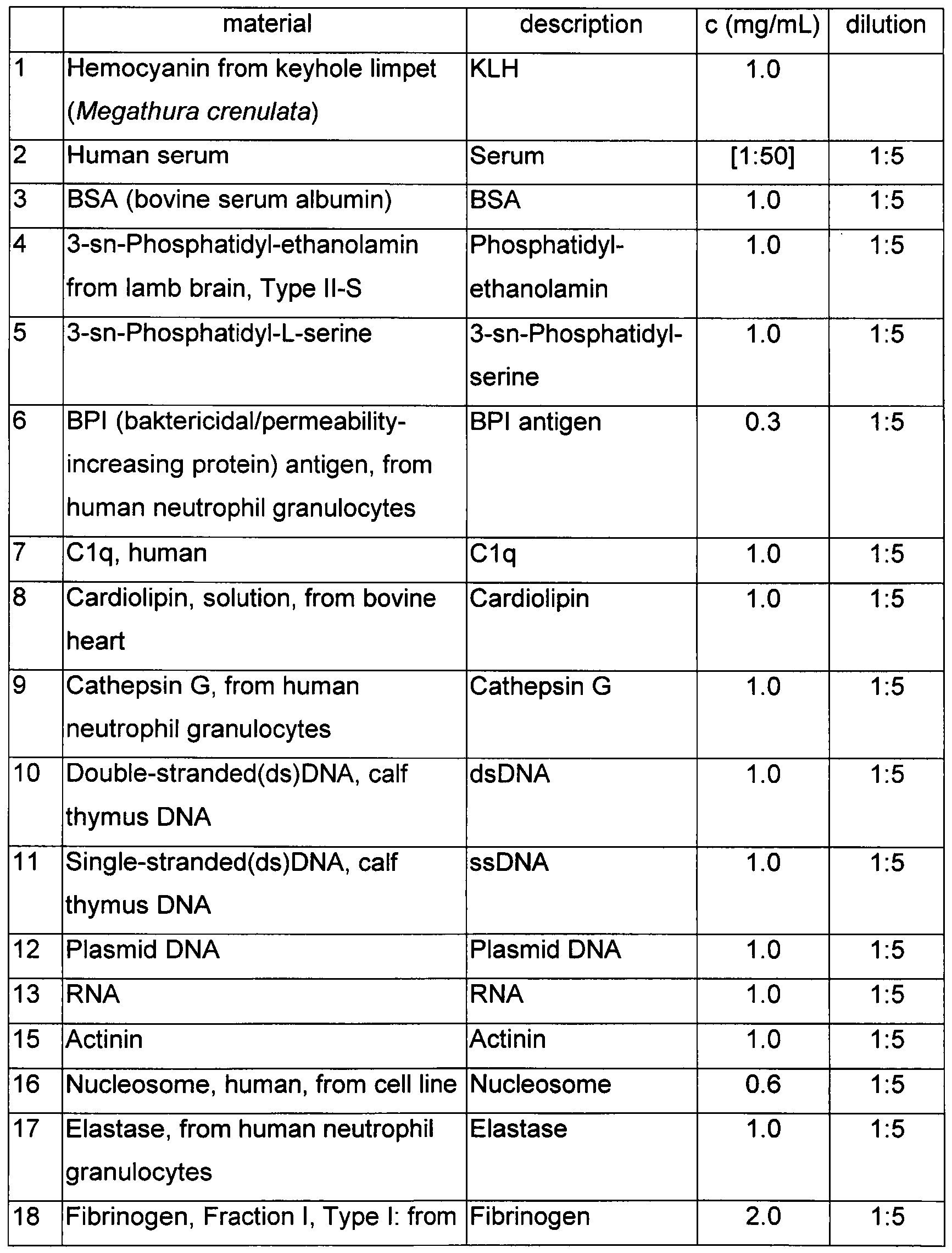


Example 7
Liquid flow changes in microcapillaries treated with various IgG subclasses, in the ' case of using whole blood
Background: The myeloid cells bind to lgG3, and not to lgG2 on the microarrays. By applying these two IgGs of different functions, it can be determined whether in arrays made from microcapillaries, cell adhesion changes the liquid flow rate or not.
Implementation:
1) Capillaries: a. Vitrotubes, round capillary, Cat No.: CV1525, 0.15IDx0.25 OD, Lot Code: 3140Z1 (www.vitrocom.com)
b. Vitrotubes, square capillary, Cat No.: 8505-050, 0.050IDxO.025mm Wall, Lot Code: 0990C1 (www.vitrocom.com)
2) Three capillaries of different assemblies are made (A, B, C), two from each for the parallel measurements (I and II).
A) Capillary without inner constriction
a. treatment with 0.25M EDTA for 30 minutes, drying
B) A capillary furnished with an inner constricting capillary and treated with nitrocellulose and lgG2
a. treatment with 0.25M EDTA for 30 minutes, drying
b. 0.2% Nitrocellulose solution (ethanol: diethyl-ether, 1 :1), 5 min, drying c. Immersion into 0.5mg/ml hlgG2 solution, for 20 minutes, drying (human lgG2, Sigma, I 5404-1 MG, 058K6116)
d. trimming 2cm at the edge, which will then be used for constriction
e. inserting the constricting element into the outer capillary
C) Capillary treated with nitrocellulose and lgG3, furnished with an inner constricting capillary
a. treatment with 0.25M EDTA for 30 minutes, drying
b. 0.2% nitrocellulose solution (ethanol: diethyl-ether, 1 :1), 5 min., drying c. Immersion into 0.5mg/ml hlgG3 solution, for 20 minutes, drying (human lgG3, Sigma I 5654-1 MG, 059K6007)
d. trimming 2cm at the edge, which will then be used for constriction
e. inserting the constricting element into the outer capillary
3) Put one drop of blood drawn from the fingertip into the narrowed inlet of the capillaries
results:
The results are shown in Figure 17. The running of sample 'B' started 7 seconds later than the running of sample 'Α'. The times given next to the figures apply to sample TV, the 's' stands for seconds and 'min' for minutes.
The results show that
- blood runs quickly along a capillary which does not have constriction, and
- blood proceeds faster in a capillary containing a constricting capillary treated with lgG2 than in a capillary comprising a constricting capillary treated with lgG3
Example 8
HBsAg test in capillaries Purpose of the experiment
Detecting an antigen-specific antibody response in a delayed flow test, in capillaries, using hepatitis B antigen
Materials
Slide: VWR ECN 631-1553
Embedding material for outer capillary: PDMS, Dow Corning, Sylgard 184
Outer capillary: Vitrocom CV1525
Inner capillary: Vitrocom 8505-050
Hepatitis antigen (HBsAg): HBV Surface Antigen adw protein; Biorbyt, orb82659
Bovine albumin: Sigma A-3059
Silane: Sigma-Aldrich, A3648
Preparation of outer capillaries
Six capillaries are arranged accurately side by side, leaving 5mm protruding ends for forming the inlet for the blood drops, and then 1ml PDMS solution is layered on the slide, uniformly distributing same on a horizontal surface, followed by baking at
80 °C for one hour.
Preparation of inner capillaries
One end of each capillary is sealed by gas flame.
The open end is immersed into 2% silane/acetone solution, dried, then immersed in water and dried again.
The open end is placed into HBsAg solution of 0.2mg/ml concentration at 3cm depth (plastic sheath of medical syringe) and in the meantime negative control capillaries are placed into 5% BSA solution in the same way, stored at 37°C for one hour, to form the antigen coating.
The unbound antigen is removed by immersing the capillary into the following solutions consecutively: PBS, PBS, 5% BSA solution in PBS, PBS, water. The capillary is dried.
Forming the capillary trap and reader
The open antigen coated ends of inner capillaries are inserted 2cm deep into the outer capillaries fixed to the slides. 3 inner capillaries coated with HBsAg antigen provide the measuring system, and 3 capillaries coated with BSA provide the reference negative control. The capillaries are fixed to one another by an adhesive tape, creating an inlet gate for the blood drop.
Measurement
Capillary blood drawn from fingertip is supplemented to 20mM EDTA concentration to prevent coagulation. 25 microliters of blood is pipetted into the inlet gate, uniformly distributing the blood on the six capillaries. The blood flow is tracked and registered in time.
The structure of HBsAg test is shown in Figure 18. In Figure 18 inner capillary channels 30 and 32 are divided into two groups, according to the Figure, in outer capillary channels 31 arranged on slide 34. In the present embodiment the outer surface of the inner capillary channels 30 is coated with HBsAg antigen, while that of the inner capillary channels 32 is coated with BSA. In Figure 18 spot 29 indicates the place where the sample, i.e. preferably blood drop has to be dripped.
Results
The distance covered by the blood flowing in the capillaries is shown in Table 3, measured from the end of the trap, in mm.
Table 3

The chart of the results of the experiment performed is shown in Figure 19.
Evaluation
In the capillaries coated with HBsAg antigen, the blood flow induced (moved) by capillary forces significantly slows down, when the blood of a subject having known HBsAg reactivity is used. The result of the experiment is shown in Figure 20.
Example 9
The effect of human lgG3, lgG2 and serum albumin on the blood flow in microcapillaries (Figure 14)
Purpose
The myeloid cells adhere to lgG3, but not to lgG2 and albumin, thus the experiment serves for proving that the blood flow rate changes as a result of cell adhesion.
Method
Microcapillaries:
a. Vitrotubes, round capillary, Cat No.: CV1525, 0.15IDx0.25OD, Lot Code: 3140Z1 (www. vitrocom . com)
b. Vitrotubes, square capillary, Cat No.: 8505-050,
0.050IDx0.025mm Wall, Lot Code: 0990C1 (www.vitrocom.com) The experiment was carried out as described in Example 7, with the difference that beyond lgG3 and lgG2, albumin was also used for coating an inner capillary, and that an empty outer capillary was not used.
Conclusion
An inner capillary coated with lgG3 slows down the blood flow in the outer capillary. The result of the experiment is shown in Figure 14. t Example 10
Purpose
Measuring the adhesion of neutrophil granulocytes on an antigen array Method
1) Antigen arrays comprising IgG subclasses, PBS and anti-human IgG antibodies (ahlgG) are made on a nitrocellulose surface
2) Whole blood or lysed blood freed from red blood cells is added to rehydrated arrays, the arrays are incubated for 1 hour at 37°C and the
adherent cells are labelled by anti-human CD66abce-FITC (Dako Cat.:F7112 Lot 00034553) antibodies.
After washing, the arrays were scanned by a laser reader.
Result
The neutrophil granulocytes adhere (bind) to the subclasses lgG3 and lgG1. The experimental result is shown in Figure 26.
Example 11
Retardation (slowing-down) of blood flow by surface-bound antibody in a microfluidic system (capillary channel system) Purpose:
Two microfluidic channels (capillary channels) differing in their coatings are compared to prove retardation of blood flow by an antibody that interacts with Fc receptors on neutrophil granulocytes Principle:
Two materials are compared in the test: bovine serum albumin (BSA) is inert with respect to FcR binding, serving as a negative control antigen, human lgG3 antibody is highly active at binding FcRs of cells, serving as positive control. Neutrophil granulocytes in the blood are expected to bind to lgG3, adhere to the channel wall, constrict passage through the microfluidic channel and thereby slow down the flow of blood.
Generation of the PDMS microfluidic structure
The macroscopic design for the microfluidic operational unit is shown in Figure 27, and the microscopic design thereof in Figure 28.
Experiments were conducted with channels that were 15 pm high. The devices were generated using soft lithography. PDMS (Sylgard 184 PDMS prepolymer, Dow Corning, Midland, Ml) was cast on the silicon disc master on which the positive relief was formed by SU8 photoresist. The PDMS was then cured at 80 °C in a convection oven for 60 minutes. The cured PDMS was peeled off the master, cut to appropriate size to fit the Nexterion slides.
Preparation of the surface serving as a basis for the microfluidic system
1) Nexterion 1 pad slide is inserted into ProPlate 16 well chamber
Slide: SCHOTT Nexterion Slide H; Hydrogel coated;
CatNo: 1070936; LotNo:4-2882
Arrangement is shown in Figure 30A. Only chambers marked with numbers 3, 4, 11 and 12 (shown in figure 30A with median-grey and dark-grey colouring) were treated, the others were left empty. First the chamber was rinsed with water then air pressure was used to remove liquid.
2) Chambers were filled with 100μΙ of solution, and incubated at 37°C for 1h
Sub-arrays 4&11 : 50pg/ml lgG3
Sub-arrays 3&12: 50 g/ml BSA
lgG3 was first dialyzed against PBS, the stock concentration was
0.78mg/ml
Manufacturer Cat.No. Lot.No. Full name
Sigma A3059- 100M1767V Albumin, bovine serum, fraction V,
100G Approx. 99% Protease-free,
Essentially gamma-globulin free Sigma 15654-1 MG 091 M6211 HUMAN IGG3, KAPPA, FROM
HUMAN PLASMA
3) Washing 2x5min PBS (in 16 pad chambers) 100pl/each chamber
4) Disassembly of chambers
5) Blocking: incubation in 100mM Tris (pH8) at room temperature for 1h
6) Washing 3x5min PBS
7) Rinsing with MilliQ water then centrifugation to remove liquid
Mounting of the microfluidic system (capillary channel system)
1) capillary system:
i. PDMS block (14.2 m height) is air plasma treated
800W microwave, vacuum<1mBar).
2) Blood (collected in two different ways):
i. 5x diluted-blood
1. Blood was collected into eppendorf tube that
already contained PBS until 5x dilution was reached ii. Citrate-blood
1. Blood was collected (drop by drop) into eppendorf tube that already contained 3.8% Na-citrate solution (ratio of blood and 3.8% sodium-citrate solution:
9:1)
3) Capillary channel system was prepared on the day of the measurement and was incubated for 2h at room temperature before 3μΙ blood was added to it.
Slide Part I Part II
10067683 5xdiluted-blood (Cap1) Citrate-blood (Cap2)
Capillary channel system 1 (Cap1)
- The flow over the BSA-coated chamber was faster, it was less than 3min
The result obtained with Capillary channel system 1 is shown in Figure 30B.
Capillary channel system 2 (Cap2):
- The flow over the BSA-coated chamber was faster, it was less than 3min
The result obtained with Capillary channel system 2 is shown in Figure 30C. f Blood flow reaching the junction of the two channels effectively stops liquid flow the other channel.
Example 12
Testing human neutrophil binding to BSA-lgG3-coated PDMS device Method:
1) Materials:
Materials of concentration of 0.5mg/ml were used for coating, dilution was carried out in PBS.
Note: The lgG3 was dialyzed against PBS
Manufacturer Cat.No. Full name
BSA Sigma A3059-100G Albumin, bovine serum
human lgG3 Sigma HUMAN IGG3, KAPPA, FROM
15654-1 MG HUMAN PLASMA
2) Chip map:
Nexterion slides were coated (SCHOTT Nexterion Slide H; Hydrogel coated; CatNo: 1070936; LotNo: 4-2882). According to Figure 30D, to the areas indicated by arrow 3-3μΙ of BSA solution and lgG3 solution,
respectively, were added by pipette.
3) Protein immobilization: Incubation at 37°C for 30min
4) Blocking: Incubation in 100mM Tris (pH8) at room temperature for 1h
5) Washing 3x5min in PBS, rinsing in Milli Q water and centrifuging for 1min to dry
6) PDMS blocks with 14μηη height channels were air plasma treated and
inserted onto the coated slides. A coated slide is shown in Figure 30H.
7) Incubation in humidified chamber at room temperature for overnight.
8) Neutrophil granulocytes:
a. Neutrophils were isolated on Ficoll gradient from human whole blood. b. 1.5*107 cell/ml concentration was set in RMPI 10% FCS medium. 9) 3μΙ of cell suspenion was introduced to the sample gate of the microfluidic device (capillary channel system) and the events were checked under microscope.
Results:
Human neutrophil granulocytes bind to the lgG3-coated section of the capillary channel. The result observed by microscope is shown in Figure 30E.
Example 13
U937 BSA-lqG3
Figure 30F: capillary coated with lgG3
Figure 30G: capillary coated with BSA
Testing U937 cell binding to BSA-lgG3 coated PDMS device Method:
1) Materials:
Materials of concentration of 0.5mg/ml were used for coating, dilution was carried out in PBS.
NOTE: The lgG3 was dialyzed against PBS
Manufacturer Cat. No. Full name
BSA Sigma A3059- Albumin, bovine serum
100G
human Sigma HUMAN IGG3, KAPPA, FROM lgG3 15654-1 MG HUMAN PLASMA
2) Chip map:
Nexterion slides were coated (SCHOTT Nexterion Slide H; Hydrogel coated; CatNo: 1070936; LotNo: 4-2882). According to Figure 30D, to the areas indicated by arrow 3-3μΙ of BSA solution and lgG3 solution, respectively, were added by pipette.
3) Protein immobilization: Incubation at 37°C for 30min
4) Blocking: Incubation in 100mM Tris (pH8) at room temperature for 1h
5) Washing 3x5min in PBS, rinsing in Milli Q water and centrifuging for 1min to dry
6) PDMS blocks with 1 pm height channels were air plasma treated and
inserted onto the coated slides. A coated slide is shown schematically in Figure 30H.
7) Incubation in humidified chamber at room temperature for 2h.
8) Preparation of a suspension of a concentration of 5*106 U937 cell/ml in RMPI 10% FCS medium.
9) 3μΙ of cell suspension was introduced to the sample gate of the microfluidic device (capillary channel system) and the events were checked under microscope.
Results:
U937 cells bind to the lgG3-coated section of the capillary channel. The results are shown in Figures 30F and 30G. In Figure 30F microscopic image of the section coated by lgG3 of the capillary channel is shown. Large number of U937 cells were bound (adhered) to the section analysed. In Figure 30G microscopic image of the section coated by BSA of the capillary channel is shown.
In the section analysed the U937 cells do not adhere to the extent as on the section coated by lgG3 of the capillary channel.
Claims
1. A capillary channel, characterised by comprising a trap section (1 , 22, 38, 40, 42, 44, 46, 47, 48) having a coating, and further comprising a collector section (2, 20) having a volume which is at least as large as that of the trap section (1, 22, 38, 40, 42, 44, 46, 47, 48), and a reader section (3, 18) adjacent to the collector section (2, 20), wherein the reader section (3, 18) optionally comprises one or more physical and/or chemical sensors adapted for measuring a flow rate and/or one or more markings for visual reading of a liquid front position, and wherein the collector section (2, 20) is located in the part between the trap section (1 , 22, 38, 40, 42, 44, 46, 47, 48) and the reader section (3, 18) of the capillary channel.
2. The capillary channel according to claim 1 , wherein the coating comprises an antigen.
3. The capillary channel according to claim 1 or 2, characterised in that the volume of the collector section (2, 20) is as large as that of the trap section (1 , 22, 38, 40, 42, 44, 46, 47, 48).
4. The capillary channel according to any of claims 1 to 3, characterised in that the trap section (1 , 22, 38, 40, 42, 44, 46, 47, 48) comprises at least one passage having a traversable cross-sectional area with a characteristic size of 1 to 50 micrometer.
5. The capillary channel according to claim 4, characterised in that the characteristic size is 20 micrometers or less.
6. The capillary channel according to claim 4 or 5, characterised in that the trap section (1 , 22, 38, 40, 42, 44, 46, 47, 48) comprises a constricting element, and the traversable cross-sectional area of the at least one passage is formed by means of the at least one constricting element.
7. The capillary channel according to any of claims 1 to 6, characterised in that the reader section (3, 18) has a transparent wall.
8. A capillary channel system, which comprises a measuring channel (106), which measuring channel (106) is a capillary channel according to any of claims 1 to 7, wherein the coating comprises an antigen, and the system comprises a negative control channel (10, 104) and optionally a positive control channel (16), and optionally a sample distributing part (102) which communicates with the channels.
9. The capillary channel system according to claim 8, characterised in that the negative control channel (10, 104) has a structural configuration identical with that of the measuring channel (106) and it is provided with a coating which differs from that of the measuring channel (106) or it is formed without coating.
10. The capillary channel system according to claim 8 or 9, characterised by comprising a negative control channel (10, 04) which - in case of flowing the same sample - in its part downstream from the trap section (1 , 22, 38, 40, 42, 44, 46, 47, 48) provides identical or higher flow rate than that in the measuring channel.
11. The capillary channel system according to any of claims 8 to 10, characterised by comprising a positive control channel (16) having a structural configuration identical with that of the measuring channel (106) and comprising a coating which differs from that of the measuring channel.
12. The capillary channel system according to any of claims 8 to 11 , characterised by comprising a positive control channel (16), which - in case of flowing the same sample - in its part downstream from the trap section (1, 22, 38, 40, 42, 44, 46, 47, 48) provides identical or lower flow rate than that in the measuring channel.
13. The capillary channel system according to any of claims 8 to 12, characterised by comprising more than one measuring channels of identical or different structural configurations.
14. The capillary channel system according to any of claims 8 to 13, characterised by comprising more than one negative control channels of identical or different structural configurations.
15. The capillary channel system according to any of claims 8 to 14, characterised by comprising more than one positive control channels of identical or different structural configurations.
16. The capillary channel system according to any of claims 8 to 15, characterised by comprising a single measuring channel (106) and a single negative control channel (104), and the end points of the measuring channel and the negative control channel (104) are connected to a common draining capillary channel.
17. A method for the analysis of immunoreactivity comprising introducing a sample into a capillary channel system according to any of claims 8 to 16, and after the liquid front of the sample in the negative control channel having reached the reader section, and, preferably, after both the liquid front of the sample in the antigen-containing capillary channel(s) and the liquid front of the sample in the negative control channel having reached the reader section, observing or measuring on the reader sections of the individual channels a feature of the flow of the sample, preferably the flow rate of the sample or a quantity proportional thereto.
18. A method for the analysis of immunoreactivity on an antigen array, comprising
contacting with a sample an antigen array comprising one or more antigens and optionally one or more reference substances; and
detecting by cells the complexes formed by the one or more antigens and by one or more components of the sample which may be captured by the antigens and, wherein for the detecting by cells the sample serves as a source of the cells, or the cells are from a source other than the sample.
19. The method according to claim 18, wherein the detecting by cells comprises contacting the array with cells.
20. The method according to claim 18 or 19, wherein the detecting by cells comprises one or more of the following steps i) to v), thereby obtaining data for the areas of the individual features of the array:
i) revealing the cells adhered on the area of the individual features of the antigen array,
ii) determining the amount of cells adhered on the area of the individual features of the antigen array,
iii) measuring or observing a quantity proportional to the amount of cells adhered on the area of the individual features of the antigen array,
iv) measuring cell adhesion on the area of the individual features of the antigen array,
v) measuring cell activation of the cells adhered on the area of the individual features of the antigen array.
21. The method according to any of claims 17 to 20, wherein the analysis of immunoreactivity comprises antibody test or immune profiling.
22. A kit comprising an antigen array, said antigen array comprises one or more antigens and optionally one or more reference substances; and the kit optionally further comprises a cellular detection agent.
23. The method according to any of claims 18 to 21 , or the kit according to claim 22, wherein the antigen array is an antigen microarray.
24. The method according to any of claims 18 to 21 , or the kit according to claim 22 or 23, wherein the antigen array is a capillary channel system according to any of claims 8 to 16.
25. The method according to any of claims 18 to 21 , or the kit according to claim 22 or 23, wherein the antigen array comprises a mixture of distinguishable beads.
26. The kit according to any of claims 22 to 25, which is a diagnostic kit, preferably for use in the diagnosis of a condition accompanied by the presence of one or more specific antibodies.
27. The method, kit, capillary channel or capillary channel system according to any of the preceding claims, wherein the one or more antigens are selected from the group consisting of parasites, microbes, viruses, prions, components thereof, proteins, peptides, glycoproteins, lipoproteins, lipids, glycolipids, nucleic acids, carbohydrates, small molecules, allergens and autoantigens.
28. Use of the method or kit according to any of claims 17 to 27, or of the capillary channel system according to any of claims 8 to 16 in the diagnosis of a condition or a disorder accompanied by the presence of one or more specific antibodies, preferably in the diagnosis of systemic lupus erythematosus, hepatitis B infection or immunity.
29. The method according to claim 17, wherein the analysis of immunoreactivity is aimed at the diagnosis of a condition or disorder accompanied by the presence of one or more specific antibodies, and the trap section of at least one measuring channel comprises an antigen capable to bind to at least one of said specific antibodies, and the method optionally further comprises comparing the flow features observed or measured in the individual channels, wherein the flow features are preferably the flow rates.
30. The method according to any of claims 18 to 21 or 23 to 28, wherein the analysis of immunoreactivity is aimed at the diagnosis of a condition or disorder accompanied by the presence of one or more specific antibodies, and the array comprises at least one antigen capable to bind to said one or more specific antibodies, and optionally the method further comprises comparing the data observed or measured for the areas of the individual antigens and for the areas of the individual reference substances.
31. The capillary channel, capillary channel system, method or kit according to any of the preceding claims, wherein the one or more antigens are selected from those listed in Table 1.
32. The method, kit or capillary channel system according to any of claims 10 to 31 , wherein the sample comprises at least one component that may be captured by the antigens present in the antigen array, and said at least one component is selected from the group consisting of immunoreactive components, preferably antibodies and complement components.
33. The method, kit or capillary channel system according to any of claims 10 to 32, wherein the sample is selected from the group consisting of biological samples, such as clinical samples, whole blood, serum, plasma, biological fluid, cultured cells, cell supernatants, cell lysates, and pure or enriched samples derived from any of these.
34. The method, kit or capillary channel system according to any of claims 10 to 33, wherein the sample is whole blood.
35. Use of cells for the analysis of immunoreactivity on an antigen array, for the detection of immune complexes on an antigen array, or for the qualitative and/or semi-quantitative analysis of the composition of immune complexes on an antigen array.
36. The method, kit or use according to any of claims 18 to 35, wherein the cells are selected from the group consisting of adherent cells such as monocytes, macrophages, dendritic cells and neutrophil granulocytes.
37. The method, kit or use according to any of claims 18 to 36, wherein the cells are detectably labelled cells.
38. The method, kit or use according to any of claims 18 to 37, wherein the antigen array contains reference substances to which detection cells adhere.
39. The method according to any of claims 18 to 21 , 23 to 25, 27 to 28, 30 to 34 or 36 to 38, wherein the cells are detectably labelled cells and the detecting by cells comprises measuring the amount of cells that adhered to immune complexes formed by the components of the sample and the antigens of the antigen array, or measuring a physico-chemical attribute proportional to said amount.
40. The method according to any of claims 18 to 21 , 23 to 25, 27 to 28, 30 to 34 or 36 to 38, wherein the detecting by cells, the step of revealing the adhered cells, the determination of the number of cells adhered and/or the measurement of the activation of adhered cells comprises measuring cellular response, wherein the cellular response is preferably selected from: production of reactive intermediers, increase in intracellular ion concentrations, phosphorylation of signaling molecules, and altered expression of surface markers, all induced by the contact between detection cells and immune complexes formed by components of the sample and antigens of the antigen array.
41. The method according to any of claims 18 to 21 , 23 to 25, 27 to 28, 30 to 34 or 36 to 38, wherein the detecting by cells, the step of revealing the adhered cells, the determination of the number of cells adhered and/or the measurement of the activation of adhered cells comprises measuring cellular responses using genetically engineered cells, chemically modified cells, primary cells isolated from a donor or immortalised cell lines.
42. The method according to any of claims 18 to 21 , 23 to 25, 27 to 28, 30 to 34, 36, 38 or 40 to 41 , wherein the detecting by cells, the step of revealing the adhered cells, the determination of the number of cells adhered and/or the measurement of the activation of adhered cells comprises a labelling-free measurement of cell adhesion or cell activation through exploiting physical, optical, electrical, mass or magnetic alterations induced by the adhesion or activation of the detection cell.
43. The method, kit, capillary channel, capillary channel system or use according to any of the preceding claims, wherein the antigen is other than connective tissue protein and other than superantigen.
44. Microfluidic device comprising a capillary channel according to any of claims 1 to 7, or a capillary channel system according to any of claims 8 to 16, 27 or 31 to 34, wherein the capillary channel or the capillary channel system is formed in the material or materials constituting the device.
45. Use of the capillary channel according to any of claims 1 to 7, the capillary channel system according to any of claims 8 to 16, 27, 31 to 34 or 43, or the microfluidic device according to claim 44 as rapid diagnostic device for point-of- care serological tests.
Priority Applications (1)
| Application Number | Priority Date | Filing Date | Title |
|---|---|---|---|
| EP14792541.6A EP3030903B1 (en) | 2013-08-07 | 2014-08-07 | Method for the analysis of immunoreactivity, and a device suitable for carrying out the method |
Applications Claiming Priority (2)
| Application Number | Priority Date | Filing Date | Title |
|---|---|---|---|
| HUP1300474 | 2013-08-07 | ||
| HU1300474A HUP1300474A2 (en) | 2013-08-07 | 2013-08-07 | Method for analyzing immunological reactivity and device therefor |
Publications (3)
| Publication Number | Publication Date |
|---|---|
| WO2015019120A2 true WO2015019120A2 (en) | 2015-02-12 |
| WO2015019120A3 WO2015019120A3 (en) | 2015-07-09 |
| WO2015019120A8 WO2015019120A8 (en) | 2016-02-18 |
Family
ID=89991221
Family Applications (1)
| Application Number | Title | Priority Date | Filing Date |
|---|---|---|---|
| PCT/HU2014/000068 WO2015019120A2 (en) | 2013-08-07 | 2014-08-07 | Method for the analysis of immunoreactivity, and a device suitable for carrying out the method |
Country Status (3)
| Country | Link |
|---|---|
| EP (1) | EP3030903B1 (en) |
| HU (1) | HUP1300474A2 (en) |
| WO (1) | WO2015019120A2 (en) |
Cited By (2)
| Publication number | Priority date | Publication date | Assignee | Title |
|---|---|---|---|---|
| WO2020005455A1 (en) * | 2018-06-29 | 2020-01-02 | Abbott Point Of Care Inc. | Cartridge device with bypass channel for mitigating drift of fluid samples |
| US10816550B2 (en) | 2012-10-15 | 2020-10-27 | Nanocellect Biomedical, Inc. | Systems, apparatus, and methods for sorting particles |
Citations (6)
| Publication number | Priority date | Publication date | Assignee | Title |
|---|---|---|---|---|
| US5545806A (en) | 1990-08-29 | 1996-08-13 | Genpharm International, Inc. | Ransgenic non-human animals for producing heterologous antibodies |
| US5569825A (en) | 1990-08-29 | 1996-10-29 | Genpharm International | Transgenic non-human animals capable of producing heterologous antibodies of various isotypes |
| US5625126A (en) | 1990-08-29 | 1997-04-29 | Genpharm International, Inc. | Transgenic non-human animals for producing heterologous antibodies |
| US6140045A (en) | 1995-03-10 | 2000-10-31 | Meso Scale Technologies | Multi-array, multi-specific electrochemiluminescence testing |
| US6197599B1 (en) | 1998-07-30 | 2001-03-06 | Guorong Chin | Method to detect proteins |
| US20070054326A1 (en) | 2002-11-07 | 2007-03-08 | Ruo-Pan Huang | Antibody-based protein array system |
Family Cites Families (5)
| Publication number | Priority date | Publication date | Assignee | Title |
|---|---|---|---|---|
| US5204525A (en) * | 1985-08-05 | 1993-04-20 | Biotrack | Capillary flow device |
| GB0116384D0 (en) * | 2001-07-04 | 2001-08-29 | Diagnoswiss Sa | Microfluidic chemical assay apparatus and method |
| GB0306098D0 (en) * | 2003-03-18 | 2003-04-23 | Platform Diagnostics Group Ltd | Sample testing device |
| GB0806771D0 (en) * | 2008-04-12 | 2008-05-14 | Spd Swiss Prec Diagnostics Gmb | Assay devices comprising bubble-forming means |
| EP2208531A1 (en) * | 2008-12-30 | 2010-07-21 | Atonomics A/S | Distribution of particles in capillary channel by application of magnetic field |
-
2013
- 2013-08-07 HU HU1300474A patent/HUP1300474A2/en unknown
-
2014
- 2014-08-07 WO PCT/HU2014/000068 patent/WO2015019120A2/en active Application Filing
- 2014-08-07 EP EP14792541.6A patent/EP3030903B1/en active Active
Patent Citations (6)
| Publication number | Priority date | Publication date | Assignee | Title |
|---|---|---|---|---|
| US5545806A (en) | 1990-08-29 | 1996-08-13 | Genpharm International, Inc. | Ransgenic non-human animals for producing heterologous antibodies |
| US5569825A (en) | 1990-08-29 | 1996-10-29 | Genpharm International | Transgenic non-human animals capable of producing heterologous antibodies of various isotypes |
| US5625126A (en) | 1990-08-29 | 1997-04-29 | Genpharm International, Inc. | Transgenic non-human animals for producing heterologous antibodies |
| US6140045A (en) | 1995-03-10 | 2000-10-31 | Meso Scale Technologies | Multi-array, multi-specific electrochemiluminescence testing |
| US6197599B1 (en) | 1998-07-30 | 2001-03-06 | Guorong Chin | Method to detect proteins |
| US20070054326A1 (en) | 2002-11-07 | 2007-03-08 | Ruo-Pan Huang | Antibody-based protein array system |
Non-Patent Citations (18)
| Title |
|---|
| "Current Protocols in Immunology", 1995, WILEY |
| BELOV,L.; I.DE, V; C.G.DOS REMEDIOS; S.P.MULLIGAN; R.I.CHRISTOPHERSON: "Immunophenotyping of leukaemias using a cluster of differentiation antibody microarray", CANCER RES, vol. 61, 2001, pages 4483 - 4489 |
| BRINKMANN, V. ET AL.: "Neutrophil extracellular traps kill bacteria", SCIENCE, vol. 303, 2004, pages 1532 - 1535, XP002469080, DOI: doi:10.1126/science.1092385 |
| CLIME, L. ET AL.: "Self-priming of liquids in capillary autonomous microfluidic systems", MICROFLUID NANOFLUID, vol. 12, 2012, pages 371 |
| EHLERS,M.R.: "CR3: a general purpose adhesion-recognition receptor essential for innate immunity", MICROBES INFECT., vol. 2, 2000, pages 289 - 294, XP002207262, DOI: doi:10.1016/S1286-4579(00)00299-9 |
| HARLOW; LANE: "Antibodies: A Laboratory Manual", 1989, COLD SPRING HARBOR |
| J.PRECHL; K.PAPP; A.ERDEI: "Antigen microarrays: descriptive chemistry or functional immunomics?", TRENDS IMMUNOL., vol. 31, 2010, pages 133 - 137, XP026989438 |
| JUNCKER, D. ET AL.: "Autonomous microfluidic capillary system", ANAL. CHEM., vol. 74, 2002, pages 6139 - 6144 |
| KOHLER ET AL., NATURE, vol. 256, 1975, pages 495 - 97 |
| LEGENDRE, BIOTECHNIQUES, vol. 9, 1990, pages 788 - 805 |
| MEZZASOMA ET AL., CLIN. CHEM., vol. 48, 2002, pages 121 - 130 |
| NIMRICHTER,L.; A.GARGIR; M.GORTLER; R.T.ALTSTOCK; A.SHTEVI; O.WEISSHAUS; E.FIRE; N.DOTAN; R.L.SCHNAAR: "Intact cell adhesion to glycan microarrays", GLYCOBIOLOGY, vol. 14, 2004, pages 197 - 203 |
| OKOCHI,M.; S.NOMURA; C.KAGA; H.HONDA: "Peptide array-based screening of human mesenchymal stem cell-adhesive peptides derived from fibronectin type III domain", BIOCHEM BIOPHYS RES COMMUN, vol. 371, 2008, pages 85 - 89, XP022639251, DOI: doi:10.1016/j.bbrc.2008.04.019 |
| SHALON ET AL., GENOME RESEARCH, vol. 6, 1996, pages 639 - 645 |
| SOEN,Y.; D.S.CHEN; D.L.KRAFT; M.M.DAVIS; P.O.BROWN: "Detection and characterization of cellular immune responses using peptide-MHC microarrays", PLOS BIOL, vol. 1, 2003, pages E65, XP002380731, DOI: doi:10.1371/journal.pbio.0000065 |
| VIDARSSON,G.; J.G.VAN DE WINKEL: "Fc receptor and complement receptor-mediated phagocytosis in host defence", CURR. OPIN. INFECT. DIS., vol. 11, 1998, pages 271 - 278 |
| ZIMMERMANN M ET AL.: "Valves for autonomous capillary systems", MICROFLUID NANOFLUID, vol. 5, 2008, pages 395, XP055455029, DOI: doi:10.1007/s10404-007-0256-2 |
| ZIMMERMANN, M. ET AL.: "Capillary pumps for autonomous capillary systems", LAB CHIP, vol. 7, 2007, pages 119, XP055000157, DOI: doi:10.1039/B609813D |
Cited By (5)
| Publication number | Priority date | Publication date | Assignee | Title |
|---|---|---|---|---|
| US10816550B2 (en) | 2012-10-15 | 2020-10-27 | Nanocellect Biomedical, Inc. | Systems, apparatus, and methods for sorting particles |
| WO2020005455A1 (en) * | 2018-06-29 | 2020-01-02 | Abbott Point Of Care Inc. | Cartridge device with bypass channel for mitigating drift of fluid samples |
| CN112638532A (en) * | 2018-06-29 | 2021-04-09 | 雅培医护站股份有限公司 | Cartridge device with bypass channel for mitigating drift of fluid sample |
| US11198123B2 (en) | 2018-06-29 | 2021-12-14 | Abbott Point Of Care Inc. | Cartridge device with bypass channel for mitigating drift of fluid samples |
| CN112638532B (en) * | 2018-06-29 | 2023-08-11 | 雅培医护站股份有限公司 | Cartridge device with bypass channel for reducing fluid sample drift |
Also Published As
| Publication number | Publication date |
|---|---|
| EP3030903B1 (en) | 2019-02-20 |
| EP3030903A2 (en) | 2016-06-15 |
| WO2015019120A3 (en) | 2015-07-09 |
| HUP1300474A2 (en) | 2015-03-02 |
| WO2015019120A8 (en) | 2016-02-18 |
Similar Documents
| Publication | Publication Date | Title |
|---|---|---|
| US9176152B2 (en) | Methods and apparatuses for detection of positional freedom of particles in biological and chemical analyses and applications in immunodiagnostics | |
| JP7403447B2 (en) | Cartridges and systems for body fluid analysis | |
| US9395365B2 (en) | Detection of infectious disease in a human or animal by measuring specific phagocytosis in a thin film sample of their anticoagulated blood | |
| US10408842B2 (en) | Subcellular western blotting of single cells | |
| WO2019196270A1 (en) | Micro-nano particles detection system and method thereof | |
| JP6116701B2 (en) | Method and apparatus for monitoring sedimentation parameters of a fluid medium sample | |
| WO2020160472A1 (en) | High-sensitivity assay | |
| AU2007298839B2 (en) | Blood typing | |
| EP3030903B1 (en) | Method for the analysis of immunoreactivity, and a device suitable for carrying out the method | |
| US20080280378A1 (en) | Grading of Immune Responses | |
| WO2017009926A1 (en) | Fixing method for specific binding substance | |
| US20230221319A1 (en) | A Method, A System, An Article, A Kit And Use Thereof For Biomolecule, Bioorganelle, Bioparticle, Cell And Microorganism Detection | |
| US20210025866A1 (en) | Devices, Systems, and Methods for Cell Analysis in Microgravity | |
| US20030044879A1 (en) | Method and device for forming a concentration gradient for chemotactic evaluation through use of laminar flow | |
| WO2006075965A1 (en) | A method for detecting an at least bivalent analyte using two affinity reactants | |
| Peter et al. | Lab-on-a-Chip proteomic assays for psychiatric disorders | |
| CA2938935C (en) | Crossmatching blood samples | |
| JP2017508961A (en) | Red blood cell detection | |
| JPH04208836A (en) | Reaction container kit | |
| WO2017130829A1 (en) | Microarray, method for manufacturing microarray, inspection method, and inspection kit | |
| US20210172943A1 (en) | Membrane carrier for liquid sample test kit, liquid sample test kit, and membrane carrier | |
| RU86090U1 (en) | BIOCHIP FOR DETERMINING SURFACE CELL MOLECULES | |
| EP1497652A2 (en) | Method, system and kit for detecting an analyte in a sample |
Legal Events
| Date | Code | Title | Description |
|---|---|---|---|
| 121 | Ep: the epo has been informed by wipo that ep was designated in this application |
Ref document number: 14792541 Country of ref document: EP Kind code of ref document: A2 |
|
| NENP | Non-entry into the national phase |
Ref country code: DE |
|
| WWE | Wipo information: entry into national phase |
Ref document number: 2014792541 Country of ref document: EP |