本発明の方法で対象となる患者は、抗CD4抗体を有効成分とする抗がん剤、抑制性の免疫チェックポイント分子に対するアンタゴニストを有効成分とする抗がん剤、及び共刺激性の免疫チェックポイント分子に対するアゴニストを有効成分とする抗がん剤から選択される少なくとも1種の抗がん剤の投与によるがん療法を受けている患者である。以下、本明細書において、抗CD4抗体を有効成分とする抗がん剤を「抗CD4抗がん剤」ということがある。また、抑制性の免疫チェックポイント分子に対するアンタゴニストを有効成分とする抗がん剤、及び共刺激性の免疫チェックポイント分子に対するアゴニストを有効成分とする抗がん剤を、「免疫チェックポイント抗がん剤」と呼ぶことがある。
Patients targeted by the method of the present invention are anticancer agents containing an anti-CD4 antibody as an active ingredient, anticancer agents containing an antagonist to an inhibitory immune checkpoint molecule as an active ingredient, and costimulatory immune check. It is a patient who has undergone cancer therapy by administration of at least one anticancer agent selected from anticancer agents having an agonist for a point molecule as an active ingredient. Hereinafter, in the present specification, an anticancer agent containing an anti-CD4 antibody as an active ingredient may be referred to as an “anti-CD4 anticancer agent”. In addition, an anticancer agent containing an antagonist for an inhibitory immune checkpoint molecule as an active ingredient and an anticancer agent containing an agonist for a costimulatory immune checkpoint molecule as an active ingredient are referred to as “immune checkpoint anticancer agent”. Sometimes referred to as “agent”.
本発明の方法では、上記した少なくとも1種の抗がん剤の投与を受けた患者由来の試料を用いて、以下の(1)~(3)の細胞表面分子のT細胞上での発現を調べる。
(1) 少なくとも1種の免疫チェックポイント受容体
(2) CD8
(3) CD44及びCD45ROからなる群より選択される少なくとも1種の細胞表面分子
In the method of the present invention, expression of the following cell surface molecules (1) to (3) on T cells is performed using a sample derived from a patient who has been administered at least one anticancer agent. Investigate.
(1) At least one immune checkpoint receptor
(2) CD8
(3) At least one cell surface molecule selected from the group consisting of CD44 and CD45RO
(1)の免疫チェックポイント受容体が陽性であり、CD8陽性であり、かつ、CD44高発現及び/又はCD45RO高発現であるT細胞集団の誘導が検出された場合、当該患者において抗がん剤が治療効果を現していると判定することができる。なお、T細胞は全てCD3を発現しているので、本発明の方法において抗がん剤の治療効果の指標となるT細胞集団も、当然ながらCD3陽性である。
When the immune checkpoint receptor of (1) is positive, is CD8 positive, and induction of a T cell population that is high in CD44 and / or high in CD45RO is detected, an anticancer drug in the patient Can be determined to have a therapeutic effect. Since all T cells express CD3, the T cell population that serves as an index of the therapeutic effect of the anticancer agent in the method of the present invention is naturally CD3 positive.
「免疫チェックポイント分子」という語には、免疫チェックポイントとして機能する受容体とリガンドの両者が包含される。免疫チェックポイントとは、免疫系が自己の体を攻撃しないための免疫逃避機構である。T細胞上には免疫チェックポイント受容体が存在し、抗原提示細胞上に発現している免疫チェックポイントリガンドと相互作用する。T細胞はMHC分子上に提示された抗原を認識して活性化し、免疫反応を起こすが、並行して生じる免疫チェックポイント受容体-リガンドの相互作用によりT細胞の活性化が調節を受ける。免疫チェックポイント受容体には共刺激性のものと抑制性のものがあり、両者のバランスによってT細胞の活性化及び免疫反応が調節を受けている。
The term “immune checkpoint molecule” includes both receptors and ligands that function as immune checkpoints. An immune checkpoint is an immune escape mechanism that prevents the immune system from attacking its own body. There are immune checkpoint receptors on T cells that interact with immune checkpoint ligands expressed on antigen-presenting cells. T cells recognize and activate the antigen presented on the MHC molecule and cause an immune response, but the activation of T cells is regulated by the immune checkpoint receptor-ligand interaction that occurs in parallel. Immune checkpoint receptors are co-stimulatory and suppressive, and the balance between the two regulates T cell activation and immune response.
免疫チェックポイント抗がん剤の対象分子が免疫チェックポイント受容体である場合、(1)の免疫チェックポイント受容体は、当該抗がん剤が対象とする分子と同一であってもよいし、異なっていてもよい。
When the target molecule of the immune checkpoint anticancer agent is an immune checkpoint receptor, the immune checkpoint receptor of (1) may be the same as the molecule targeted by the anticancer agent, May be different.
(1)の免疫チェックポイント受容体の具体例としては、PD-1、CD137、TIM-3、CTLA-4、BTLA、LAG-3、OX40、及びGITRからなる群より選択される少なくとも1種を挙げることができる。好ましい具体例として、PD-1、CD137及びTIM-3からなる群より選択される少なくとも1種を挙げることができる。上記のうちのいずれか2種以上、又は3種以上の発現を調べてもよい。(1)の免疫チェックポイント受容体はいずれも、陽性である場合に治療効果ありと判定することができる。CD137はヒトにおいても腫瘍反応性CD8+ T細胞に発現することが報告されており、またTIM-3はT細胞刺激時にCD137と同様の発現動態をとることが報告されている。従って、本発明では、CD137と同様にTIM-3も(1)の免疫チェックポイント受容体の好ましい具体例として挙げることができる。なお、当該分野におけるこれまでの報告はすべてがん組織へ浸潤した細胞の解析であり、治療効果を血中レベルで明確に表現できるという報告はない。
Specific examples of the immune checkpoint receptor of (1) include at least one selected from the group consisting of PD-1, CD137, TIM-3, CTLA-4, BTLA, LAG-3, OX40, and GITR. Can be mentioned. Preferable specific examples include at least one selected from the group consisting of PD-1, CD137 and TIM-3. Any two or more of the above, or three or more of the expression may be examined. If any of the immune checkpoint receptors in (1) is positive, it can be determined that there is a therapeutic effect. CD137 is also reported to be expressed in tumor-reactive CD8 + T cells in humans, and TIM-3 has been reported to have the same expression kinetics as CD137 upon T cell stimulation. Therefore, in the present invention, like CD137, TIM-3 can also be mentioned as a preferred specific example of the immune checkpoint receptor of (1). In addition, all the reports so far in this field are analysis of cells infiltrating cancer tissues, and there is no report that the therapeutic effect can be clearly expressed at the blood level.
マウスのCD3+、CD8+かつCD44hiのT細胞集団はエフェクターまたはメモリー細胞として知られている。また、ヒトのエフェクターまたはメモリー細胞となるT細胞の亜集団を同定するに際して、マウスのCD44はヒトCD45ROを代替するという意味で、カウンターパート分子ととらえられることはよく知られている。従って、ヒト患者を対象とする場合、CD44に代えてCD45ROの発現のみを調べてもよい。もっとも、ヒトにおいて、CD44のみを調べてもよいし、CD44とCD45ROの両者を調べてもよい。
Murine CD3 + , CD8 + and CD44 hi T cell populations are known as effector or memory cells. It is also well known that mouse CD44 can be regarded as a counterpart molecule in the sense that it replaces human CD45RO in identifying subpopulations of T cells that serve as human effector or memory cells. Therefore, when targeting human patients, only the expression of CD45RO may be examined instead of CD44. However, in humans, only CD44 may be examined, or both CD44 and CD45RO may be examined.
CD8+ T細胞には、ナイーブ細胞、エフェクター細胞、メモリー細胞の3種類が存在し、メモリー細胞はさらにセントラルメモリー細胞とエフェクターメモリー細胞の2つに分けられる。セントラルメモリーCD8+ T細胞およびエフェクターメモリーCD8+ T細胞は、抗原特異的に活性化するので、即時に細胞傷害性を発揮するが、その作用は後者の方がより強い。従って、本発明においては、上記(1)の免疫チェックポイント受容体が陽性であるエフェクターメモリーCD8+ T細胞集団が誘導されているか否かを調べることが特に好ましい。
There are three types of CD8 + T cells: naive cells, effector cells, and memory cells. Memory cells are further divided into two types: central memory cells and effector memory cells. Central memory CD8 + T cells and effector memory CD8 + T cells are activated in an antigen-specific manner and thus exhibit immediate cytotoxicity, but the latter is more potent. Therefore, in the present invention, it is particularly preferable to examine whether or not an effector memory CD8 + T cell population that is positive for the immune checkpoint receptor of (1) above is induced.
エフェクターメモリーCD8+ T細胞ではCCR7やCD62Lなどの接着因子の発現が低下することが知られており、また、エフェクターメモリーCD8+ T細胞ではCD45RAが陰性であることも知られている(内山ら、新潟大学医学部保健学科紀要 10(3), 19-28, 2013-03、及びHiroshi Takata and Masafumi Takiguchi, Journal of Immunology, 2006; 177:4330-4340など)。従って、本発明においては、CD45RA、CD62L、及びCCR7のうちのいずれか1種以上のT細胞上での発現をさらに調べてもよい。CD45RAは、陰性であるT細胞集団の誘導が検出された場合、抗がん剤の治療効果ありと判定できる。CD62Lは、低発現であるT細胞集団の誘導が検出された場合、抗がん剤の治療効果ありと判定できる。CCR7は、陰性であるT細胞集団の誘導が検出された場合、抗がん剤の治療効果ありと判定できる。いずれか2種(すなわち、CD45RA及びCD62L、CD45RA及びCCR7、あるいはCD62L及びCCR7)の発現を調べてもよいし、3種全ての発現を調べてもよい。
The effector memory CD8 + T cells are known to decrease the expression of adhesion factors, such as CCR7 and CD62L, also, the effector memory CD8 + T cells are also known CD45RA is negative (Uchiyama et al., Bulletin of Department of Health Science, Niigata University 10 (3), 19-28, 2013-03, and Hiroshi Takata and Masafumi Takiguchi, Journal of Immunology, 2006; 177: 4330-4340). Therefore, in the present invention, expression on any one or more T cells of CD45RA, CD62L, and CCR7 may be further examined. CD45RA can be determined to have a therapeutic effect of an anticancer drug when induction of a negative T cell population is detected. CD62L can be determined to have a therapeutic effect of an anticancer agent when the induction of a low-expression T cell population is detected. CCR7 can be determined to have a therapeutic effect of an anticancer drug when induction of a negative T cell population is detected. Expression of any two kinds (that is, CD45RA and CD62L, CD45RA and CCR7, or CD62L and CCR7) may be examined, or expression of all three kinds may be examined.
患者由来の試料としては、血液試料を好ましく用いることができる。血液試料は、組織試料と比べてサンプルの均一性をより高く保証できるため好ましい。もっとも、本発明の方法においては、生検等により採取された腫瘍組織を試料として用いても差し支えない。
A blood sample can be preferably used as the patient-derived sample. A blood sample is preferred because it can guarantee a higher sample uniformity than a tissue sample. However, in the method of the present invention, tumor tissue collected by biopsy or the like may be used as a sample.
特定の細胞表面分子発現パターンを有するT細胞集団が患者体内で増殖しているか否かは、例えば以下のような手法で調べることができる。
(a) 患者由来試料のフローサイトメトリー解析
(b) 抗CD8抗体を固定化した支持体を用いて患者由来試料中のCD8+ 細胞を捕捉、洗浄の後、測定対象の細胞表面分子に対する複数の抗体にそれぞれ異なるシグナルを発する標識物質を結合させた複数の標識抗体と同時に反応させ、シグナルを区別して同時に測定・解析を行なう。
(c) 患者由来試料をプロテアーゼ等の適当な酵素で前処理して細胞表面分子をステム部分から切断し、遊離した細胞表面分子群をマルチプルELISAにて測定する。
(d) 細胞表面分子のmRNAをRT-PCRにてマルチプレックス測定する。
Whether or not a T cell population having a specific cell surface molecule expression pattern is proliferating in a patient can be examined, for example, by the following method.
(a) Flow cytometric analysis of patient-derived samples
(b) Capturing CD8 + cells in a patient-derived sample using a support on which an anti-CD8 antibody is immobilized, washing, and then binding labeled substances that emit different signals to multiple antibodies against the cell surface molecules to be measured The reaction is performed simultaneously with the plurality of labeled antibodies, and the signals are distinguished and simultaneously measured and analyzed.
(c) A patient-derived sample is pretreated with an appropriate enzyme such as a protease to cleave cell surface molecules from the stem portion, and the released cell surface molecule group is measured by multiple ELISA.
(d) Cell surface molecule mRNA is multiplex measured by RT-PCR.
(a)のフローサイトメトリー解析は、解析手法自体は周知の常法であり、下記実施例にも具体的に記載されている。上記した(1)~(3)の細胞表面分子に対する抗体も公知であり、市販品も存在するので、そのような公知の抗体を用いてフローサイトメトリー解析を実施することができる。
The flow cytometry analysis of (a) is a well-known conventional method of analysis, and is specifically described in the following examples. Antibodies against the cell surface molecules (1) to (3) described above are also known, and there are commercially available products. Therefore, flow cytometry analysis can be performed using such known antibodies.
具体的には、例えば、患者から採取した血液試料から比重遠心法等の常法によりリンパ球を回収し、該リンパ球を検査対象の細胞表面分子に対する標識抗体(通常は蛍光標識が用いられる)と反応させ、次いで反応後のリンパ球をフローサイトメーターにて解析すればよい。フローサイトメトリー用抗体の蛍光色素として、同一の励起波長で異なる波長の蛍光を発する種々の蛍光色素が開発され市販されており、そのような蛍光色素で標識された抗体を利用すれば、複数の細胞表面分子を同時に検出することができる。
Specifically, for example, lymphocytes are collected from a blood sample collected from a patient by a conventional method such as specific gravity centrifugation, and the lymphocytes are labeled antibodies against cell surface molecules to be examined (usually fluorescent labels are used). Then, the lymphocytes after the reaction may be analyzed with a flow cytometer. As fluorescent dyes for antibodies for flow cytometry, various fluorescent dyes that emit fluorescence at different wavelengths at the same excitation wavelength have been developed and marketed. If antibodies labeled with such fluorescent dyes are used, a plurality of fluorescent dyes can be used. Cell surface molecules can be detected simultaneously.
なお、リンパ球を分離せず、抗凝固剤が添加され採取した血液を、そのまま標識抗体で染色して、その後で溶血して赤血球を除いたのちに、リンパ球をフローサイトメーターで解析してもよい。
In addition, without separating lymphocytes, blood collected with anticoagulant added is stained with labeled antibody as it is, then hemolyzed to remove red blood cells, and then analyzed with a flow cytometer. Also good.
細胞表面分子の発現は、細胞、および分化ステージや活性化段階の違いにより変化するが、免疫担当細胞には、それぞれの分子の発現強度が、狭い範囲に固定された、比較的均一な細胞集団(サブポピュレーション)が多く存在する。発現が全くない、すなわち、発現強度がバックグランドレベルの場合、その細胞は発現が陰性(あるいは-)と表記される。たとえば、CD8-などである。また、陰性細胞よりも明らかに発現強度の強い細胞は陽性(あるいは+)と表記される。たとえば、CD8+などである。
The expression of cell surface molecules changes depending on the cell and the differentiation stage and activation stage, but immunocompetent cells have a relatively uniform cell population in which the expression intensity of each molecule is fixed within a narrow range. There are many (sub-populations). If there is no expression, ie, the expression intensity is at the background level, the cell is expressed as negative (or-). For example, CD8 -, and the like. In addition, cells that are clearly stronger in expression intensity than negative cells are marked as positive (or +). For example, CD8 + .
一方、複数のサブポピュレーションが混ざった細胞をフローサイトメーターで解析する場合、同じ細胞表面分子が発現していても、複数の発現強度が混在することがある。これらは一次元のプロットでは重なり合い区別はつかないが、二次元に展開してプロットすると、それぞれのサブポピュレーションがお互いに区別できる。
On the other hand, when a cell mixed with a plurality of subpopulations is analyzed with a flow cytometer, a plurality of expression intensities may be mixed even if the same cell surface molecule is expressed. These can not be distinguished from each other in a one-dimensional plot, but if they are expanded and plotted in two dimensions, each subpopulation can be distinguished from each other.
最も発現強度の高いサブポピュレーションと、陰性のサブポピュレーションの中間の発現強度を持つサブポピュレーションがプロットされる場合、最初のものをhigh(hi、高発現)と表記し、最後のものはlow(lo、低発現)と表記される。たとえば、CD44hi、CD44loである。なお、lowと陰性のものが機能的に異ならない場合は、慣習的に両者を含めてLowと呼ぶこともある。たとえば、CD44の発現が陰性のものはCD44-であるが、CD44hi、CD44loは発現が陽性なので、両方ともCD44+である。
When a subpopulation with the highest expression intensity and a subpopulation with an expression intensity intermediate that of a negative subpopulation is plotted, the first is labeled high (hi) and the last is Expressed as low (lo, low expression). For example, CD44 hi and CD44 lo . Note that if low and negative are not functionally different, they are customarily called Low, including both. For example, CD44 − is negative for CD44 expression, but CD44 hi and CD44 lo are both positive for CD44 + because they are positive for expression.
一方、それぞれの分子の発現強度が狭い範囲に固定された均一な細胞集団が多く存在し、その機能との関連付けが一対一で対応させることができる場合、公式に認定されたサブポピュレーションとなる。たとえば、CD44hiのCD8+ T細胞はCD8+メモリーT細胞で、CD44loのT細胞はナイーブT細胞である。さらに、複数の細胞表面分子を組み合わせてフローサイトメトリーで解析する場合、より複雑な多くのサブポピュレーションの分類が可能となる。
On the other hand, if there are many uniform cell populations in which the expression intensity of each molecule is fixed in a narrow range and the association with the function can be made to correspond one-to-one, it becomes an officially recognized subpopulation. . For example, CD8 hi CD8 + T cells are CD8 + memory T cells and CD44 lo T cells are naive T cells. Furthermore, when a plurality of cell surface molecules are combined and analyzed by flow cytometry, it is possible to classify more complicated subpopulations.
本発明において、各種細胞表面分子の発現に関する「陽性」「陰性」「高発現」「低発現」とは、上記の意味で用いられている。
In the present invention, “positive”, “negative”, “high expression”, and “low expression” relating to the expression of various cell surface molecules are used in the above meaning.
(b)の方法も、上述の通り(1)~(3)の細胞表面分子に対する抗体が各種公知であるので、適当な標識物質を用いて実施することができる。標識物質としては、例えば、放出波長の異なる発色団又は蛍光色素を挙げることができ、市販のQdot(登録商標)なども好ましく使用可能である。励起波長が異なる色素を用いる場合には、異なる波長の励起光を同時に照射して同時に計測することも可能である。抗CD8抗体を固定化する支持体は、公知の免疫測定手法においてB/F分離を容易にするために一般的に用いられる、抗体又は抗原を固定化する支持体を用いることができ、例えばプレートや磁性ビーズ等を挙げることができるが、これらに限定されない。患者由来試料は血液試料を好ましく用いることができる。
The method (b) can also be carried out using an appropriate labeling substance since various antibodies against the cell surface molecules (1) to (3) are known as described above. Examples of the labeling substance include chromophores or fluorescent dyes having different emission wavelengths, and commercially available Qdot (registered trademark) can also be preferably used. When dyes having different excitation wavelengths are used, it is also possible to simultaneously measure by simultaneously irradiating excitation light of different wavelengths. As the support for immobilizing the anti-CD8 antibody, a support for immobilizing an antibody or an antigen generally used for facilitating B / F separation in a known immunoassay method can be used, for example, a plate Examples thereof include, but are not limited to, magnetic beads and the like. As the patient-derived sample, a blood sample can be preferably used.
(c)の方法では、患者由来試料について、測定対象の細胞表面分子を細胞表面から切断する前処理が必要であるが、各細胞表面分子を細胞から切断できる適当な酵素を選択して用いればよい。例えば、CD44はメタロプロテアーゼであるADAM17にて切断することができる。前処理後の試料をマルチプルELISAに付せばよい。患者由来試料は、当該方法でも血液試料を好ましく用いることができる。
In the method (c), a pretreatment for cleaving the cell surface molecule to be measured from the cell surface is necessary for the patient-derived sample, but if an appropriate enzyme capable of cleaving each cell surface molecule from the cell is selected and used. Good. For example, CD44 can be cleaved with ADAM17, a metalloprotease. The pretreated sample may be subjected to multiple ELISA. As the patient-derived sample, a blood sample can be preferably used in this method.
(d)の方法も、マルチプレックスRT-PCR自体は公知であり、また本願で測定対象とする細胞表面分子はいずれもmRNA配列が公知であるので、適宜プライマーを設計して実施することができる。当該方法でも、患者由来試料は血液試料を好ましく用いることができる。
The method of (d) is also known for multiplex RT-PCR itself, and the cell surface molecules to be measured in this application are known for their mRNA sequences, and can be implemented by designing primers appropriately. . Also in this method, a blood sample can be preferably used as the patient-derived sample.
(b)~(d)の方法でも、患者由来試料としては血液試料を好ましく用いることができる。各種細胞表面分子の発現が陽性、陰性、高発現、低発現のいずれであるかも、各測定方法に応じて測定値に基づいて区別することができる。例えば、陽性は発現あり、陰性は発現なし(バックグラウンド程度以下)であり、また測定値の大きさに従い高発現、低発現を区別することができる。
In the methods (b) to (d), a blood sample can be preferably used as the patient-derived sample. Whether the expression of various cell surface molecules is positive, negative, high expression, or low expression can be distinguished based on the measurement value depending on each measurement method. For example, positive is expression, negative is no expression (less than background), and high expression and low expression can be distinguished according to the magnitude of the measured value.
特定の細胞表面分子発現パターンを有するT細胞集団が誘導される、あるいはそのようなT細胞集団が増加するとは、抗がん剤投与前の該患者と比較して、抗がん剤投与後の該患者においてより多くのそのようなT細胞集団が検出されることをいう。従って、本発明の実施においては通常、抗CD4抗がん剤及び免疫チェックポイント抗がん剤から選択される少なくとも1種の抗がん剤を投与する前の患者からも試料を採取してT細胞集団の解析が行われ得る。抗がん剤の投与開始後は、定期的に患者から試料を採取し、解析を行なってよい。
A T cell population having a specific cell surface molecule expression pattern is induced, or an increase in such a T cell population is compared to the patient before the administration of the anticancer agent. More such T cell populations are detected in the patient. Therefore, in the practice of the present invention, a sample is usually collected from a patient before administration of at least one anticancer agent selected from an anti-CD4 anticancer agent and an immune checkpoint anticancer agent. Analysis of the cell population can be performed. After the start of administration of the anticancer agent, a sample may be periodically collected from the patient and analyzed.
本発明の方法で対象となる患者は、哺乳動物であり、典型的にはヒトである。
The patient targeted by the method of the present invention is a mammal, typically a human.
抗CD4抗体を有効成分とする抗がん剤は、本願発明者らが鋭意研究の結果開発した固形がん用の抗がん剤である。当該抗がん剤は、免疫抑制に係るCD4陽性細胞の除去により固形がんにおける免疫不全環境を解除し、CD8陽性CTL(T細胞)によるがん細胞の破壊を促進して治療効果を得るものであり、さらには固形がんの転移や再発をも防止することができる。本発明において、「抗がん剤」との語には、がんの発生(初発、転移、再発)の抑制及び増殖の抑制が包含される。従って、「抗がん剤」には、がんの治療剤、予防剤、転移抑制剤、及び再発抑制剤が包含される。
An anticancer agent comprising an anti-CD4 antibody as an active ingredient is an anticancer agent for solid cancer developed by the inventors of the present invention as a result of earnest research. The anti-cancer drug eliminates the immune deficiency environment in solid cancer by removing CD4 positive cells related to immunosuppression, and promotes the destruction of cancer cells by CD8 positive CTL (T cells) to obtain therapeutic effects Furthermore, metastasis and recurrence of solid cancer can be prevented. In the present invention, the term “anticancer agent” includes suppression of cancer occurrence (initial, metastasis, recurrence) and growth. Therefore, “anticancer agents” include cancer therapeutic agents, preventive agents, metastasis suppressors, and recurrence inhibitors.
抗CD4抗体を有効成分とする抗がん剤は、具体的には、以下のいずれかを有効成分とする。両者を組み合わせて用いたものであってもよい。以下、本明細書において、(i)及び(ii)の有効成分を総称して「抗CD4成分」と呼ぶことがある。
(i) 高い細胞傷害活性を有する抗CD4抗体
(ii) 細胞毒成分が結合された、抗CD4抗体又はその抗原結合性断片
Specifically, an anticancer agent containing an anti-CD4 antibody as an active ingredient has any of the following as an active ingredient. A combination of both may be used. Hereinafter, in this specification, the active ingredients (i) and (ii) are sometimes collectively referred to as “anti-CD4 ingredients”.
(i) Anti-CD4 antibody having high cytotoxic activity
(ii) Anti-CD4 antibody or antigen-binding fragment thereof to which a cytotoxic component is bound
本発明の試験方法において対象となる患者がヒトである場合、(i)と(ii)のいずれでも、抗CD4抗体は、典型的にはヒトCD4に対する抗体であり、ヒト型キメラ抗体、ヒト化抗体(非ヒト由来抗体のCDR領域をヒト抗体の相当する領域に移植したもの)、又はヒト抗体(非ヒト動物又はヒト細胞株を用いて製造される、ヒトの体内で産生されるものと同じ抗体)である。
When the subject patient in the test method of the present invention is a human, in both (i) and (ii), the anti-CD4 antibody is typically an antibody against human CD4, and is a human chimeric antibody, humanized An antibody (a non-human-derived antibody CDR region transplanted to a corresponding region of a human antibody) or a human antibody (produced using a non-human animal or human cell line, the same as that produced in the human body) Antibody).
抗体が有する細胞傷害活性には、抗体依存性細胞傷害活性(ADCC活性)と補体依存性細胞傷害活性(CDC活性)がある。抗CD4成分が上記(1)の場合、抗CD4抗体はADCC活性とCDC活性のいずれを有するものであってもよいが、CD4陽性細胞に対し十分に高い殺傷能力を発揮できる、高い細胞傷害活性を有する抗体を用いる必要がある。
The cytotoxic activity of antibodies includes antibody-dependent cytotoxic activity (ADCC activity) and complement-dependent cytotoxic activity (CDC activity). When the anti-CD4 component is (1) above, the anti-CD4 antibody may have either ADCC activity or CDC activity, but has high cytotoxic activity that can exhibit sufficiently high killing ability against CD4 positive cells It is necessary to use an antibody having
「高い細胞傷害活性」とは、ADCC活性の場合、公知の測定方法を用いてCD4発現細胞に対するADCC活性を測定したときに、ADCC活性を有することが知られている公知の抗CD4抗体6G5やCE9.1よりも高いADCC活性を有することをいう。また、CDC活性の場合、公知の測定方法を用いて、同一の補体を用いた実験系でCD4発現細胞に対するCDC活性を測定したときに、CDC活性を有することが知られている公知の抗CD4抗体OKT4よりも強いCDC活性を示すことをいう。
In the case of ADCC activity, “high cytotoxic activity” refers to known anti-CD4 antibody 6G5, which is known to have ADCC activity, when ADCC activity against CD4-expressing cells is measured using a known measurement method. Having higher ADCC activity than CE9.1. Further, in the case of CDC activity, when a CDC activity against a CD4-expressing cell is measured in an experimental system using the same complement using a known measurement method, a known anti-cancer activity known to have CDC activity is known. It means that the CDC activity is stronger than the CD4 antibody OKT4.
抗体のADCC活性やCDC活性を測定方法は、Cancer Immunol. Immunother., 36, 373 (1993)等に記載され公知であり、また市販のキット類も存在する。そのような市販のキットを用いて、公知の抗CD4抗体よりも細胞傷害活性が高いかどうかを評価してよい。市販のキットを用いた細胞傷害活性の測定方法の具体例が下記実施例に記載されている。あるいは、ヒト末梢血単核球と抗CD4抗体を混合して37℃で数時間反応させ、フローサイトメトリー解析により反応液中のCD8+ 細胞に対するCD3+ 細胞の割合を測定し、得られた測定値を、ADCC活性を有しない抗CD4抗体や上記した公知の抗CD4抗体を用いた場合の測定値と比較することにより、抗CD4抗体のADCC活性の強さを評価することができる。
Methods for measuring ADCC activity and CDC activity of antibodies are described in Cancer Immunol. Immunother., 36, 373 (1993), etc., and there are also commercially available kits. Such a commercially available kit may be used to evaluate whether the cytotoxic activity is higher than that of a known anti-CD4 antibody. Specific examples of methods for measuring cytotoxic activity using commercially available kits are described in the following examples. Alternatively, human peripheral blood mononuclear cells and anti-CD4 antibody are mixed and reacted at 37 ° C for several hours, and the ratio of CD3 + cells to CD8 + cells in the reaction mixture is measured by flow cytometric analysis, and the measurement obtained The strength of the ADCC activity of the anti-CD4 antibody can be evaluated by comparing the value with the measured value when the anti-CD4 antibody not having ADCC activity or the above-mentioned known anti-CD4 antibody is used.
好ましくは、高い細胞傷害活性を有する抗CD4抗体は、公知の抗CD4抗体6G5やCE9.1の10倍以上、より好ましくは100倍以上のADCC活性を有するか、公知の抗CD4抗体OKT4の10倍以上、より好ましくは100倍以上のCDC活性を有する。ここでいう「10倍以上」とは、例えば、一定量の細胞に対して細胞傷害活性を示す抗体濃度の最小値が、公知の上記抗体のそれの1/10以下であることを意味する。なお、抗CD4抗体のCD4に対するアフィニティーについては、抗体結合活性KDが1×10-9 M程度以下であればよい。
Preferably, the anti-CD4 antibody having high cytotoxic activity has an ADCC activity 10 times or more, more preferably 100 times or more that of the known anti-CD4 antibody 6G5 or CE9.1, or 10 times that of the known anti-CD4 antibody OKT4. It has a CDC activity that is at least twice, more preferably at least 100 times. Here, “10 times or more” means, for example, that the minimum value of the antibody concentration exhibiting cytotoxic activity against a certain amount of cells is 1/10 or less of that of the known antibody. Note that the affinity for CD4 anti-CD4 antibody, antibody binding activity with a K D may be at most about 1 × 10 -9 M.
高い細胞傷害活性を有する抗CD4抗体は、例えば、公知の手法により作出したモノクローナル抗CD4抗体又は既に確立されている公知の抗CD4抗体から、この分野で公知の手法によりその細胞傷害活性を高めることによって作出することができる。あるいは、細胞表面に発現するCD4を特異的に認識し、かつ強力な細胞傷害活性を有する抗CD4抗体が公知であれば、そのような抗体を本発明の剤の有効成分として利用してもよい。例えば、WO 2010/074266には、従来の抗CD4抗体よりもADCC活性が高められた抗CD4抗体が開示されている。
An anti-CD4 antibody having high cytotoxic activity can be obtained by, for example, increasing its cytotoxic activity from a monoclonal anti-CD4 antibody produced by a known method or a known anti-CD4 antibody already established by a known method in this field. Can be created by. Alternatively, if an anti-CD4 antibody that specifically recognizes CD4 expressed on the cell surface and has strong cytotoxic activity is known, such an antibody may be used as an active ingredient of the agent of the present invention. . For example, WO 2010/074266 discloses an anti-CD4 antibody having ADCC activity higher than that of a conventional anti-CD4 antibody.
モノクローナル抗体の作製方法自体はこの分野で周知の常法である。例えば、周知のハイブリドーマ法により作製する場合、CD4タンパク質ないしはその適当な断片(細胞外領域、例えばCD4のN末より394番目までの領域)を免疫原として用いて動物(ヒトを除く)に免疫し、該動物から脾細胞やリンパ球のような抗体産生細胞を採取し、これをミエローマ細胞と融合させてハイブリドーマを調製し、CD4タンパク質と結合する抗体を産生するハイブリドーマをスクリーニングし、これを増殖させて培養上清から抗CD4モノクローナル抗体を得ることができる。CD4の遺伝子配列、アミノ酸配列及び立体構造等の情報は、公的データベースに登録されており、例えばNCBIのGenBankにはM12807のアクセッション番号で登録されている。免疫原として用いるCD4タンパク質ないしはその適当な断片は、このような配列情報に基づいて周知の遺伝子工学的手法により容易に調製することができる。
The monoclonal antibody production method itself is a conventional method well known in this field. For example, when a known hybridoma method is used, animals (except humans) are immunized using CD4 protein or an appropriate fragment thereof (extracellular region, for example, the region from CDN N-terminal to 394th region) as an immunogen. Collecting antibody-producing cells such as spleen cells and lymphocytes from the animal, fusing them with myeloma cells to prepare hybridomas, screening for hybridomas producing antibodies that bind to CD4 protein, and proliferating them. Thus, an anti-CD4 monoclonal antibody can be obtained from the culture supernatant. Information such as the gene sequence, amino acid sequence, and three-dimensional structure of CD4 is registered in a public database, and for example, registered in NCBI GenBank with an accession number of M12807. CD4 protein used as an immunogen or an appropriate fragment thereof can be easily prepared by a well-known genetic engineering technique based on such sequence information.
キメラ抗体、ヒト化抗体及びヒト抗体の作製方法も、この分野で周知の方法として確立している。例えば、抗CD4ヒト抗体は、CD4認識を担保するCDR配列断片をカセット改変法にて調製することができる。
Methods for producing chimeric antibodies, humanized antibodies and human antibodies have also been established as well-known methods in this field. For example, for an anti-CD4 human antibody, a CDR sequence fragment that guarantees CD4 recognition can be prepared by a cassette modification method.
抗体の細胞傷害活性を高める手法も公知であり、いずれの手法を用いてもよい。公知の手法の一例を以下に記載する。
Techniques for increasing the cytotoxic activity of antibodies are also known, and any technique may be used. An example of a known technique is described below.
ADCC活性を増強する方法の一つとして、抗体のFc部分に存在している糖鎖に含まれるフコース(コアフコース)を除去するポテリジェント(登録商標)技術が挙げられる(Yamane-Ohnuki N, Satoh M, Production of therapeutic antibodies with controlled fucosylation, MAbs2009; 1: 230-236.)。このコアフコースを付加する酵素はFucT-8(Fut-8)と称される遺伝子にコードされているので、Fut-8をノックアウトした動物細胞内で組換え抗体をコードする遺伝子を発現させることにより、ADCC活性が増強された抗体分子を得ることができる(Yamane-Ohnuki N, et al., Establishment of FUT8 knockout Chinese hamster ovary cells: an ideal host cell line for producing completely defucosylated antibodies with enhanced antibody-dependent cellular cytotoxicity, Biotechnol Bioeng 2004; 87: 614-622.)。
One of the methods for enhancing ADCC activity is the Potergent (registered trademark) technology that removes fucose (core fucose) contained in the sugar chain present in the Fc portion of an antibody (Yamane-Ohnuki N, Satoh M). , Production of therapeutic antibodies with controlled fucosylation, MAbs2009; 1: 230-236.). Since the enzyme that adds this core fucose is encoded by a gene called FucT-8 (Fut-8), by expressing a gene encoding a recombinant antibody in an animal cell knocked out of Fut-8, Antibody molecules with enhanced ADCC activity can be obtained (Yamane-Ohnuki N, et al., Establishment of FUT8 knockout Chinese hamster ovary cells: an ideal host cell line for producing completely defbosylated antibodiestoxicwith endependentity body Biotechnol Bioeng 2004; 87: 614-622.).
ADCC活性を増強する他の方法として、フコース基質供与を遮ってしまう手法も知られている。しかしながら、当該手法ではコアフコースに限らず全てのフコースを除去してしまうため、コアフコース特異的な手法ではなく、上記したポテリジェント(登録商標)技術がより好ましい。
As another method for enhancing ADCC activity, a method of blocking fucose substrate donation is also known. However, since this method removes not only core fucose but all fucose, the above-described Poterigent (registered trademark) technique is more preferable than the core fucose-specific method.
ADCC活性を増強するさらなる他の方法として、抗体のFc部位に存在する糖鎖を変換する方法が挙げられる。当該方法では、アンテナ型分岐糖鎖部のGlcNAcをGnT-III遺伝子操作で導入することによりコアフコース付加を回避する(M. Schuster et al., Improved effector functions of a therapeutic monoclonal Lewis Y-specific antibody by glycoform engineering, Cancer Res 2005; 65: 7934-7941.)。このような手法により作出されたADCC活性が増強された抗CD4抗体を用いてもよい。
As yet another method for enhancing ADCC activity, there is a method of converting a sugar chain present in the Fc site of an antibody. In this method, by adding GlcNAc of the antenna-type branched sugar chain part by GnT-III gene manipulation, core fucose addition is avoided (M. Schuster et al., Iproved effector functions of a therapeutic monoclonal Lewis Y-specific antibody by glycoform engineering, Cancer Res 2005; 65: 7934-7941.). An anti-CD4 antibody with enhanced ADCC activity produced by such a technique may be used.
CDC活性を増強する方法としては、例えば、アイソタイプIgG1の一部にアイソタイプIgG3の配列を組み合わせてCDC活性を高めるコンプリジェント(登録商標)技術が知られている(Natsume A, In M, Takamura H, et al. Engineered antibodies of IgG1/IgG3 mixed isotype with enhanced cytotoxic activities, Cancer Res. 2008; 68: 3863-3872.)。
As a method for enhancing CDC activity, for example, Complement (registered trademark) technology for increasing CDC activity by combining a part of isotype IgG1 with a sequence of isotype IgG3 (Natsume A, In M, Takamura H, et al. Engineered antibodies of IgG1 / IgG3 mixed isotype with enhanced cytotoxic activities, Cancer Res. 2008; 68: 3863-3872.).
さらに、上記したポテリジェント(登録商標)技術とコンプリジェント(登録商標)技術を組み合わせて抗体の細胞傷害活性を強力に高めるアクリタマブ(登録商標)技術も知られている(Natsume A, et al., Improving effector functions of antibodies for cancer treatment: Enhancing ADCC and CDC, Drug Des Devel Ther. 2009; 3: 7-16)。このような手法でADCC活性及びCDC活性の両者を高めた抗CD4抗体を用いてもよい。
Furthermore, the Aclitamab (registered trademark) technology that enhances the cytotoxic activity of the antibody by combining the above-mentioned Potergent (registered trademark) technology and the complementary (registered trademark) technology is also known (Natsume A, et al., Improving effector functions of antibodies for cancer treatment: Enhancing ADCC and CDC, Drug Des Devel Ther. 2009; 3: 7-16). An anti-CD4 antibody in which both ADCC activity and CDC activity are enhanced by such a technique may be used.
抗CD4抗体に細胞毒成分を結合して用いる場合、該抗体は高い細胞傷害活性を有していなくてよく、細胞毒成分によってCD4陽性細胞が傷害される。CD4との結合性を維持した抗体断片(抗原結合性断片)に細胞毒成分を結合させたものを用いてもよい。
When a cytotoxic component is bound to an anti-CD4 antibody, the antibody may not have high cytotoxic activity, and CD4 positive cells are damaged by the cytotoxic component. An antibody fragment (antigen-binding fragment) that maintains the binding property to CD4 may be used in which a cytotoxic component is bound.
本発明において、細胞毒成分とは、生細胞を破壊する活性を有する物質をいい、生物由来の毒物、化学物質、放射性物質等が包含される。
In the present invention, the term “cytotoxic component” refers to a substance having an activity of destroying living cells, and includes biologically derived toxic substances, chemical substances, radioactive substances and the like.
抗原結合性断片は、もとの抗体の対応抗原に対する結合性(抗原抗体反応性)を維持している限り、いかなる抗体断片であってもよい。具体例としては、Fab、F(ab')2、scFv等を挙げることができるが、これらに限定されない。FabやF(ab')2は、周知の通り、モノクローナル抗体をパパインやペプシンのようなタンパク分解酵素で処理することにより得ることができる。scFv(single chain fragment of variable region、単鎖抗体)の作製方法も周知であり、例えば、上記の通りに作製したハイブリドーマのmRNAを抽出し、一本鎖cDNAを調製し、免疫グロブリンH鎖及びL鎖に特異的なプライマーを用いてPCRを行なって免疫グロブリンH鎖遺伝子及びL鎖遺伝子を増幅し、これらをリンカーで連結し、適切な制限酵素部位を付与してプラスミドベクターに導入し、該ベクターで大腸菌を形質転換してscFvを発現させ、これを大腸菌から回収することにより、scFvを得ることができる。
The antigen-binding fragment may be any antibody fragment as long as the binding property of the original antibody to the corresponding antigen (antigen-antibody reactivity) is maintained. Specific examples include, but are not limited to, Fab, F (ab ′) 2 , scFv, and the like. As is well known, Fab and F (ab ′) 2 can be obtained by treating a monoclonal antibody with a proteolytic enzyme such as papain or pepsin. A method for producing scFv (single chain fragment of variable region) is also well known. For example, mRNA of a hybridoma produced as described above is extracted, single-stranded cDNA is prepared, and immunoglobulin H chain and L PCR is carried out using primers specific to the chain to amplify the immunoglobulin H chain gene and L chain gene, and these are ligated with a linker, added with an appropriate restriction enzyme site, and introduced into a plasmid vector. ScFv can be obtained by transforming E. coli to express scFv and recovering it from E. coli.
上述した通り、本発明の試験方法は、抗CD4抗がん剤及び免疫チェックポイント抗がん剤から選択される少なくとも1種の抗がん剤によるがん療法の治療効果の評価に用いられる。すなわち、抗CD4抗がん剤又は免疫チェックポイント抗がん剤の単剤投与、又は複数の免疫チェックポイント抗がん剤の併用投与、又は抗CD4抗がん剤と1種以上の免疫チェックポイント抗がん剤との併用投与によるがん療法の治療効果の評価に用いられる。もっとも、本発明において対象となる患者は、さらに他のがん療法との併用治療を受けていてもよい。例えば、細胞性免疫を賦活する活性又はNK細胞を活性化する作用を有する物質、及び免疫細胞療法から選択される少なくとも1種をさらに組み合わせて投与されていてもよい。
As described above, the test method of the present invention is used for evaluating the therapeutic effect of cancer therapy with at least one anticancer agent selected from an anti-CD4 anticancer agent and an immune checkpoint anticancer agent. That is, single administration of an anti-CD4 anticancer agent or immune checkpoint anticancer agent, or combined administration of multiple immune checkpoint anticancer agents, or an anti-CD4 anticancer agent and one or more immune checkpoints It is used to evaluate the therapeutic effect of cancer therapy by combined administration with anticancer drugs. But the patient who becomes object in the present invention may have received combination treatment with other cancer therapy further. For example, at least one selected from substances that have an activity to activate cellular immunity or an action to activate NK cells and at least one selected from immune cell therapy may be administered in combination.
がん細胞は、抑制性の免疫チェックポイント受容体に対するリガンドを発現し、該受容体を利用して細胞傷害性T細胞による破壊から逃避している。従って、抑制性の受容体に対するアンタゴニストを投与することで、がん細胞による免疫チェックポイント機構の利用を妨害し、CD8+ T細胞によるがん細胞の殺傷を促進することができる。また、共刺激性の免疫チェックポイント受容体に対するアゴニストを投与することで、免疫反応を促進し、それによりCD8+ T細胞によるがん細胞の殺傷を促進することも可能である。抗PD-1抗体や抗PD-L1抗体など、製造承認を受けている免疫チェックポイント抗がん剤が既に存在し、公知である。
Cancer cells express a ligand for the suppressive immune checkpoint receptor and utilize the receptor to escape from destruction by cytotoxic T cells. Therefore, administration of an antagonist to an inhibitory receptor can interfere with the use of immune checkpoint mechanisms by cancer cells and promote killing of cancer cells by CD8 + T cells. It is also possible to promote immune response by administering an agonist for a costimulatory immune checkpoint receptor, thereby promoting the killing of cancer cells by CD8 + T cells. There are already known immune checkpoint anticancer agents that have received manufacturing approval, such as anti-PD-1 antibody and anti-PD-L1 antibody.
「アンタゴニスト」という語には、受容体とリガンドとの結合による受容体の活性化を妨害する各種の物質が包含される。例えば、受容体に結合して受容体-リガンド間の結合を妨害する物質、及びリガンドに結合して受容体-リガンド間の結合を妨害する物質を挙げることができる。
The term “antagonist” includes various substances that interfere with the activation of the receptor by binding the receptor to the ligand. For example, a substance that binds to a receptor and interferes with the binding between the receptor and the ligand, and a substance that binds with the ligand and interferes with the binding between the receptor and the ligand can be exemplified.
例えば、「抑制性の免疫チェックポイント分子に対するアンタゴニスト」は、抑制性の免疫チェックポイント分子(抑制性の受容体又は該受容体のリガンド)と結合するアンタゴニスト性抗体、抑制性の免疫チェックポイントリガンドに基づいて設計された、受容体を活性化しない可溶性のポリペプチド、又は該ポリペプチドを発現可能なベクター等であり得る。対象となる抑制性の免疫チェックポイント分子として、受容体としてはPD-1、CTLA-4、LAG-3、TIM-3、BTLA等を挙げることができ、リガンドとしてはPD-L1(PD-1のリガンド)、PD-L2(PD-1のリガンド)、CD80(CTLA-4のリガンド)、CD86(CTLA-4のリガンド)、GAL9(TIM-3のリガンド)、HVEM(BTLAのリガンド)等を挙げることができる。抗体の製造方法、化学合成又は遺伝子工学的手法によるポリペプチドの製造方法は、この分野で周知の常法であり、当業者であれば上記のような抑制性の免疫チェックポイント分子に対するアンタゴニストを常法により調製することができる。
For example, “an antagonist to an inhibitory immune checkpoint molecule” refers to an antagonistic antibody that binds to an inhibitory immune checkpoint molecule (an inhibitory receptor or a ligand of the receptor), an inhibitory immune checkpoint ligand, It may be a soluble polypeptide that does not activate the receptor, or a vector that can express the polypeptide. Examples of the suppressive immune checkpoint molecule of interest include PD-1, CTLA-4, LAG-3, TIM-3, and BTLA as receptors, and PD-L1 (PD-1) as a ligand. Ligand), PD-L2 (PD-1 ligand), CD80 (CTLA-4 ligand), CD86 (CTLA-4 ligand), GAL9 (TIM-3 ligand), HVEM (BTLA ligand), etc. Can be mentioned. Antibody production methods, polypeptide synthesis methods by chemical synthesis or genetic engineering techniques are conventional methods well known in the art, and those skilled in the art will normally employ antagonists to the suppressive immune checkpoint molecules described above. It can be prepared by the method.
「共刺激性の免疫チェックポイント分子に対するアゴニスト」は、共刺激性の免疫チェックポイント受容体と結合する、アゴニスト活性を有する抗体、共刺激性の免疫チェックポイントリガンドに基づいて設計された、受容体を活性化する作用を有する可溶性のポリペプチド、又は該ポリペプチドを発現可能なベクター等であり得る。対象となる共刺激性の免疫チェックポイント分子として、受容体としてはCD137、OX40、GITR等を挙げることができ、リガンドとしてはCD137L(CD137のリガンド)、OX40L(OX40のリガンド)、TNFSF18(GITRのリガンド)等を挙げることができる。
“Agonist for costimulatory immune checkpoint molecule” is an antibody having agonist activity that binds to a costimulatory immune checkpoint receptor, a receptor designed based on a costimulatory immune checkpoint ligand It may be a soluble polypeptide having an action of activating or a vector capable of expressing the polypeptide. Examples of co-stimulatory immune checkpoint molecules of interest include CD137, OX40, and GITR. Receptors include CD137L (CD137 ligand), OX40L (OX40 ligand), TNFSF18 (GITR Ligand).
抗CD4成分と免疫チェックポイント分子に対する抗体とを併用する場合、上記したアンタゴニスト抗体の好ましい具体例としては、例えば、受容体に結合して該受容体へのリガンドの結合を阻害する、抗PD-1抗体、抗CTLA-4抗体、抗LAG-3抗体、抗TIM-3抗体、抗BTLA抗体を挙げることができ、アゴニスト抗体の好ましい具体例としては、例えば、受容体に結合して下流のシグナル経路を作動させる活性を有する、抗CD137抗体、抗OX40抗体、抗GITR抗体を挙げることができる。さらには、抑制性の免疫チェックポイント受容体に対するリガンドに結合して該リガンドの受容体への結合を阻害する、抗PD-L1抗体、抗PD-L2抗体、抗CD80抗体、抗CD86抗体、抗GAL9抗体、抗HVEM抗体を好ましい具体例として挙げることができる。抗CD4成分と併用する免疫チェックポイント分子に対する抗体(免疫チェックポイント抗体)の数は特に限定されない。抗CD4成分と1種類の免疫チェックポイント抗体を併用してもよいし、抗CD4成分と2種類の免疫チェックポイント抗体を併用してもよく、さらには抗CD4成分と3種類又はそれ以上の免疫チェックポイント抗体を併用してもよい。
When an anti-CD4 component and an antibody against an immune checkpoint molecule are used in combination, preferred specific examples of the antagonist antibody described above include, for example, anti-PD-, which binds to a receptor and inhibits the binding of a ligand to the receptor. 1 antibody, anti-CTLA-4 antibody, anti-LAG-3 antibody, anti-TIM-3 antibody, and anti-BTLA antibody. Preferred specific examples of the agonist antibody include, for example, a receptor-bound downstream signal. Examples thereof include anti-CD137 antibody, anti-OX40 antibody, and anti-GITR antibody that have the activity of activating the pathway. Furthermore, anti-PD-L1, anti-PD-L2, anti-CD80, anti-CD86, anti-CD86, anti-PD-L1, anti-CD80, anti-CD86, anti-CD86, anti-CD86, anti-CD86, anti-CD86, Preferable specific examples include GAL9 antibody and anti-HVEM antibody. The number of antibodies (immune checkpoint antibodies) against the immune checkpoint molecule used in combination with the anti-CD4 component is not particularly limited. Anti-CD4 component and one type of immune checkpoint antibody may be used in combination, anti-CD4 component and two types of immune checkpoint antibody may be used in combination, and anti-CD4 component and three or more types of immunity A checkpoint antibody may be used in combination.
上記した抗体の中でも、抗CD4成分と好ましく用いることができる抗体として、アンタゴニスト性抗PD-1抗体、アンタゴニスト性抗CTLA-4抗体、アンタゴニスト性抗LAG-3抗体、アンタゴニスト性抗TIM-3抗体、アンタゴニスト性抗BTLA抗体、抗PD-L1抗体、抗PD-L2抗体、アゴニスト性抗CD137抗体、アゴニスト性抗OX40抗体、及びアゴニスト性抗GITR抗体からなる群より選択される少なくとも1種を、より好ましくは、アンタゴニスト性抗PD-1抗体、アンタゴニスト性抗CTLA-4抗体、抗PD-L1抗体、抗PD-L2抗体、アゴニスト性抗CD137抗体、及びアゴニスト性抗OX40抗体からなる群より選択される少なくとも1種、あるいはアンタゴニスト性抗LAG-3抗体、アンタゴニスト性抗TIM-3抗体、アンタゴニスト性抗BTLA抗体、及びアゴニスト性抗GITR抗体からなる群より選択される少なくとも1種を挙げることができる。
Among the antibodies described above, as an antibody that can be preferably used as an anti-CD4 component, an antagonistic anti-PD-1 antibody, an antagonistic anti-CTLA-4 antibody, an antagonistic anti-LAG-3 antibody, an antagonistic anti-TIM-3 antibody, More preferably, at least one selected from the group consisting of an antagonistic anti-BTLA antibody, an anti-PD-L1 antibody, an anti-PD-L2 antibody, an agonistic anti-CD137 antibody, an agonistic anti-OX40 antibody, and an agonistic anti-GITR antibody. Is at least selected from the group consisting of antagonistic anti-PD-1 antibody, antagonistic anti-CTLA-4 antibody, anti-PD-L1 antibody, anti-PD-L2 antibody, agonistic anti-CD137 antibody, and agonistic anti-OX40 antibody One or a small number selected from the group consisting of antagonistic anti-LAG-3 antibodies, antagonistic anti-TIM-3 antibodies, antagonistic anti-BTLA antibodies, and agonistic anti-GITR antibodies There can be at least one type.
とりわけ好ましい例として、アンタゴニスト性抗PD-1抗体、抗PD-L1抗体、及び抗PD-L2抗体からなる群より選択される少なくとも1種を挙げることができる。抗CD4成分とアゴニスト性抗PD-1抗体、抗PD-L1抗体、及び抗PD-L2抗体から選択される少なくとも1種とを併用するのみでも非常に顕著な抗がん作用を得ることができるが、さらに他の免疫チェックポイントアンタゴニスト又はアゴニスト等(好ましい例として、アゴニスト性抗CD137抗体、アゴニスト性抗OX40抗体、及びアンタゴニスト性抗CTLA-4抗体等)の1種又は2種以上も併用することで、さらに高い治療効果を得ることができる。
Particularly preferred examples include at least one selected from the group consisting of antagonistic anti-PD-1 antibody, anti-PD-L1 antibody, and anti-PD-L2 antibody. Even when only an anti-CD4 component is used in combination with at least one selected from an agonistic anti-PD-1 antibody, an anti-PD-L1 antibody, and an anti-PD-L2 antibody, a very remarkable anticancer effect can be obtained. However, one or more of other immune checkpoint antagonists or agonists (preferably, agonistic anti-CD137 antibody, agonistic anti-OX40 antibody, antagonistic anti-CTLA-4 antibody, etc.) should be used in combination. Thus, a higher therapeutic effect can be obtained.
あるいは、抗CD4成分と併用するとりわけ好ましい抗体のさらなる例として、アンタゴニスト性抗CTLA-4抗体を挙げることができる。抗CD4成分とアンタゴニスト性抗CTLA-4抗体のみを併用してもよいし、さらに他の免疫チェックポイントアンタゴニスト又はアゴニスト等(好ましい例として、アンタゴニスト性抗PD-1抗体、抗PD-L1抗体、抗PD-L2抗体、アゴニスト性抗CD137抗体、及びアゴニスト性抗OX40抗体等)の1種又は2種以上を併用してもよく、これによりさらに高い治療効果を得ることができる。
Alternatively, an antagonistic anti-CTLA-4 antibody can be mentioned as a further example of a particularly preferable antibody used in combination with the anti-CD4 component. Only the anti-CD4 component and the antagonistic anti-CTLA-4 antibody may be used in combination, or other immune checkpoint antagonists or agonists (preferred examples include antagonistic anti-PD-1 antibody, anti-PD-L1 antibody, anti-antibody One or more of PD-L2 antibody, agonistic anti-CD137 antibody, and agonistic anti-OX40 antibody, etc.) may be used in combination, whereby a higher therapeutic effect can be obtained.
あるいはまた、抗CD4抗体と併用するとりわけ好ましい抗体のさらなる例として、アゴニスト性抗CD137抗体を挙げることができる。抗CD4成分とアゴニスト性抗CD137抗体のみを併用してもよいし、さらに他の免疫チェックポイントアンタゴニスト又はアゴニスト等(好ましい例として、アンタゴニスト性抗PD-1抗体、抗PD-L1抗体、抗PD-L2抗体、及びアンタゴニスト性抗CTLA-4抗体等)の1種又は2種以上を併用してもよく、これによりさらに高い治療効果を得ることができる。
Alternatively, an agonistic anti-CD137 antibody can be mentioned as a further example of a particularly preferable antibody used in combination with the anti-CD4 antibody. Only an anti-CD4 component and an agonistic anti-CD137 antibody may be used in combination, or other immune checkpoint antagonists or agonists (preferred examples include antagonistic anti-PD-1 antibody, anti-PD-L1 antibody, anti-PD- One type or two or more types of L2 antibody and antagonistic anti-CTLA-4 antibody may be used in combination, whereby a higher therapeutic effect can be obtained.
一部の免疫チェックポイント分子については既に抗体の開発が進んでおり、そのような公知の抗体は特に好ましく使用し得る。好ましい抗体の組み合わせの具体例として、例えば、抗CD4成分と、アンタゴニスト性抗PD-1抗体と、アンタゴニスト性抗CTLA-4抗体の3種の組み合わせや、抗CD4成分と、抗PD-L1抗体と、アンタゴニスト性抗CTLA-4抗体の3種の組み合わせを挙げることができるが、抗体の組み合わせはこれに限定されない。
For some immune checkpoint molecules, the development of antibodies has already progressed, and such known antibodies can be particularly preferably used. Specific examples of preferred antibody combinations include, for example, three combinations of an anti-CD4 component, an antagonistic anti-PD-1 antibody, and an antagonistic anti-CTLA-4 antibody, an anti-CD4 component, and an anti-PD-L1 antibody. In addition, three combinations of antagonistic anti-CTLA-4 antibodies can be mentioned, but the combination of antibodies is not limited thereto.
抗CD4成分と組み合わせて使用することができるその他の物質としては、IFN-α/β、IL-12やGM-CSF、各種ケモカイン類(CCL10, CCL5, RANTES, MIP-1等)などの、細胞性免疫を賦活する活性又はNK(ナチュラルキラー)細胞を活性化する作用を有する物質を挙げることができる。これらの物質を抗CD4成分と併用することで、免疫系によるがん細胞の破壊をさらに促進することができる。
Other substances that can be used in combination with anti-CD4 components include cells such as IFN-α / β, IL-12, GM-CSF, and various chemokines (CCL10, CCL5, RANTES, MIP-1, etc.) The substance which has the activity which activates sexual immunity, or the action which activates NK (natural killer) cell can be mentioned. By using these substances in combination with anti-CD4 components, destruction of cancer cells by the immune system can be further promoted.
免疫細胞療法は、自己の免疫細胞を用いてがん細胞を攻撃する治療法のことである。がん患者から採取された血液や摘出されたがん組織から免疫細胞を取り出し、生体外で培養して免疫細胞を増殖ないしは活性化させ、これを回収して同患者に投与することにより、患者体内のがん細胞を攻撃する。抗CD4成分と組み合わせて使用可能な免疫細胞療法は特に限定されず、がんの治療に用いられている公知の細胞療法のいずれであってもよい。例えば、腫瘍組織に存在するリンパ球(腫瘍内浸潤リンパ球)を分離、増殖させて投与するTIL療法、NK細胞を中心としたリンパ球を患者から採取、増殖させて投与するLAK療法、患者から採取したリンパ球とがん細胞を用いてリンパ球を刺激し、患者の癌細胞に特異的なCTLを増殖させて投与するCTL療法、遺伝子改変で作成するT細胞(chimeric antigen receptor; CAR-T)移入療法などを挙げることができるが、これらに限定されない。抗CD4成分と免疫細胞療法との併用によっても、さらに高い治療効果を得ることができる。
Immune cell therapy is a treatment that attacks cancer cells using their own immune cells. By removing immune cells from blood collected from cancer patients or removed cancer tissues, culturing them in vitro, proliferating or activating immune cells, collecting them and administering them to the patient, Attacks cancer cells in the body. The immune cell therapy that can be used in combination with the anti-CD4 component is not particularly limited, and may be any known cell therapy used for cancer treatment. For example, TIL therapy that isolates, proliferates and administers lymphocytes (tumor infiltrating lymphocytes) present in tumor tissue, LAK therapy that collects, proliferates, and administers lymphocytes, mainly NK cells, from patients CTL therapy that stimulates lymphocytes using collected lymphocytes and cancer cells, and proliferates and administers CTL specific to the patient's cancer cells, T cells created by genetic modification (chimeric antigen receptor; CAR-T ) Transfer therapy can be mentioned, but is not limited thereto. A higher therapeutic effect can also be obtained by the combined use of an anti-CD4 component and immune cell therapy.
ある有効成分ないしは薬剤を「併用する」、あるいは「組み合わせて用いる」とは、複数の有効成分を患者に対し同時に、順次に、又は別々に投与することを意味する。組み合わせて用いられる複数の有効成分は、別々の製剤として提供されてもよいし、同時に投与される場合には、同一の製剤中に複数の有効成分が含有された形態であってもよい。
“Use together” or “use in combination” with a certain active ingredient or drug means that a plurality of active ingredients are administered to a patient simultaneously, sequentially or separately. The plurality of active ingredients used in combination may be provided as separate preparations, or may be in the form of containing a plurality of active ingredients in the same preparation when administered simultaneously.
抗CD4成分の投与経路は、経口投与でも非経口投与でもよいが、筋肉内投与、皮下投与、静脈内投与、動脈内投与等の非経口投与が好ましい。固形がん組織の近傍に局所投与してもよく、あるいは固形がん近傍の所属リンパ節に投与してもよいが、全身投与で用いることが好ましい。抗CD4成分と組み合わせて用いる他の物質の投与経路も同様である。
The administration route of the anti-CD4 component may be oral administration or parenteral administration, but parenteral administration such as intramuscular administration, subcutaneous administration, intravenous administration and intraarterial administration is preferred. It may be administered locally in the vicinity of the solid cancer tissue, or may be administered to regional lymph nodes in the vicinity of the solid cancer, but is preferably used for systemic administration. The route of administration of other substances used in combination with the anti-CD4 component is the same.
抗CD4成分の投与量は、対象となる固形がんの治療に有効な量であればよい。有効量は、腫瘍の大きさや症状、患者の年齢や体重等に応じて適宜選択される。特に限定されないが、抗CD4成分の投与量は、対象の患者に対し1日当たりの有効量として体重1kg当たり0.001mg/kg~1000mg/kg程度、例えば0.01mg/kg~100mg/kg程度であり得る。1日の投与は1回でもよく、数回に分けて投与してもよい。また、治療期間中の抗CD4成分の投与は1回でもよく、あるいは数日間毎日、又は数日、数週若しくは数月おきに複数回投与してもよい。
The dose of the anti-CD4 component may be an amount effective for treatment of the target solid cancer. The effective amount is appropriately selected according to the size and symptoms of the tumor, the age and weight of the patient, and the like. Although not particularly limited, the dose of the anti-CD4 component can be about 0.001 mg / kg to 1000 mg / kg per kg body weight, for example, about 0.01 mg / kg to 100 mg / kg as the effective daily dose for the subject patient. . The daily administration may be once or may be divided into several times. In addition, the anti-CD4 component may be administered once during the treatment period, or may be administered every day for several days, or multiple times every several days, weeks, or months.
免疫チェックポイント分子に対するアンタゴニストの投与量も、腫瘍の大きさや症状等に応じて適宜選択される。通常、抗CD4成分よりも投与総量及び投与回数を多くすることで望ましい併用の効果が得られる。免疫チェックポイント分子に対する抗体をアンタゴニストとして用いる場合、例えば、1回の投与量を抗CD4成分の1/5~5倍とし、投与回数を3倍~10倍ないしはそれ以上に増やして投与することができる。アンタゴニストの投与は長期間継続して行なってもよい。抗CD4成分の単回投与とアンタゴニストの数回投与を組み合わせる場合、アンタゴニストの投与は、抗CD4成分の投与の前に、同時に、又は後に開始することができる。免疫チェックポイント分子に対するアゴニストを抗CD4成分と併用する場合も、その投与量等はアンタゴニストと同様でよい。
The dose of the antagonist for the immune checkpoint molecule is also appropriately selected according to the size and symptoms of the tumor. Usually, the desired combined effect can be obtained by increasing the total dose and the number of administrations compared to the anti-CD4 component. When an antibody against an immune checkpoint molecule is used as an antagonist, for example, one dose may be 1/5 to 5 times that of the anti-CD4 component, and the administration frequency may be increased to 3 to 10 times or more. it can. Administration of the antagonist may be continued for a long period of time. When a single administration of the anti-CD4 component is combined with several administrations of the antagonist, administration of the antagonist can be initiated before, simultaneously with, or after administration of the anti-CD4 component. When an agonist for an immune checkpoint molecule is used in combination with an anti-CD4 component, the dosage and the like may be the same as for an antagonist.
抗CD4成分と組み合わせて用いられ得るその他の物質・療法は、それらを単独でがん治療に使用する場合と同様に使用してもよいが、抗CD4成分との併用により効果が高まるため、薬剤の投与量や投与回数、投与期間等を減じることも可能である。
Other substances and therapies that can be used in combination with the anti-CD4 component may be used in the same manner as when used alone for cancer treatment, but the effect is enhanced by the combined use with the anti-CD4 component. It is also possible to reduce the dose, the frequency of administration, the administration period, and the like.
抗CD4成分及びこれと組み合わせて使用され得る他の物質は、各投与経路に適した、薬剤的に許容される担体、希釈剤、賦形剤等の添加剤と適宜混合して製剤することができる。製剤形態としては、錠剤、カプセル剤、顆粒剤、散剤、シロップ剤などの経口剤や、吸入剤、注射剤、座剤、液剤などの非経口剤などを挙げることができる。製剤方法及び使用可能な添加剤は、医薬製剤の分野において周知であり、いずれの方法及び添加剤をも用いることができる。
The anti-CD4 component and other substances that can be used in combination with it may be formulated by appropriately mixing with pharmaceutically acceptable carriers, diluents, excipients, and other additives suitable for each administration route. it can. Examples of the dosage form include oral preparations such as tablets, capsules, granules, powders, and syrups, and parenteral preparations such as inhalants, injections, suppositories, and liquids. Formulation methods and usable additives are well known in the field of pharmaceutical formulations, and any method and additive can be used.
本発明の方法により治療効果が得られていることが確認できた場合には、当該患者に対して行なっていた抗がん剤投与を継続することで、がんを効果的に治療し、転移を防止し、又は再発を防止することができる。「治療効果」との語には、転移防止効果及び再発防止効果が包含される。例えば、患者が抗CD4抗がん剤と免疫チェックポイント抗がん剤との併用投与を受けている場合には、併用を継続することで望ましい治療効果を得ることができる。
When it can be confirmed that the therapeutic effect is obtained by the method of the present invention, the cancer is effectively treated by continuing the administration of the anticancer agent that has been given to the patient, and metastasis is achieved. Can be prevented, or recurrence can be prevented. The term “therapeutic effect” includes a metastasis preventing effect and a recurrence preventing effect. For example, when a patient is receiving a combination administration of an anti-CD4 anticancer agent and an immune checkpoint anticancer agent, a desired therapeutic effect can be obtained by continuing the combination.
治療効果が確認できなかった場合には、抗がん剤の投与量の見直しや、他の治療手段との併用、あるいは他の治療手段への切り替えなどを検討することができる。例えば、患者が抗CD4抗がん剤の投与のみ受けていた場合には免疫チェックポイント抗がん剤の併用を開始したり、あるいは既に併用しているケースであれば併用する免疫チェックポイント抗がん剤の種類や用量を変更する、等の検討をすることができる。抗がん剤による治療の開始後、一旦は治療効果が確認されたものの、その後特定のT細胞集団の誘導が不十分になった場合にも、同様に投与量の見直し等の検討をすることができる。
If the therapeutic effect cannot be confirmed, review of the dose of the anticancer agent, combined use with other treatment means, or switching to another treatment means can be considered. For example, if the patient has only been administered anti-CD4 anticancer drugs, start the combined use of immune checkpoint anticancer drugs, or if the patient has already been combined with the immune checkpoint It is possible to consider changing the type and dose of cancer drugs. Even after the treatment with anticancer drugs has been confirmed, once the therapeutic effect has been confirmed, but the induction of a specific T cell population becomes insufficient, the dosage should be reviewed in the same way. Can do.
以下、本発明を実施例に基づきより具体的に説明する。もっとも、本発明は下記実施例に限定されるものではない。
Hereinafter, the present invention will be described more specifically based on examples. However, the present invention is not limited to the following examples.
1.高いADCC活性を有する抗CD4ヒト化抗体の作製
WO 2010/074266に記載された方法により、ADCC活性が増強された抗ヒトCD4ヒト化抗体IT1208(可変領域としてWO 2010/074266に記載のHV2及びLV0を含む、サブタイプはIgG1)を作製した。Biacore T100を用いて測定された抗体結合活性はKD(nM)<0.009であり、高い結合活性を有していた。
1. Production of anti-CD4 humanized antibody having high ADCC activity By the method described in WO 2010/074266, anti-human CD4 humanized antibody IT1208 having enhanced ADCC activity (HV2 and LV0 described in WO 2010/074266 as a variable region) The subtype was IgG1). The antibody binding activity measured using Biacore T100 was K D (nM) <0.009 and had high binding activity.
IT1208のADCC活性の測定は、プロメガ社が市販している、ADCC活性測定キットのプロトコールに従って次の条件で実施した。健常人由来PBMC12500個、anti-CD4mAb(IT1208)、およびプロメガキットに含まれているADCC Bioassay Effector cellの75000個を加え穏やかによく混ぜたのち、CO2インキュベーター中で37℃、6時間培養し、発光試薬Bio-Glo reagentを加え、室温で20分培養し、発光プレートリーダーで化学発光を測定した。
The ADCC activity of IT1208 was measured under the following conditions according to the protocol of ADCC activity measurement kit commercially available from Promega. Add 125,000 healthy PBMCs, anti-CD4 mAb (IT1208), and 75000 ADCC Bioassay Effector cells included in the Promega kit, mix gently, and then incubate in a CO 2 incubator at 37 ° C for 6 hours. A luminescence reagent, Bio-Glo reagent, was added, incubated at room temperature for 20 minutes, and chemiluminescence was measured with a luminescence plate reader.
その結果を図1に示す。IT1208は1nM以上でADCC活性を示し、濃度依存的に増強し50nMで最大値に達している。コントロール抗体として使用したリツキシマブ(antiCD20)は、ADCC活性がみられ始めた濃度が10nM以降で、かつ、最大に達する濃度も1μM以降であった。
The result is shown in FIG. IT1208 exhibits ADCC activity at 1 nM or more, and increases in a concentration-dependent manner, reaching a maximum value at 50 nM. Rituximab (antiCD20) used as a control antibody had ADCC activity starting at a concentration of 10 nM or higher and reaching a maximum concentration of 1 μM or higher.
2.抗CD4抗体単独、免疫チェックポイント抗体単独、又は抗CD4抗体+免疫チェックポイント抗体併用での抗腫瘍効果の作用機序
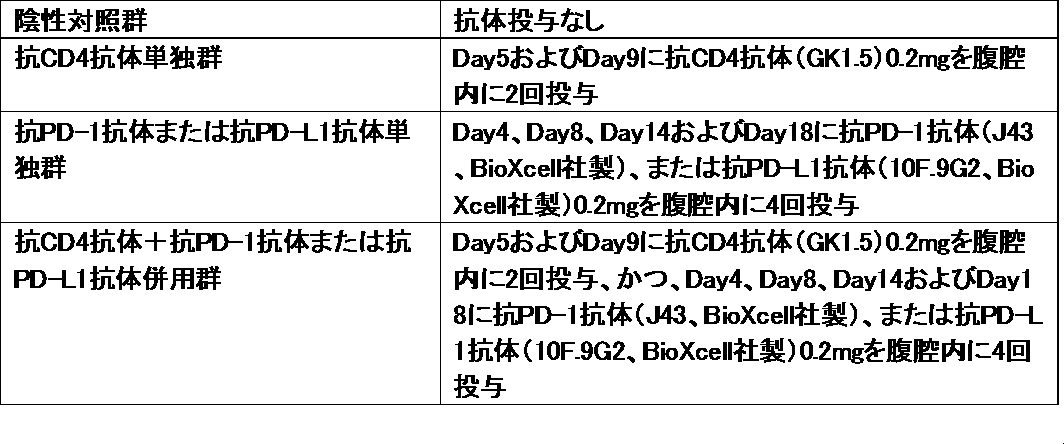
C57BL/6系統マウス(雌、7週齢)の右側腹部にマウスメラノーマ細胞株B16F10(5x105 cells/mouse)を皮下移植した後、下記の通りに抗体投与を行なった(Day0=がん細胞移植日)。
2. Mechanism of anti-tumor effect of anti-CD4 antibody alone, immune checkpoint antibody alone, or anti-CD4 antibody + immune checkpoint antibody combination Mouse melanoma cell line on the right flank of C57BL / 6 mice (female, 7 weeks old) After subcutaneous implantation of B16F10 (5 × 10 5 cells / mouse), antibody administration was performed as follows (Day 0 = day of cancer cell transplantation).
図2は、腫瘍細胞を移植したC57BL/6マウスの各群(抗CD4単独群、免疫チェックポイント抗体単独群、及び両者の併用群)について、Day16の時点で固形腫瘍径を測定、腫瘍体積を計算(短径x短径x長径xπ/6)した結果である。
Fig. 2 shows the measurement of solid tumor diameter at day 16 for each group of C57BL / 6 mice transplanted with tumor cells (anti-CD4 alone group, immune checkpoint antibody alone group, and a combination of both), and tumor volume. It is the result of calculation (minor axis x minor axis x major axis xπ / 6).
抗CD4抗体はB16メラノーマの固形腫瘍の増殖を、対照群のそれの約1/3までに有意に抑制した(Dunnett, 有意水準p<0.01)。ここで単剤としての腫瘍増殖抑制効果を観察すると、抗CD4抗体が他の免疫チェックポイント抗体単剤の抑制効果よりも明白に優れている事が示されている。
Anti-CD4 antibody significantly inhibited the growth of solid tumors of B16 melanoma by about 1/3 that of the control group (Dunnett, significant level p <0.01). Observing the tumor growth inhibitory effect as a single agent here shows that the anti-CD4 antibody is clearly superior to the inhibitory effect of other immune checkpoint antibodies alone.
抗PD-L1抗体、抗PD-L2抗体、抗OX40抗体、または抗CTLA-4抗体の単独投与は、抗CD4抗体より弱いが、対照群のそれに対して、それぞれ有意に抑制した(Dunnett, 有意水準p<0.01)。さらに、抗CD4抗体と、抗PD-L1抗体、抗PD-L2抗体、抗OX40抗体、または抗CTLA-4抗体とを併用すると、B16メラノーマの固形腫瘍の増殖は、抗CD4抗体を投与しない単独投与群よりも強く抑制され、免疫チェックポイント抗体単独投与群と抗CD4抗体併用投与群との平均値との差は有意であり(Dunnett, 有意水準p<0.05、あるいはp<0.01)、併用効果が明らかになった。特に、抗CD4抗体と抗PD-L1抗体との併用群は、抗CD4抗体単独群との間で、腫瘍体積の平均値はどちらも有意(有意水準5%、Dunnet)に差があり、抗CD4抗体と抗PD-L1抗体の併用による相乗効果が顕著に表れた。
Single administration of anti-PD-L1 antibody, anti-PD-L2 antibody, anti-OX40 antibody, or anti-CTLA-4 antibody was weaker than anti-CD4 antibody, but significantly suppressed it compared to that in the control group (Dunnett, significant Level p <0.01). Furthermore, when an anti-CD4 antibody is used in combination with an anti-PD-L1 antibody, an anti-PD-L2 antibody, an anti-OX40 antibody, or an anti-CTLA-4 antibody, the growth of a solid tumor of B16 melanoma alone is not administered with an anti-CD4 antibody. Suppressed more strongly than the administration group, and the difference between the mean value of the immune checkpoint antibody single administration group and the anti-CD4 antibody combination administration group was significant (Dunnett, significance level p <0.05 or p <0.01). Became clear. In particular, the combination group of anti-CD4 antibody and anti-PD-L1 antibody was significantly different from the anti-CD4 antibody alone group in the mean value of tumor volume (significant level 5%, Dunnet), The synergistic effect of the combination of CD4 antibody and anti-PD-L1 antibody was prominent.
図3は、抗CD4抗体と抗CD8抗体の併用についての検討結果である。腫瘍体積の計算は上記と同様に行なった。図3BはDay15での腫瘍体積を比較した結果である。
FIG. 3 shows the results of studies on the combined use of anti-CD4 antibody and anti-CD8 antibody. Tumor volume was calculated as described above. FIG. 3B shows the results of comparing tumor volumes on Day 15.
図3に示すように、抗CD4抗体とともに抗CD8抗体を投与すると、抗CD4抗体の抗腫瘍作用はすべて消失した。抗CD4抗体の作用機序にはCD8陽性T細胞、すなわち、細胞傷害性T細胞(CTL)が大きく寄与していることが明らかになった。
As shown in FIG. 3, when the anti-CD8 antibody was administered together with the anti-CD4 antibody, all of the anti-tumor action of the anti-CD4 antibody disappeared. It was clarified that CD8 positive T cells, that is, cytotoxic T cells (CTL), contributed greatly to the mechanism of action of the anti-CD4 antibody.
B16F10細胞株を移植したC57BL/6マウスの各群について、Day14の時点で犠牲死させ、腫瘍を切除した。腫瘍組織の一部については、腫瘍内リンパ球を分離し、フローサイトメーターで解析し、残りの一部で組織切片を作製した。手順を以下に記載する。
Each group of C57BL / 6 mice transplanted with the B16F10 cell line was sacrificed at Day 14 and the tumor was excised. For a part of the tumor tissue, intratumoral lymphocytes were separated and analyzed with a flow cytometer, and a tissue section was prepared with the remaining part. The procedure is described below.
マウスに抗CD45.2抗体を静脈内注射し、3分後に腫瘍組織を分離した。腫瘍組織をはさみでミンチし、コラゲナーゼ処理した後に、比重遠心法で腫瘍内リンパ球を取得した。
Mice were injected intravenously with anti-CD45.2 antibody, and the tumor tissue was isolated 3 minutes later. The tumor tissue was minced with scissors and treated with collagenase, and then intratumoral lymphocytes were obtained by specific gravity centrifugation.
腫瘍内リンパ球を、抗CD45抗体、抗CD11b抗体、抗CD19抗体、抗NK1.1抗体、および抗CD8抗体で染色し、腫瘍実質組織内リンパ球(CD45+ IVS CD45.2-)に含まれる、CD11b- CD19- NK1.1- CD8+の、腫瘍実質組織内CD8+ T細胞集団(以下、CD8+ T細胞と記する)をフローサイトメトリーで解析した。さらに、抗CD45抗体、抗CD11b抗体、抗CD19抗体、抗NK1.1抗体、抗CD8抗体、抗PD-1抗体、および抗CD137抗体で染色して、PD-1+ CD137+ CD8+ T細胞の解析を行った。
The intratumoral lymphocytes, anti-CD45 antibody, anti-CD11b antibodies, and stained with anti-CD19 antibody, anti-NK1.1 antibody, and anti-CD8 antibodies, tumor parenchyma in lymphocytes (CD45 + IVS CD45.2 -) contained in the , CD11b − CD19 − NK1.1 − CD8 + , a CD8 + T cell population in the tumor parenchyma (hereinafter referred to as CD8 + T cells) was analyzed by flow cytometry. In addition, staining with anti-CD45 antibody, anti-CD11b antibody, anti-CD19 antibody, anti-NK1.1 antibody, anti-CD8 antibody, anti-PD-1 antibody, and anti-CD137 antibody, PD-1 + CD137 + CD8 + T cells Analysis was performed.
また、CD8T細胞をPMAとイオノマイシンで刺激培養した後に、抗IFNγ抗体、および抗TNFα抗体で染色して、IFNγ+ TNFα+ CD8+ T細胞の解析を行なった。
In addition, CD8T cells were stimulated with PMA and ionomycin and then stained with anti-IFNγ antibody and anti-TNFα antibody to analyze IFNγ + TNFα + CD8 + T cells.
なお、上記各抗体で染色する前に抗マウスCD16/CD32抗体(clone 2.4G2, BioXcell)でFcレセプターをブロックした。測定はGallios (Beckman Coulter)で行い、FlowJo ソフトウェア (version 9.7.5; FlowJo, LLC) で解析した。死細胞は、ヨウ化プロピジウム(PI)で染色して除去している。
The Fc receptor was blocked with an anti-mouse CD16 / CD32 antibody (clone 2.4G2, BioXcell) before staining with the above antibodies. Measurements were made with Gallios® (Beckman® Coulter) and analyzed with FlowJo® software (version 9.7.5; FlowJo, LLC). Dead cells are removed by staining with propidium iodide (PI).
腫瘍組織はTissue-Tek OCT コンパウンド(Sakura Finetek)に埋設し、液体窒素下で凍結させた。6-μmの厚さの組織切片を作製し、Blocking One (ナカライテスク)で非特異反応をブロックし、抗CD8抗体、抗ΔhLNGFR(truncated form of human low-affinity nerve growth factor receptor)抗体およびヨウ化プロピジウムで染色した。
その後 Prolong Gold reagent (Life Technologies)で包埋し、SP5 共焦点顕微鏡 (Leica Microsystems)で免疫染色像を観察した。
Tumor tissue was embedded in Tissue-Tek OCT compound (Sakura Finetek) and frozen under liquid nitrogen. 6-μm thick tissue sections were prepared, blocking non-specific reactions with Blocking One (Nacalai Tesque), anti-CD8 antibody, anti-ΔhLNGFR (truncated form of human low-affinity nerve growth factor receptor) antibody and iodination Stained with propidium.
Then, it was embedded with Prolong Gold reagent (Life Technologies), and immunostained images were observed with SP5 confocal microscope (Leica Microsystems).
図4Bに示すように、抗CD4抗体は腫瘍内CD8陽性細胞を、対照群のそれの27倍までに有意に増強した(Dunnett, 有意水準p<0.01)。また、PD-1+ CD137+ CD8+ T細胞、およびIFNγ+ TNFα+ CD8+ T細胞も、それぞれ2.7倍、および3.2倍まで増加した(図4E, G)。すなわち、マウスにおいて特に抗CD4抗体によるCD4陽性細胞の枯渇に応答してCD8+ CD44hi CD62Llo PD-1+ CD137+細胞が増加した。
As shown in FIG. 4B, anti-CD4 antibody significantly enhanced intratumoral CD8 positive cells by 27 times that of the control group (Dunnett, significance level p <0.01). PD-1 + CD137 + CD8 + T cells and IFNγ + TNFα + CD8 + T cells also increased to 2.7 times and 3.2 times, respectively (FIGS. 4E and G). That is, CD8 + CD44 hi CD62L lo PD-1 + CD137 + cells increased in response to depletion of CD4 positive cells by anti-CD4 antibody in mice.
一方、腫瘍組織切片を免疫組織学的手法で解析すると、抗CD4抗体投与により腫瘍内CD8陽性細胞が明らかに増加しているのが確認された(図5)。
On the other hand, when the tumor tissue section was analyzed by an immunohistological technique, it was confirmed that the CD8 positive cells in the tumor were clearly increased by the administration of the anti-CD4 antibody (FIG. 5).
3.抗CD4抗体単独、抗PD-1抗体若しくは抗PD-L1抗体単独、又は抗CD4抗体+抗PD-1抗体若しくは抗PD-L1抗体併用での抗腫瘍効果の作用機序
C57BL/6系統マウス(雌、7週齢)の右側腹部にマウスメラノーマ細胞株B16F10(5x105 cells/mouse)を皮下移植した後、下記の通りに抗体投与を行なった(Day0=がん細胞移植日)。
3. Action mechanism of anti-tumor effect with anti-CD4 antibody alone, anti-PD-1 antibody or anti-PD-L1 antibody alone, or anti-CD4 antibody + anti-PD-1 antibody or anti-PD-L1 antibody combination C57BL / 6 strain mice ( The mouse melanoma cell line B16F10 (5 × 10 5 cells / mouse) was subcutaneously transplanted into the right flank of females (7 weeks of age), and then the antibody was administered as described below (Day 0 = day of cancer cell transplantation).
各群のマウスより、Day14の時点で血液サンプルを採取し、血液中のCD8+細胞におけるPD-1+細胞、CD44hi細胞及びCD137+細胞の割合をフローサイトメトリー解析により調べた。手順を以下に記載する。
A blood sample was collected from each group of mice on Day 14, and the ratio of PD-1 + cells, CD44 hi cells and CD137 + cells in CD8 + cells in the blood was examined by flow cytometry analysis. The procedure is described below.
マウスから採血し、比重遠心法で末梢血リンパ球を取得した。末梢血リンパ球を、抗CD45抗体、抗CD11b抗体、抗CD19抗体、抗NK1.1抗体、抗CD8抗体、抗CD44抗体(clone IM7)、および抗PD-1抗体で染色して、末梢血PD-1+ CD44hi CD8+ T細胞をフローサイトメトリーで解析した。さらに、抗CD45抗体、抗CD11b抗体、抗CD19抗体、抗NK1.1抗体、抗CD8抗体、抗CD44抗体、およびCD137抗体で染色して、末梢血CD137+ CD44+ CD8+ T細胞を解析した。
Blood was collected from mice, and peripheral blood lymphocytes were obtained by specific gravity centrifugation. Peripheral blood lymphocytes are stained with anti-CD45 antibody, anti-CD11b antibody, anti-CD19 antibody, anti-NK1.1 antibody, anti-CD8 antibody, anti-CD44 antibody (clone IM7), and anti-PD-1 antibody to obtain peripheral blood PD -1 + CD44 hi CD8 + T cells were analyzed by flow cytometry. Further, peripheral blood CD137 + CD44 + CD8 + T cells were analyzed by staining with anti-CD45 antibody, anti-CD11b antibody, anti-CD19 antibody, anti-NK1.1 antibody, anti-CD8 antibody, anti-CD44 antibody, and CD137 antibody.
なお、上記各抗体で染色する前に抗マウスCD16/CD32抗体(clone 2.4G2, BioXcell)でFcレセプターをブロックした。測定はGallios (Beckman Coulter)で行い、FlowJo ソフトウェア (version 9.7.5; FlowJo, LLC) で解析した。死細胞は、ヨウ化プロピジウムで染色して除去している。
The Fc receptor was blocked with an anti-mouse CD16 / CD32 antibody (clone 2.4G2, BioXcell) before staining with the above antibodies. Measurements were made with Gallios® (Beckman® Coulter) and analyzed with FlowJo® software (version 9.7.5; FlowJo, LLC). Dead cells are removed by staining with propidium iodide.
結果を図6に示す。抗CD4抗体単独、抗CD4抗体と抗PD-1抗体を併用して、または、抗CD4抗体と抗PD-L1抗体(clone 10F.9G2)を併用して投与すると、PD-1+ CD44hi CD8+ T細胞、CD137+ CD44hi CD8+ T細胞、及びPD-1+ CD137+ CD8+ T細胞が増加することが顕著に示された。
The results are shown in FIG. PD-1 + CD44 hi CD8 when administered together with anti-CD4 antibody alone, anti-CD4 antibody and anti-PD-1 antibody, or anti-CD4 antibody and anti-PD-L1 antibody (clone 10F.9G2) It was markedly increased in + T cells, CD137 + CD44 hi CD8 + T cells, and PD-1 + CD137 + CD8 + T cells.
また、各群のマウスからDay14の時点で腫瘍を採取し、腫瘍内mRNAを抽出し、定量RT-PCRにより下記の遺伝子の発現量を調べた。
TNF-α(Tnf)、IFN-γ(Ifng)、Cxcl10、Cd274、Fasl、Prf1、グランザイム(Gzmb)
In addition, tumors were collected from mice of each group at Day 14, the tumor mRNA was extracted, and the expression levels of the following genes were examined by quantitative RT-PCR.
TNF-α (Tnf), IFN-γ (Ifng), Cxcl10, Cd274, Fasl, Prf1, Granzyme (Gzmb)
結果を図7に示す。抗CD4抗体投与によって、IFN-γやグランザイムなど、細胞傷害性T細胞をはじめとするエフェクター細胞が産生する液性分子が多く発現することが確認された。これにより、CD4陽性細胞除去→CD8陽性細胞の活性化・組織浸潤増加→活性化エフェクター細胞(CTLなど)による抗腫瘍細胞効果、といった作用機序が強く示唆された。
Results are shown in FIG. It was confirmed that the administration of anti-CD4 antibody expressed many humoral molecules produced by effector cells including cytotoxic T cells such as IFN-γ and granzyme. This strongly suggested a mechanism of action such as removal of CD4-positive cells → activation of CD8-positive cells / increase in tissue invasion → anti-tumor cell effect by activated effector cells (CTL, etc.).