WO2016153039A1 - Training device for endoscopic endonasal skull base surgery - Google Patents
Training device for endoscopic endonasal skull base surgery Download PDFInfo
- Publication number
- WO2016153039A1 WO2016153039A1 PCT/JP2016/059622 JP2016059622W WO2016153039A1 WO 2016153039 A1 WO2016153039 A1 WO 2016153039A1 JP 2016059622 W JP2016059622 W JP 2016059622W WO 2016153039 A1 WO2016153039 A1 WO 2016153039A1
- Authority
- WO
- WIPO (PCT)
- Prior art keywords
- skull base
- carotid artery
- internal carotid
- training
- flow path
- Prior art date
Links
Images


Classifications
-
- G—PHYSICS
- G09—EDUCATION; CRYPTOGRAPHY; DISPLAY; ADVERTISING; SEALS
- G09B—EDUCATIONAL OR DEMONSTRATION APPLIANCES; APPLIANCES FOR TEACHING, OR COMMUNICATING WITH, THE BLIND, DEAF OR MUTE; MODELS; PLANETARIA; GLOBES; MAPS; DIAGRAMS
- G09B23/00—Models for scientific, medical, or mathematical purposes, e.g. full-sized devices for demonstration purposes
- G09B23/28—Models for scientific, medical, or mathematical purposes, e.g. full-sized devices for demonstration purposes for medicine
- G09B23/30—Anatomical models
- G09B23/303—Anatomical models specially adapted to simulate circulation of bodily fluids
-
- B—PERFORMING OPERATIONS; TRANSPORTING
- B33—ADDITIVE MANUFACTURING TECHNOLOGY
- B33Y—ADDITIVE MANUFACTURING, i.e. MANUFACTURING OF THREE-DIMENSIONAL [3-D] OBJECTS BY ADDITIVE DEPOSITION, ADDITIVE AGGLOMERATION OR ADDITIVE LAYERING, e.g. BY 3-D PRINTING, STEREOLITHOGRAPHY OR SELECTIVE LASER SINTERING
- B33Y10/00—Processes of additive manufacturing
-
- B—PERFORMING OPERATIONS; TRANSPORTING
- B33—ADDITIVE MANUFACTURING TECHNOLOGY
- B33Y—ADDITIVE MANUFACTURING, i.e. MANUFACTURING OF THREE-DIMENSIONAL [3-D] OBJECTS BY ADDITIVE DEPOSITION, ADDITIVE AGGLOMERATION OR ADDITIVE LAYERING, e.g. BY 3-D PRINTING, STEREOLITHOGRAPHY OR SELECTIVE LASER SINTERING
- B33Y50/00—Data acquisition or data processing for additive manufacturing
- B33Y50/02—Data acquisition or data processing for additive manufacturing for controlling or regulating additive manufacturing processes
-
- B—PERFORMING OPERATIONS; TRANSPORTING
- B33—ADDITIVE MANUFACTURING TECHNOLOGY
- B33Y—ADDITIVE MANUFACTURING, i.e. MANUFACTURING OF THREE-DIMENSIONAL [3-D] OBJECTS BY ADDITIVE DEPOSITION, ADDITIVE AGGLOMERATION OR ADDITIVE LAYERING, e.g. BY 3-D PRINTING, STEREOLITHOGRAPHY OR SELECTIVE LASER SINTERING
- B33Y80/00—Products made by additive manufacturing
-
- G—PHYSICS
- G09—EDUCATION; CRYPTOGRAPHY; DISPLAY; ADVERTISING; SEALS
- G09B—EDUCATIONAL OR DEMONSTRATION APPLIANCES; APPLIANCES FOR TEACHING, OR COMMUNICATING WITH, THE BLIND, DEAF OR MUTE; MODELS; PLANETARIA; GLOBES; MAPS; DIAGRAMS
- G09B23/00—Models for scientific, medical, or mathematical purposes, e.g. full-sized devices for demonstration purposes
- G09B23/28—Models for scientific, medical, or mathematical purposes, e.g. full-sized devices for demonstration purposes for medicine
- G09B23/285—Models for scientific, medical, or mathematical purposes, e.g. full-sized devices for demonstration purposes for medicine for injections, endoscopy, bronchoscopy, sigmoidscopy, insertion of contraceptive devices or enemas
-
- G—PHYSICS
- G09—EDUCATION; CRYPTOGRAPHY; DISPLAY; ADVERTISING; SEALS
- G09B—EDUCATIONAL OR DEMONSTRATION APPLIANCES; APPLIANCES FOR TEACHING, OR COMMUNICATING WITH, THE BLIND, DEAF OR MUTE; MODELS; PLANETARIA; GLOBES; MAPS; DIAGRAMS
- G09B23/00—Models for scientific, medical, or mathematical purposes, e.g. full-sized devices for demonstration purposes
- G09B23/28—Models for scientific, medical, or mathematical purposes, e.g. full-sized devices for demonstration purposes for medicine
- G09B23/30—Anatomical models
- G09B23/34—Anatomical models with removable parts
-
- G—PHYSICS
- G09—EDUCATION; CRYPTOGRAPHY; DISPLAY; ADVERTISING; SEALS
- G09B—EDUCATIONAL OR DEMONSTRATION APPLIANCES; APPLIANCES FOR TEACHING, OR COMMUNICATING WITH, THE BLIND, DEAF OR MUTE; MODELS; PLANETARIA; GLOBES; MAPS; DIAGRAMS
- G09B9/00—Simulators for teaching or training purposes
-
- B—PERFORMING OPERATIONS; TRANSPORTING
- B29—WORKING OF PLASTICS; WORKING OF SUBSTANCES IN A PLASTIC STATE IN GENERAL
- B29C—SHAPING OR JOINING OF PLASTICS; SHAPING OF MATERIAL IN A PLASTIC STATE, NOT OTHERWISE PROVIDED FOR; AFTER-TREATMENT OF THE SHAPED PRODUCTS, e.g. REPAIRING
- B29C64/00—Additive manufacturing, i.e. manufacturing of three-dimensional [3D] objects by additive deposition, additive agglomeration or additive layering, e.g. by 3D printing, stereolithography or selective laser sintering
- B29C64/10—Processes of additive manufacturing
- B29C64/141—Processes of additive manufacturing using only solid materials
- B29C64/153—Processes of additive manufacturing using only solid materials using layers of powder being selectively joined, e.g. by selective laser sintering or melting
-
- B—PERFORMING OPERATIONS; TRANSPORTING
- B29—WORKING OF PLASTICS; WORKING OF SUBSTANCES IN A PLASTIC STATE IN GENERAL
- B29L—INDEXING SCHEME ASSOCIATED WITH SUBCLASS B29C, RELATING TO PARTICULAR ARTICLES
- B29L2031/00—Other particular articles
- B29L2031/753—Medical equipment; Accessories therefor
- B29L2031/7546—Surgical equipment
Definitions
- the present invention relates to a training device for transnasal endoscopic surgery for a skull base lesion.
- the sphenoid bone located approximately in the center of the skull base of the human body has a depression called a turkey fold in the approximate center, and the pituitary gland, which is an endocrine organ that secretes hormones, is housed in the depression.
- the pituitary gland which is an endocrine organ that secretes hormones, is housed in the depression.
- transsphenoidal sinus surgery has been performed in which the upper gums (upper groin) behind the upper lip are incised and the tumor is approached from the incision under a microscope.
- transnasal endoscopic surgery surgical tools such as endoscopes and scalpels are inserted from both nostrils into the nasal cavity that is the intranasal cavity and the paranasal sinus that is present in the bone adjacent to the nasal cavity. This is an operation for approaching a lesion through the nasal cavity, and is originally performed by an otolaryngologist for an intranasal or sinus lesion. Therefore, in transnasal endoscopic surgery (hereinafter referred to as “transnasal endoscopic skull base surgery”) for skull base lesions, the nasal cavity structure and paranasal sinus structure are generally understood, and intranasal lesions and sinus intranasal structures are generally understood.
- the sinuses include the frontal sinus in the frontal bone, the ethmoid sinus in the ethmoid, the maxillary sinus in the maxilla, and the sphenoid sinus in the sphenoid bone.
- the surgical instrument In the initial stage of transnasal endoscopic skull base surgery, that is, the approach stage in the nasal cavity and paranasal sinus, the surgical instrument is operated by an otolaryngologist.
- the otolaryngologist operates the endoscope, while the brain surgeon operates other surgical tools to remove tissue such as bone, brain dura incision, Tumor removal and the like are performed.
- the brain dura mater After removal of the tumor, the brain dura mater is closed in collaboration with a brain surgeon and an otolaryngologist. Then, the treatment of the sinuses and nasal cavity is performed by the otolaryngologist, and the operation is completed.
- the internal carotid artery located behind the outer wall of the nasal cavity may have to be exposed to the surgical space depending on the position and size of the lesion.
- the internal carotid artery is a pair of left and right blood vessels that pass through both sides of the pituitary gland and is a very important blood vessel in the head and neck.
- the surgical space in transnasal endoscopic skull base surgery is a narrow space such as the nasal cavity and paranasal sinuses, and there is a lot of bleeding from the internal carotid artery, so use a cleaning tool inserted into the surgical space together with the endoscope.
- the hemostasis operation performed while cleaning the endoscope lens is extremely difficult for a brain surgeon or an otolaryngologist.
- a surgical training particularly a training for coping with damage to the internal carotid artery.
- This training may be performed using the cadaver head, but using the cadaver head is very restrictive from an ethical point of view, and even when the cadaver head can be used. Therefore, there is no blood flow, and therefore it is difficult to perform coping training when the internal carotid artery is damaged.
- the present invention has been made in view of the above-mentioned facts, and its main technical problem is to provide a device for performing training for transnasal endoscopic skull base surgery, particularly training for coping with damage to the internal carotid artery. Is to provide.
- a training device for nasal endoscopic skull base surgery composed of a human head model and liquid circulation means.
- the human head model includes an anterior portion including a nose having a nostril, at least a portion of a nasal septum, at least a portion of an outer wall of the nasal cavity, at least a portion of a posterior sphenoid sinus wall, and an internal carotid artery.
- the liquid circulating means is disposed between a storage tank for storing a non-transparent colored liquid, and the storage tank and at least a part of the internal carotid artery.
- Training apparatus nasal endoscopic skull base surgery and symptoms are provided.
- the human head model includes a rear part including a nasal bone, and the main part is formed separately from the front part and the rear part and is detachably attached to the rear part.
- the front part is formed separately from the rear part and the main part and is detachably attached to the rear part to which the main part is attached.
- the human head model preferably includes a base that detachably accommodates the rear portion to which the main portion is attached.
- the main part is preferably formed based on three-dimensional image data generated from tomographic information of the human head.
- the main part is conveniently formed from a powder sintered material.
- the front part is preferably formed from an elastomer.
- the training device for transnasal endoscopic skull base surgery provided by the present invention, when the nasal septum, the outer wall of the nasal cavity and the posterior wall of the sphenoid sinus are cut by the cutting tool inserted into the nasal cavity from the nostril, the internal carotid artery is When exposed to the nasal cavity and the internal carotid artery is damaged by the cutting tool, a non-transparent colored liquid is ejected, so that coping training can be performed when the internal carotid artery is damaged.
- FIG. 1 is a hydraulic circuit diagram illustrating a training device for transnasal endoscopic skull base surgery constructed in accordance with the present invention.
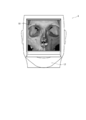
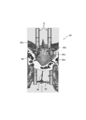
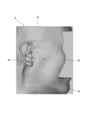
- FIG. The perspective view of a human head model. The front view of a human head model in the state where a front part and a wedge were removed. Front view of the front. The left side view of a front part. FIG. The top view of a rear part. Rear left side view. Sectional drawing seen from the AA direction in FIG. Sectional drawing seen from the BB direction in FIG. The front view of the principal part. The rear view of the principal part. The top view of the principal part. The left view of the main part. The perspective view of a base main body. The left view of a base main body. The perspective view of a middle upper frame. The perspective view of a middle lower frame. The perspective view of a wedge.
- the training apparatus for transnasal endoscopic skull base surgery comprises a human head model 4 and liquid circulation means 6.
- the human head model 4 includes a front portion 8, a rear portion 10, a base 12, and a wedge 14 as shown in FIGS. 2 and 3.
- the front portion 8 on which at least a part of the human body face is reproduced is disposed on the front surface of the human head model 4, and it is important to include the nose portion 16 in which the nostril 16a is formed.
- the front portion 8 formed in a square shape in front view separately from the rear portion 10 and a main portion 40 to be described later is a nose portion 16 located on the center side in the left-right direction in the front view and on the lower side in the up-down direction.
- a frontal head 18 positioned above the nose 16 and extending in the left-right direction; right and left orbits 20 positioned below each of the left and right ends of the frontal head 18;
- the left and right orbital lower portions 22 positioned below, the upper jaw portion 24 positioned below the nose portion 16, the rectangular upper piece 26 extending rearward from the upper end of the frontal head 18, and extending rearward from the lower end of the upper jaw portion 24.
- a rectangular lower piece 28 The front surface of the nose portion 16 protrudes forward, and left and right nostrils 16 a penetrating the front portion 8 are formed in the lower portion of the nose portion 16.
- the rear surface of the front portion 8 is formed corresponding to the front shape of the rear portion 10, for example, the rear surface of the nose portion 16 is recessed forward, and the rear surface of each orbital portion 20 protrudes rearward.
- the anterior portion 8 is attached to the posterior portion 10
- the rear surface is accommodated in the orbit 32 of the rear portion 10 described later.
- the front portion 8 is preferably formed by injection molding from an elastomer that approximates the flexibility and elasticity of the human skin based on three-dimensional image data generated from tomographic information of the human head.
- the rear part 10 where at least a part of the human skull is reproduced is arranged behind the front part 8.
- it is formed in a quadrangular shape when viewed from the front, and the upper surface, the lower surface, and the left and right side surfaces can come into surface contact with the flat surface.
- the rear surface is also formed so as to be in surface contact with the flat surface.
- the posterior part 10 is formed with a nasal cavity 30 located on the center side in the left-right direction in the front view and on the lower side in the up-down direction, and left and right eye sockets 32 located above the nasal cavity 30 and recessed backward.
- the posterior part 10 is formed in a fan-shaped front skull base 34 located on the front side in a plan view, a butterfly-shaped middle skull fossa 36 located behind the front skull base 34, and behind the middle skull fossa 36.
- a trapezoidal posterior cranial fossa 38 is formed.
- a main portion 40 formed separately from the rear portion 10 is detachably attached to the rear portion 10. In the present embodiment, the main portion 40 is located approximately at the center in the left-right direction in plan view as shown in FIG.
- the nasal cavity 30 is located behind the nostril 16a when the front part 8 is mounted on the front surface of the rear part 10, and extends rearward from the nostril 16a, and is formed as a cavity in the nostril 16a.
- the nasal cavity 30 is covered with a nasal cavity outer wall 42 and is partitioned into two chambers on the left and right by a nasal septum 44.
- the nasal cavity outer wall 42 includes a nasal bone 46 located between the orbits 32, a frontal bone 48 located above the nasal bone 46, a maxilla bone 50 located behind the nasal bone 46, and a posterior side of the maxilla bone 50.
- the sphenoid sinus front wall 60 is a portion of the sphenoid bone 58 that covers the anterior side of the sphenoid sinus 64 located behind the nasal cavity 30.
- the sphenoid sinus 64 is one of the paranasal sinuses and is a cavity existing in the sphenoid bone 58.
- the nasal septum 44 includes a nasal septal cartilage 68 located at the front end, a vertical plate 52 c of the ethmoid bone 52 located behind the nasal septal cartilage 68, and a rib located below the nasal septal cartilage 68. 70.
- the main portion 40 includes at least a part of the outer nasal wall 42, at least a part of the nasal septum 44, at least a part of the sphenoid sinus posterior wall 66 covering the posterior side of the sphenoid sinus 64, and at least a part of the internal carotid artery 72. It is important to include In the illustrated embodiment, the main portion 40 has a rear side of the outer nasal cavity wall 42, specifically, as shown in FIG. 11, a rear side of the upper turbinate 52a, a rear side of the middle turbinate 52b, and a rear side of the lower turbinate 54.
- the main portion 40 includes the rear side of the nasal septum 44, specifically, the rear side of the vertical plate 52c of the ethmoid 52 and the rear side of the rib 70.
- the sphenoid sinus posterior wall 66 of the main portion 40 has a Turkish heel 66 a located in the center of the middle cranial fossa 36 and recessed downward, and both left and right sides of the Turkish heel 66 a.
- the internal carotid artery 72 of the main portion 40 has the tilting table 66d.
- the internal carotid artery 72 is formed as a pair of left and right sides and includes both the paraclinal portion 72a and the cavernous sinus portion 72b, but may be one of the left and right sides. Alternatively, it may be either the paraclinal part 72a or the cavernous sinus part 72b.
- the internal carotid artery 72 is hollow.
- the rear part 10 and the main part 40 may contain all of each of said each structure
- the inner and outer surfaces of the nasal cavity outer wall 42 and the surface of the nasal septum 44 may be covered with an elastic membrane simulating the mucous membrane of the human body.
- the elastic film can be formed from an appropriate rubber material such as silicon rubber.
- the rear part 10 and the main part 40 are preferably formed from a powder sintered material by a powder sintering additive manufacturing method based on three-dimensional image data generated from tomographic information of the human head. Since the powder sintering additive manufacturing method itself is a known modeling method, the detailed description thereof is omitted in this specification.
- the powder sintered material for forming the rear portion 10 and the main portion 40 polyamide (nylon), polycarbonate, polyester, polyacetal, polyethylene, polypropylene, polyvinyl chloride, polystyrene, polybutylene, ABS resin, cellulose resin, acrylic resin Synthetic resin powders having relatively rigidity such as epoxy resin and fluororesin can be mentioned.
- the posterior portion 10 and the main portion 40 include a nasal septal cartilage 68 and an internal carotid artery 72, which are tissues other than bone.
- bone portions such as the sphenoid bone 58 are mainly cut during training.
- the powder sintered material for forming the rear portion 10 and the main portion 40 preferably has a machinability similar to that of a human bone, and is therefore preferably composed of a synthetic resin powder and an inorganic filler powder.
- inorganic fillers examples include talc, calcium carbonate, glass beads, silica, clay, kaolin, barium sulfate, wollastonite, mica, titanium oxide, diatomaceous earth, hydroxyapatite and metal powder, but human bone machinability. Glass beads are particularly advantageous because they are more approximate.
- the ratio of the synthetic resin powder to the inorganic filler is preferably 30 to 90% by weight of the synthetic resin powder and 10 to 70% by weight of the inorganic filler from the viewpoint of machinability similar to human bones.
- the synthetic resin powder is 50 to 80% by weight and the inorganic filler is 20 to 50% by weight.
- nasal septal cartilage 68 and the internal carotid artery 72 may be formed separately from the rear portion 10 and the main portion 40 from a rubber material or a resin material such as silicon rubber or polyvinyl alcohol. Further, the internal carotid artery 72 is formed separately from the main portion 40 from a rubber material or a resin material such as silicon rubber or polyvinyl alcohol inside a tubular portion formed hollow from a powder sintered material together with the main portion 40. A tubular blood vessel member may be inserted.
- the base 12 includes a base main body 74 (see FIGS. 15 and 16) that houses the front portion 8 and the rear portion 10 to which the main portion 40 is mounted, and the front portion 8, the rear portion 10, and the main portion. It is comprised from the middle upper frame 76 (refer FIG. 17) and the middle lower frame 78 (refer FIG. 18) accommodated in the base main body 74 with the part 40.
- the base body 74 includes a rear wall 80 that is formed flat on both the inner and outer surfaces, an upper surface wall 82 that extends forward from the upper end of the rear wall 80, and a front surface that extends from the lower end of the rear wall 80.
- the bottom wall 84 extends, the right side wall 86 extends forward from the right end of the rear wall 80, and the left side wall 88 extends forward from the left end of the rear wall 80.
- a rectangular housing recess is formed by the inner surface of each wall. It is prescribed.
- a pair of through-openings 82a and 84a are formed in the upper surface wall 82 and the lower surface wall 84, respectively, at intervals in the width direction.
- the inner surfaces of the top wall 82, the right side wall 86, and the left side wall 88 are formed substantially perpendicular to the inner surface of the rear wall 80.
- the inner surface of the lower wall 84 is formed to be inclined upward from the front end toward the rear end.
- the outer surfaces of the upper wall 82 and the lower wall 84 are formed flat.
- the base body 74 is preferably formed from a powder sintered material by a powder sintering additive manufacturing method.
- the middle and upper frame 76 is bonded to the rectangular upper surface plate 90, the rectangular rear surface plate 92 extending downward from the rear end of the upper surface plate 90, and the inner surfaces of the upper surface plate 90 and the rear surface plate 92.
- a pair of through openings 90a are formed in the upper surface plate 90 at intervals in the width direction, and a pair of through openings 94a are formed in the brain 94 following each through opening 90a of the upper surface plate.
- the width dimension of each of the upper surface plate 90 and the rear surface plate 92 is formed corresponding to the width dimension of the housing recess of the base body 74.
- the upper surface and the rear surface of the brain 94 are formed flat corresponding to the inner surfaces of the upper surface plate 90 and the rear surface plate 92.
- the width dimension of the brain 94 is formed corresponding to the width dimension of the upper surface plate 90 and the rear surface plate 92, and the right side surface and the left side surface of the brain 94 are formed flat.
- the middle and lower frame 78 includes a rectangular lower surface plate 96, a rectangular right side surface plate 98 extending upward from the right end of the lower surface plate 96, and a rectangular left side surface plate 100 extending upward from the left end of the lower surface plate 96. It has.
- the bottom plate 96 is formed with a pair of circular through openings 96a spaced apart in the width direction.
- a bone portion 96b formed in a shape capable of being in surface contact with the rear surface of the rear portion 10 is erected.
- the upper surface plate 90 and the rear surface plate 92 of the middle and upper frame 76 and the middle and lower frame 78 are preferably formed from a powder sintered material by a powder sintering additive manufacturing method, like the rear portion 10 and the main portion 40.
- the brain 94 of the middle upper frame 76 is preferably made of an elastomer, like the front portion 8.
- the wedge 14 is for fixing the rear portion 10 to which the front portion 8 and the main portion 40 are mounted in the base 12, and as shown in FIG. 19, the thickness gradually decreases from the front end toward the rear end. Is formed.
- a pair of U-shaped notches 14a are formed at the rear end at intervals in the width direction.
- the upper piece 26 of the front portion 8 is positioned between the upper surface wall 82 of the base body 74 and the upper surface plate 90 of the middle upper frame 76. Further, the lower piece 28 of the front portion 8 is positioned between the lower surface wall 84 of the base body 74 and the lower surface plate 96 of the middle lower frame 78. Then, by inserting the wedge 14 between the lower piece 28 and the lower surface wall 84 of the base body 74, the front part 8, the rear part 10 and the main part 40 are fixed in the base 12 (see FIG. 2). .) As with the rear portion 10 and the main portion 40, the wedge 14 is conveniently formed from a powder sintered material by a powder sintering additive manufacturing method.
- the liquid circulation means 6 includes a storage tank 102, a supply flow path 104, a return flow path 106, and a circulation pump 108 as shown in FIG.
- the storage tank 102 stores a non-transparent colored liquid, for example, a bloody red liquid.
- the supply flow path 104 is disposed between the storage tank 102 and the lower end of the paraclinal base portion 72 a of the internal carotid artery 72, and the through opening 84 a of the lower surface wall 84 of the base main body 74 and the middle lower frame 78.
- the through hole 96 a of the lower surface plate 96 is inserted.
- the return flow path 106 is disposed between the upper end of the cavernous sinus 72 b of the internal carotid artery 72 and the storage tank 102, and the through opening 94 a of the brain 94 and the through opening of the upper surface plate 90 of the middle upper frame 76. 90a and the through-opening 82a of the upper surface wall 82 of the base main body 74 are inserted.
- the liquid in the storage tank 102 is circulated through the supply flow path 104, the internal carotid artery 72 and the return flow path 106 by the circulation pump 108.
- the procedure when coping training is performed when the internal carotid artery 72 is damaged using the nasal endoscope skull base surgery training apparatus 2 as described above will be described.
- surgical tools such as an endoscope, a cutting tool, a suction tool, a cleaning tool, and a scalpel (all of which are not shown) are inserted into the nasal cavity 30 as a surgical space from both nostrils 16a of the nose 16. Is to insert.
- the second part of the procedure is mainly to cut the posterior side of the radius 70 of the main part 40 and the anterior wall of the sphenoid sinus 60 with a cutting tool and expand the surgical space to approach the sphenoid sinus sinus wall 66. It is.
- the sphenoid sinus posterior wall 66 of the main part 40 is mainly cut with a cutting tool, and the paraclinal part 72a and / or the cavernous sinus 72b of the internal carotid artery 72 is exposed to the operation space. It is to let you.
- the generated cutting waste is appropriately sucked by the suction tool, and when the cutting waste adheres to the endoscope lens, the cleaning liquid is removed from the cleaning tool by the endoscope. Ejecting toward the lens and cleaning the endoscope lens.
- the fourth procedure is to damage the parastomatic pedestal 72a and / or the cavernous sinus 72b with a cutting tool, and to eject liquid from the parastomatic pedestal 72a and / or the cavernous sinus 72b.
- the fifth of the procedures is to adjust and control the liquid ejection from the paraclinal part 72a and / or the cavernous sinus part 72b while cleaning the endoscope lens with a cleaning tool to secure a visual field, It is to stop the eruption.
- the rear side of the radius 70 of the main portion 40, the anterior wall of the sphenoid sinus 60 and the butterfly are cut by a cutting tool inserted into the nasal cavity 30 from the nostril 16a.
- the osseous posterior wall 66 is cut, the paraclinal portion 72a and / or the cavernous sinus portion 72b of the internal carotid artery 72 is exposed in the nasal cavity 30, and further, the paracondylar portion 72a and / or the cavernous sinus portion.
- 72b is damaged by the cutting tool, a non-transparent colored liquid is ejected. Therefore, a countermeasure training can be performed in the case where the paraclinal portion 72a or / and the cavernous sinus portion 72b are damaged.
- the parasparous pedestal 72a is damaged when, for example, most of the paraseptal pedestal 72a of the internal carotid artery is exposed in the surgical space.
- Coping training can be performed. In this training, the operation of exposing the periphery of the damaged site in the parastable platform portion 72a to the surgical space during the liquid ejection is omitted, and the endoscope lens is washed with a cleaning tool during the liquid ejection to secure a visual field.
- the main training content is to control bleeding.
- the training for dealing with the damage to the paraclinal part 72a can be performed.
- the sphenoid sinus posterior wall 66 and the like are cut with a cutting tool while the endoscope lens is being cleaned with a cleaning tool while the liquid is being ejected, and the periphery of the portion (damaged part) in the paraclinal platform 72a is operated.
- the main training content is to control bleeding after being exposed to space.
- the main part 40 to be cut during training is formed separately from the rear part 10 and is detachably attached to the rear part 10, so that the trainee can cut the main part.
- the understanding of the three-dimensional structure of the nasal cavity and paranasal sinuses can be further deepened, and only the main portion 40 needs to be replaced after one training is performed.
- the replacement cost is suppressed.
- the insertion portion of the surgical instrument inserted from the nostril is formed with a length corresponding to this length and the patient. It is so thin that it can be inserted into the nasal cavity from the nostril (generally, an endoscope has an outer diameter of about 2.8 to 4 mm, and other surgical tools have an outer diameter of about 1 to 2 mm).
- an endoscope has an outer diameter of about 2.8 to 4 mm, and other surgical tools have an outer diameter of about 1 to 2 mm.
- precise work is required to prevent complications such as internal carotid artery damage. The requirement for precise work is the same for other operations, but particularly high accuracy is required for the operation.
- the surgeon's hand tremor In the actual operation, the operator abuts the surgical instrument against the nostril wall of the patient, or the fourth finger and the fifth Hand tremor is prevented by placing a finger on the patient's face.
- the front part 8 is formed of an elastomer that approximates the flexibility and elasticity of the human skin, so that the trainee can perform the operation inserted through the nostril 16a, as in the actual operation. Shake of the hand can be prevented by placing the tool against the wall of the nostril 16a or placing the fourth and fifth fingers on the front surface of the front portion 8.
Landscapes
- Engineering & Computer Science (AREA)
- Physics & Mathematics (AREA)
- General Physics & Mathematics (AREA)
- Chemical & Material Sciences (AREA)
- Theoretical Computer Science (AREA)
- Health & Medical Sciences (AREA)
- Educational Technology (AREA)
- Educational Administration (AREA)
- Business, Economics & Management (AREA)
- Materials Engineering (AREA)
- Manufacturing & Machinery (AREA)
- Medical Informatics (AREA)
- Medicinal Chemistry (AREA)
- Mathematical Optimization (AREA)
- Mathematical Physics (AREA)
- Pure & Applied Mathematics (AREA)
- Computational Mathematics (AREA)
- Algebra (AREA)
- Mathematical Analysis (AREA)
- General Health & Medical Sciences (AREA)
- Radiology & Medical Imaging (AREA)
- Pulmonology (AREA)
- Instructional Devices (AREA)
- Optics & Photonics (AREA)
- Mechanical Engineering (AREA)
Abstract
Provided is a device for carrying out training for endoscopic endonasal skull base surgery.
A training device 2 for endoscopic endonasal skull base surgery is constituted of a human head model 4 and a liquid circulation means 6. The human head model 4 is provided with a front part 8 that includes a nose part 16 in which nostrils 16a are formed and a main part 40 that includes at least part of a nasal septum 44, at least part of a nasal cavity lateral wall 42, at least part of a sphenoidal sinus posterior wall 66, and at least part of an internal carotid artery 72. The liquid circulation means 6 is provided with: a retention tank 102 for retaining a nontransparent colored liquid; a supply flow path 104 disposed between the retention tank 102 and the at least part of an internal carotid artery 72; a return flow path 106 disposed between the at least part of an internal carotid artery 72 and the retention tank 102; and a circulating pump 108 for circulating the liquid in the retention tank 102 through the supply flow path 104, the at least part of an internal carotid artery 72, and the return flow path 106.
Description
本発明は、頭蓋底病変に対する経鼻内視鏡手術の訓練装置に関する。
The present invention relates to a training device for transnasal endoscopic surgery for a skull base lesion.
人体の頭蓋底略中央に位置する蝶形骨には、その略中央にトルコ鞍と呼ばれる窪みが存在し、この窪み中にホルモンを分泌する内分泌器官である脳下垂体が収容されている。脳下垂体に発生した腫瘍に対しては、従来、上唇後方の歯茎上部(上歯齦部)を切開し、顕微鏡下において切開部分から腫瘍にアプローチする経蝶形骨洞手術が行われていた。しかしながら、内視鏡の小型化等をはじめとする手術具の改良に伴い、顕微鏡下の経蝶形骨洞手術と比較して視野が広く、手術部位を明瞭に確認し得ると共に低侵襲である経鼻内視鏡手術が脳下垂体腫瘍に対して行われるようになっている。又、経鼻内視鏡手術の適応疾患は脳下垂体腫瘍のほか、従来は開頭して病変にアプローチする開頭手術が行われていた髄膜腫や脊索腫等の頭蓋底病変にも拡大しており、経鼻内視鏡手術の重要性は一層高まっている。
The sphenoid bone located approximately in the center of the skull base of the human body has a depression called a turkey fold in the approximate center, and the pituitary gland, which is an endocrine organ that secretes hormones, is housed in the depression. For tumors that have occurred in the pituitary gland, conventionally, transsphenoidal sinus surgery has been performed in which the upper gums (upper groin) behind the upper lip are incised and the tumor is approached from the incision under a microscope. However, with the improvement of surgical tools such as downsizing of endoscopes, the field of view is wider compared to transsphenoidal sinus surgery under the microscope, and the surgical site can be clearly confirmed and less invasive Transnasal endoscopic surgery has been performed on pituitary tumors. In addition to pituitary tumors, indications for transnasal endoscopic surgery have also expanded to skull base lesions such as meningiomas and chordomas, which previously had craniotomy to open and approach the lesion. Therefore, the importance of transnasal endoscopic surgery is increasing.
経鼻内視鏡手術は、両鼻孔から鼻孔内空洞である鼻腔、及び鼻腔に隣接した骨内に存在する空洞である副鼻腔に内視鏡やメス等の手術具を挿入し、鼻腔及び副鼻腔を経て病変にアプローチする手術であり、元来、鼻腔内病変や副鼻腔内病変に対して耳鼻科医により行われていた手術である。このため、頭蓋底病変に対する経鼻内視鏡手術(以下「経鼻内視鏡頭蓋底手術」という。)においては、通常、鼻腔構造及び副鼻腔構造を理解し且つ鼻腔内病変及び副鼻腔内病変に対する経鼻内視鏡手術に習熟している耳鼻科医と、頭蓋内の構造を理解し且つ頭蓋底病変に対する手術に習熟している脳外科医との共同により行われる。尚、副鼻腔には、前頭骨内の前頭洞、篩骨内の篩骨洞、上顎骨内の上顎洞及び蝶形骨内の蝶形骨洞がある。
In transnasal endoscopic surgery, surgical tools such as endoscopes and scalpels are inserted from both nostrils into the nasal cavity that is the intranasal cavity and the paranasal sinus that is present in the bone adjacent to the nasal cavity. This is an operation for approaching a lesion through the nasal cavity, and is originally performed by an otolaryngologist for an intranasal or sinus lesion. Therefore, in transnasal endoscopic surgery (hereinafter referred to as “transnasal endoscopic skull base surgery”) for skull base lesions, the nasal cavity structure and paranasal sinus structure are generally understood, and intranasal lesions and sinus intranasal structures are generally understood. This is done in collaboration with an otolaryngologist familiar with transnasal endoscopic surgery for lesions and a brain surgeon who understands the structure of the skull and is familiar with surgery for skull base lesions. The sinuses include the frontal sinus in the frontal bone, the ethmoid sinus in the ethmoid, the maxillary sinus in the maxilla, and the sphenoid sinus in the sphenoid bone.
経鼻内視鏡頭蓋底手術の初期段階、即ち、鼻腔及び副鼻腔におけるアプローチの段階では、耳鼻科医により手術具が操作される。次いで頭蓋底部の骨、脳硬膜に接近した段階では、耳鼻科医により内視鏡が操作される一方、脳外科医により他の手術具が操作され、骨等の組織切除、脳硬膜切開や腫瘍摘出等が行われる。腫瘍摘出後は、脳外科医と耳鼻科医との共同により脳硬膜閉鎖が行われる。そして、耳鼻科医により副鼻腔及び鼻腔の処置が行われ、手術が終了する。
In the initial stage of transnasal endoscopic skull base surgery, that is, the approach stage in the nasal cavity and paranasal sinus, the surgical instrument is operated by an otolaryngologist. Next, when approaching the bone and brain dura mater at the base of the skull, the otolaryngologist operates the endoscope, while the brain surgeon operates other surgical tools to remove tissue such as bone, brain dura incision, Tumor removal and the like are performed. After removal of the tumor, the brain dura mater is closed in collaboration with a brain surgeon and an otolaryngologist. Then, the treatment of the sinuses and nasal cavity is performed by the otolaryngologist, and the operation is completed.
経鼻内視鏡頭蓋底手術においては、鼻腔から病変に至る経路を確保するため、鼻腔の周囲を覆う鼻腔外壁や鼻腔を左右に仕切る鼻中隔等の組織を切削具により切除する必要がある。又、組織切除の際、病変の位置や大きさ等によって鼻腔外壁後方に位置する内頸動脈を手術空間に露出させなければならないことがある。内頸動脈は、脳下垂体の両側を通っている左右一対の血管であり、頭部及び頸部における非常に重要な血管である。そして、組織切除の際のほか、脳硬膜切開や腫瘍摘出の際等に内頸動脈が損傷してしまうと、動脈であることから多量の血液が噴出するため、致命傷となるおそれがある。従って、このような場合には早急の処置が必要であるが、内頸動脈から噴出する多量の血液によって内視鏡レンズが汚染されるため、視野が妨げられ、内視鏡によって手術空間を確認することが困難となる。又、経鼻内視鏡頭蓋底手術における手術空間は鼻腔や副鼻腔等の狭い空間であり、且つ内頸動脈からの出血が多量であるため、内視鏡と共に手術空間に挿入した洗浄具により内視鏡レンズを洗浄しながら行う止血作業は脳外科医にとっても或いは耳鼻科医にとっても極めて困難な作業である。
In transnasal endoscopic skull base surgery, in order to secure a route from the nasal cavity to the lesion, it is necessary to remove the outer wall of the nasal cavity covering the periphery of the nasal cavity and the nasal septum that partitions the nasal cavity into the left and right with a cutting tool. In tissue resection, the internal carotid artery located behind the outer wall of the nasal cavity may have to be exposed to the surgical space depending on the position and size of the lesion. The internal carotid artery is a pair of left and right blood vessels that pass through both sides of the pituitary gland and is a very important blood vessel in the head and neck. If the internal carotid artery is damaged during cerebral dural incision or tumor removal in addition to tissue resection, a large amount of blood is ejected from the artery, which may be fatal. Therefore, immediate treatment is necessary in such a case, but the endoscope lens is contaminated by a large amount of blood ejected from the internal carotid artery, so the visual field is obstructed and the operation space is confirmed by the endoscope. Difficult to do. In addition, the surgical space in transnasal endoscopic skull base surgery is a narrow space such as the nasal cavity and paranasal sinuses, and there is a lot of bleeding from the internal carotid artery, so use a cleaning tool inserted into the surgical space together with the endoscope. The hemostasis operation performed while cleaning the endoscope lens is extremely difficult for a brain surgeon or an otolaryngologist.
そこで、経鼻内視鏡頭蓋底手術を行う術者には、手術訓練、特に内頸動脈が損傷した場合の対処訓練、を行うことが望まれている。この訓練は屍体頭部が使用されて行われることがあるが、屍体頭部を使用することは倫理上の観点等から大きな制約が伴うと共に、屍体頭部を使用することができた場合でも屍体であることから血流がなく、このため内頸動脈が損傷した場合の対処訓練を行うことは困難である。尚、経鼻内視鏡頭蓋底手術の訓練を行うための装置に関する文献は見当たらない。
Therefore, it is desirable for an operator who performs a transnasal endoscopic skull base operation to perform a surgical training, particularly a training for coping with damage to the internal carotid artery. This training may be performed using the cadaver head, but using the cadaver head is very restrictive from an ethical point of view, and even when the cadaver head can be used. Therefore, there is no blood flow, and therefore it is difficult to perform coping training when the internal carotid artery is damaged. In addition, there is no literature related to a device for training transnasal endoscopic skull base surgery.
本発明は上記事実に鑑みてなされたものであり、その主たる技術的課題は、経鼻内視鏡頭蓋底手術の訓練、特に内頸動脈が損傷した場合の対処訓練、を行うための装置を提供することである。
The present invention has been made in view of the above-mentioned facts, and its main technical problem is to provide a device for performing training for transnasal endoscopic skull base surgery, particularly training for coping with damage to the internal carotid artery. Is to provide.
本発明によれば、上記技術的課題を解決する経鼻内視鏡頭蓋底手術の訓練装置として、人体頭部模型と液体循環手段とから構成された経鼻内視鏡頭蓋底手術の訓練装置にして、該人体頭部模型は、鼻孔が形成された鼻部を含む前部と、鼻中隔の少なくとも一部、鼻腔外壁の少なくとも一部、蝶形骨洞後壁の少なくとも一部及び内頸動脈の少なくとも一部を含む主部とを具備し、該液体循環手段は、非透明有色液体を貯留する貯留タンクと、該貯留タンクと該内頸動脈の少なくとも一部との間に配設された供給流路と、該内頸動脈の少なくとも一部と該貯留タンクとの間に配設された戻り流路と、該貯留タンク内の該液体を該供給流路、該内頸動脈の少なくとも一部及び該戻り流路を通して循環させるための循環ポンプとを具備する、ことを特徴とする経鼻内視鏡頭蓋底手術の訓練装置が提供される。
According to the present invention, as a training device for nasal endoscopic skull base surgery that solves the above technical problem, a training device for nasal endoscopic skull base surgery composed of a human head model and liquid circulation means is provided. The human head model includes an anterior portion including a nose having a nostril, at least a portion of a nasal septum, at least a portion of an outer wall of the nasal cavity, at least a portion of a posterior sphenoid sinus wall, and an internal carotid artery. The liquid circulating means is disposed between a storage tank for storing a non-transparent colored liquid, and the storage tank and at least a part of the internal carotid artery. A supply flow path, a return flow path disposed between at least a portion of the internal carotid artery and the storage tank, and the liquid in the storage tank for at least one of the supply flow path and the internal carotid artery And a circulation pump for circulating through the return flow path. Training apparatus nasal endoscopic skull base surgery and symptoms are provided.
好ましくは、該人体頭部模型は鼻骨を含む後部を具備し、該主部は該前部及び該後部とは別個に形成されていて該後部に着脱自在に装着されている。該前部は該後部及び該主部とは別個に形成されていて、該主部が装着された該後部に着脱自在に装着されているのが好都合である。該人体頭部模型は該主部が装着された該後部を離脱自在に収容する基台を具備するのが好適である。該主部は人体頭部の断層撮影情報から生成される3次元画像データに基づき形成されているのが好ましい。該主部は粉末焼結材料から形成されているのが好都合である。該前部はエラストマーから形成されているのが好適である。
Preferably, the human head model includes a rear part including a nasal bone, and the main part is formed separately from the front part and the rear part and is detachably attached to the rear part. Conveniently, the front part is formed separately from the rear part and the main part and is detachably attached to the rear part to which the main part is attached. The human head model preferably includes a base that detachably accommodates the rear portion to which the main portion is attached. The main part is preferably formed based on three-dimensional image data generated from tomographic information of the human head. The main part is conveniently formed from a powder sintered material. The front part is preferably formed from an elastomer.
本発明によって提供される経鼻内視鏡頭蓋底手術の訓練装置においては、鼻孔から鼻腔に挿入された切削具により鼻中隔、鼻腔外壁及び蝶形骨洞後壁が切削されると内頸動脈が鼻腔内に露出し、更に内頸動脈が切削具により損傷されると非透明有色液体が噴出するため、内頸動脈が損傷した場合の対処訓練が行われ得る。
In the training device for transnasal endoscopic skull base surgery provided by the present invention, when the nasal septum, the outer wall of the nasal cavity and the posterior wall of the sphenoid sinus are cut by the cutting tool inserted into the nasal cavity from the nostril, the internal carotid artery is When exposed to the nasal cavity and the internal carotid artery is damaged by the cutting tool, a non-transparent colored liquid is ejected, so that coping training can be performed when the internal carotid artery is damaged.
以下、本発明に従って構成された経鼻内視鏡頭蓋底手術の訓練装置の好適実施形態について、添付図面を参照しつつ詳細に説明する。
Hereinafter, a preferred embodiment of a training device for nasal endoscopic skull base surgery constructed according to the present invention will be described in detail with reference to the accompanying drawings.
図1に示すとおり、全体を符号2で示す経鼻内視鏡頭蓋底手術の訓練装置は、人体頭部模型4と、液体循環手段6とから構成されている。図示された実施形態においては、人体頭部模型4は、図2及び図3に示すとおり、前部8と、後部10と、基台12と、楔14とを具備する。
As shown in FIG. 1, the training apparatus for transnasal endoscopic skull base surgery, indicated as a whole by reference numeral 2, comprises a human head model 4 and liquid circulation means 6. In the illustrated embodiment, the human head model 4 includes a front portion 8, a rear portion 10, a base 12, and a wedge 14 as shown in FIGS. 2 and 3.
図4及び図5を参照して説明する。人体顔面の少なくとも一部が再現された前部8は、人体頭部模型4の前面に配設されるものであり、鼻孔16aが形成された鼻部16を含むのが重要である。図示された実施形態においては、後部10及び後述する主部40とは別個に正面視四角形状に形成された前部8は、正面視左右方向中央側且つ上下方向下側に位置する鼻部16と、鼻部16の上方に位置していて左右方向に延びる前頭部18と、前頭部18の左右方向両端側の各々の下方に位置する左右の眼窩部20と、各眼窩部20の下方に位置する左右の眼窩下部22と、鼻部16の下方に位置する上顎部24と、前頭部18の上端から後方に延びる矩形状上部片26と、上顎部24の下端から後方に延びる矩形状下部片28とを含む。鼻部16の前面は前方に向かって突出しており、鼻部16の下部には前部8を貫通する左右の鼻孔16aが形成されている。又、前部8の後面は、後部10の前面形状に対応して形成され、例えば鼻部16の後面は前方に向かって窪んでおり、各眼窩部20の後面は後方に向かって突出している。前部8が後部10に装着された際には、鼻部16の後面には、後述する後部10の鼻骨46、上顎骨50前上側及び鼻中隔軟骨68前側が収容され、又、各眼窩部20の後面は、後述する後部10の眼窩32に収容される。前部8は、人体頭部の断層撮影情報から生成される3次元画像データに基づき、人体の皮膚の柔軟性及び弾力性に近似したエラストマーから射出成形により形成されているのが好適である。
This will be described with reference to FIG. 4 and FIG. The front portion 8 on which at least a part of the human body face is reproduced is disposed on the front surface of the human head model 4, and it is important to include the nose portion 16 in which the nostril 16a is formed. In the illustrated embodiment, the front portion 8 formed in a square shape in front view separately from the rear portion 10 and a main portion 40 to be described later is a nose portion 16 located on the center side in the left-right direction in the front view and on the lower side in the up-down direction. A frontal head 18 positioned above the nose 16 and extending in the left-right direction; right and left orbits 20 positioned below each of the left and right ends of the frontal head 18; The left and right orbital lower portions 22 positioned below, the upper jaw portion 24 positioned below the nose portion 16, the rectangular upper piece 26 extending rearward from the upper end of the frontal head 18, and extending rearward from the lower end of the upper jaw portion 24. And a rectangular lower piece 28. The front surface of the nose portion 16 protrudes forward, and left and right nostrils 16 a penetrating the front portion 8 are formed in the lower portion of the nose portion 16. Further, the rear surface of the front portion 8 is formed corresponding to the front shape of the rear portion 10, for example, the rear surface of the nose portion 16 is recessed forward, and the rear surface of each orbital portion 20 protrudes rearward. . When the anterior portion 8 is attached to the posterior portion 10, the nasal bone 46, the maxillary bone 50 anterior upper side and the nasal septal cartilage 68 anterior side of the posterior portion 10, which will be described later, are accommodated on the rear surface of the nose portion 16. The rear surface is accommodated in the orbit 32 of the rear portion 10 described later. The front portion 8 is preferably formed by injection molding from an elastomer that approximates the flexibility and elasticity of the human skin based on three-dimensional image data generated from tomographic information of the human head.
図6乃至図10を参照して説明する。人体頭蓋骨の少なくとも一部が再現された後部10は、前部8の後方に配設されるものである。図示された実施形態においては、図6に示すとおり正面視において四角形状に形成され、上面、下面及び左右両側面が平坦面に面接触し得るようになっていると共に、図7及び図8に示すとおり後面も平坦面に面接触し得るように形成されている。後部10は、正面視左右方向中央側且つ上下方向下側に位置する鼻腔30と、鼻腔30の上方に位置していて後方に向かって窪んだ左右の眼窩32とが形成されている。又、図7に示すとおり後部10は、平面視において前側に位置する扇形状前頭蓋底34と、前頭蓋底34の後方に位置する蝶形状中頭蓋窩36と、中頭蓋窩36の後方に位置する台形状後頭蓋窩38とが形成されている。後部10には、後部10とは別個に形成された主部40が着脱自在に装着されているのが好ましい。本実施形態においては、主部40は、図7に示すとおり平面視左右方向略中央に位置している。
This will be described with reference to FIGS. The rear part 10 where at least a part of the human skull is reproduced is arranged behind the front part 8. In the illustrated embodiment, as shown in FIG. 6, it is formed in a quadrangular shape when viewed from the front, and the upper surface, the lower surface, and the left and right side surfaces can come into surface contact with the flat surface. As shown, the rear surface is also formed so as to be in surface contact with the flat surface. The posterior part 10 is formed with a nasal cavity 30 located on the center side in the left-right direction in the front view and on the lower side in the up-down direction, and left and right eye sockets 32 located above the nasal cavity 30 and recessed backward. In addition, as shown in FIG. 7, the posterior part 10 is formed in a fan-shaped front skull base 34 located on the front side in a plan view, a butterfly-shaped middle skull fossa 36 located behind the front skull base 34, and behind the middle skull fossa 36. A trapezoidal posterior cranial fossa 38 is formed. It is preferable that a main portion 40 formed separately from the rear portion 10 is detachably attached to the rear portion 10. In the present embodiment, the main portion 40 is located approximately at the center in the left-right direction in plan view as shown in FIG.
鼻腔30は、後部10の前面に前部8が装着された際に鼻孔16aの後方に位置し、鼻孔16aから後方に向かって延びていて、鼻孔16a内の空洞として形成されている。鼻腔30は、鼻腔外壁42によって周囲が覆われ、鼻中隔44によって左右2室に仕切られている。鼻腔外壁42は、図9に示すとおり、眼窩32間に位置する鼻骨46と、鼻骨46の上方に位置する前頭骨48と、鼻骨46の後方に位置する上顎骨50と、上顎骨50の後方に位置する篩骨52の上鼻甲介52a及び中鼻甲介52bと、中鼻甲介52bの下方に位置する下鼻甲介54と、中鼻甲介52b及び下鼻甲介54の後方に位置する口蓋骨56と、口蓋骨56の後方に位置する蝶形骨58の蝶形骨洞前壁60及び翼状突起62とから構成されている。蝶形骨洞前壁60は、蝶形骨58のうち、鼻腔30の後方に位置する蝶形骨洞64の前側を覆う部分である。蝶形骨洞64は、副鼻腔の1つであって蝶形骨58内に存在する空洞である。一方、鼻中隔44は、図10に示すとおり、前端に位置する鼻中隔軟骨68と、鼻中隔軟骨68の後上方に位置する篩骨52の垂直板52cと、鼻中隔軟骨68の後下方に位置する鋤骨70とから構成されている。
The nasal cavity 30 is located behind the nostril 16a when the front part 8 is mounted on the front surface of the rear part 10, and extends rearward from the nostril 16a, and is formed as a cavity in the nostril 16a. The nasal cavity 30 is covered with a nasal cavity outer wall 42 and is partitioned into two chambers on the left and right by a nasal septum 44. As shown in FIG. 9, the nasal cavity outer wall 42 includes a nasal bone 46 located between the orbits 32, a frontal bone 48 located above the nasal bone 46, a maxilla bone 50 located behind the nasal bone 46, and a posterior side of the maxilla bone 50. The upper turbinate 52a and the middle turbinate 52b, the lower turbinate 54 located below the middle turbinate 52b, and the palate 56 located behind the middle turbinate 52b and the lower turbinate 54 And the sphenoid sinus anterior wall 60 and wing-like projection 62 of the sphenoid bone 58 located behind the palate 56. The sphenoid sinus front wall 60 is a portion of the sphenoid bone 58 that covers the anterior side of the sphenoid sinus 64 located behind the nasal cavity 30. The sphenoid sinus 64 is one of the paranasal sinuses and is a cavity existing in the sphenoid bone 58. On the other hand, as shown in FIG. 10, the nasal septum 44 includes a nasal septal cartilage 68 located at the front end, a vertical plate 52 c of the ethmoid bone 52 located behind the nasal septal cartilage 68, and a rib located below the nasal septal cartilage 68. 70.
図11乃至図14を参照して説明する。主部40は、鼻腔外壁42の少なくとも一部、鼻中隔44の少なくとも一部、蝶形骨洞64の後側を覆う蝶形骨洞後壁66の少なくとも一部及び内頸動脈72の少なくとも一部を含むのが重要である。図示された実施形態においては、主部40は、鼻腔外壁42後側、詳しくは図11に示すとおり、上鼻甲介52a後側と、中鼻甲介52b後側と、下鼻甲介54後側と、口蓋骨56後側と、蝶形骨洞前壁60と、翼状突起62とを含む。又、本実施形態において主部40は、鼻中隔44後側、詳しくは篩骨52の垂直板52c後側と、鋤骨70後側とを含む。主部40の蝶形骨洞後壁66には、図12乃至図14に示すとおり、中頭蓋窩36の中央に位置して下方に向かって窪んだトルコ鞍66aと、トルコ鞍66aの左右両側に位置していて前頭蓋底34の後端から後方に突出した左右一対の前床突起66bと、トルコ鞍66aの後端から上方に突出した後床突起66cと、後床突起66cの下側から後方に向かって下方に傾斜して延びる斜台66dとが形成されているのが好都合である。(尚、トルコ鞍66a、後床突起66c及び斜台66dについては、図9及び図10にも符号を付してあるので参照されたい。)主部40の内頸動脈72は、斜台66dの左右方向外方に位置して下方から上方に向かって延びる傍斜台部72a、又は傍斜台部72aの上端から前方に向かって延びて前床突起66bの左右方向内方を通り、次いで上方に向かって延びる海綿静脈洞部72bを含むのが好ましい。図示された実施形態においては、内頸動脈72は、左右一対形成されており、且つ傍斜台部72a及び海綿静脈洞部72bの両方を含むが、左右のいずれかで一方であってもよく、或いは傍斜台部72a又は海綿静脈洞部72bのいずれか一方であってもよい。又、内頸動脈72は中空に形成されている。尚、後部10及び主部40は、上記各組織の各々の全部を含んでいてもよく、或いは上記各組織以外の組織を含んでいてもよい。又、鼻腔外壁42の内外面及び鼻中隔44の表面は人体の粘膜を模した弾性膜によって覆われていてもよい。この弾性膜は、シリコンゴムの如き適宜のゴム材料から形成され得る。
This will be described with reference to FIGS. The main portion 40 includes at least a part of the outer nasal wall 42, at least a part of the nasal septum 44, at least a part of the sphenoid sinus posterior wall 66 covering the posterior side of the sphenoid sinus 64, and at least a part of the internal carotid artery 72. It is important to include In the illustrated embodiment, the main portion 40 has a rear side of the outer nasal cavity wall 42, specifically, as shown in FIG. 11, a rear side of the upper turbinate 52a, a rear side of the middle turbinate 52b, and a rear side of the lower turbinate 54. The posterior side of the palate 56, the sphenoid sinus anterior wall 60, and the wing-like projection 62. In the present embodiment, the main portion 40 includes the rear side of the nasal septum 44, specifically, the rear side of the vertical plate 52c of the ethmoid 52 and the rear side of the rib 70. As shown in FIGS. 12 to 14, the sphenoid sinus posterior wall 66 of the main portion 40 has a Turkish heel 66 a located in the center of the middle cranial fossa 36 and recessed downward, and both left and right sides of the Turkish heel 66 a. A pair of left and right front floor protrusions 66b protruding rearward from the rear end of the front skull base 34, a rear floor protrusion 66c protruding upward from the rear end of the Turkish bowl 66a, and a lower side of the rear floor protrusion 66c. It is advantageous to form a tilting table 66d extending downward and rearward from the rear. (Please refer to FIG. 9 and FIG. 10 for the turret ridge 66a, the rear floor projection 66c, and the tilting table 66d.) The internal carotid artery 72 of the main portion 40 has the tilting table 66d. Of the left side in the left-right direction extending from the lower side to the upper side, or extending from the upper end of the side-right side table part 72a to the front, passing through the front side protrusion 66b in the left-right direction, It is preferable to include a cavernous sinus 72b extending upward. In the illustrated embodiment, the internal carotid artery 72 is formed as a pair of left and right sides and includes both the paraclinal portion 72a and the cavernous sinus portion 72b, but may be one of the left and right sides. Alternatively, it may be either the paraclinal part 72a or the cavernous sinus part 72b. The internal carotid artery 72 is hollow. In addition, the rear part 10 and the main part 40 may contain all of each of said each structure | tissue, or may contain structures other than said each structure | tissue. The inner and outer surfaces of the nasal cavity outer wall 42 and the surface of the nasal septum 44 may be covered with an elastic membrane simulating the mucous membrane of the human body. The elastic film can be formed from an appropriate rubber material such as silicon rubber.
後部10及び主部40は、人体頭部の断層撮影情報から生成される3次元画像データに基づき粉末焼結材料から粉末焼結積層造形法により形成されているのが好ましい。粉末焼結積層造形法自体は公知の造形法であるので、その詳細な説明は本明細書においては省略する。後部10及び主部40を形成するための粉末焼結材料としては、ポリアミド(ナイロン)、ポリカーボネイト、ポリエステル、ポリアセタール、ポリエチレン、ポリプロピレン、ポリ塩化ビニル、ポリスチレン、ポリブチレン、ABS樹脂、セルロース系樹脂、アクリル樹脂、エポキシ樹脂及びフッ素樹脂の如き比較的剛性の合成樹脂粉末を挙げることができるが、粉末焼結積層造形法による成形性、強度及び硬度の点から、ポリアミド、特にポリアミド11、が好適である。又、後部10及び主部40は骨以外の組織である鼻中隔軟骨68及び内頸動脈72を含むものであるが、後述するように訓練において蝶形骨58等の骨部が主に切削されることから、後部10及び主部40形成用の粉末焼結材料は、人骨に近似した切削性を有することが好適であり、このため合成樹脂粉末と無機充填剤粉末とから構成されているのが好ましい。無機充填剤としては、タルク、炭酸カルシウム、ガラスビーズ、シリカ、クレー、カオリン、硫酸バリウム、ウォラストナイト、雲母、酸化チタン、珪藻土、ヒドロキシアパタイト及び金属粉末を挙げることができるが、人骨の切削性により近似している点から、特にガラスビーズが好都合である。合成樹脂粉末と無機充填剤の比率は、人骨に近似した切削性の点から、合成樹脂粉末が30乃至90重量%であって無機充填剤が10乃至70重量%であるの好適であり、特に合成樹脂粉末が50乃至80重量%であって無機充填剤が20乃至50重量%であるのが好ましい。尚、鼻中隔軟骨68及び内頸動脈72は、ゴム材料や樹脂材料、例えばシリコンゴムやポリビニルアルコール、から後部10及び主部40とは別個に形成されていてもよい。又、内頸動脈72については、主部40と共に粉末焼結材料から中空に形成された管状部の内側にゴム材料や樹脂材料、例えばシリコンゴムやポリビニルアルコール、から主部40とは別個に形成された管状の血管部材が挿入されていてもよい。
The rear part 10 and the main part 40 are preferably formed from a powder sintered material by a powder sintering additive manufacturing method based on three-dimensional image data generated from tomographic information of the human head. Since the powder sintering additive manufacturing method itself is a known modeling method, the detailed description thereof is omitted in this specification. As the powder sintered material for forming the rear portion 10 and the main portion 40, polyamide (nylon), polycarbonate, polyester, polyacetal, polyethylene, polypropylene, polyvinyl chloride, polystyrene, polybutylene, ABS resin, cellulose resin, acrylic resin Synthetic resin powders having relatively rigidity such as epoxy resin and fluororesin can be mentioned. From the viewpoint of formability, strength and hardness by the powder sintering lamination molding method, polyamide, particularly polyamide 11 is preferable. The posterior portion 10 and the main portion 40 include a nasal septal cartilage 68 and an internal carotid artery 72, which are tissues other than bone. However, as will be described later, bone portions such as the sphenoid bone 58 are mainly cut during training. The powder sintered material for forming the rear portion 10 and the main portion 40 preferably has a machinability similar to that of a human bone, and is therefore preferably composed of a synthetic resin powder and an inorganic filler powder. Examples of inorganic fillers include talc, calcium carbonate, glass beads, silica, clay, kaolin, barium sulfate, wollastonite, mica, titanium oxide, diatomaceous earth, hydroxyapatite and metal powder, but human bone machinability. Glass beads are particularly advantageous because they are more approximate. The ratio of the synthetic resin powder to the inorganic filler is preferably 30 to 90% by weight of the synthetic resin powder and 10 to 70% by weight of the inorganic filler from the viewpoint of machinability similar to human bones. Preferably, the synthetic resin powder is 50 to 80% by weight and the inorganic filler is 20 to 50% by weight. Note that the nasal septal cartilage 68 and the internal carotid artery 72 may be formed separately from the rear portion 10 and the main portion 40 from a rubber material or a resin material such as silicon rubber or polyvinyl alcohol. Further, the internal carotid artery 72 is formed separately from the main portion 40 from a rubber material or a resin material such as silicon rubber or polyvinyl alcohol inside a tubular portion formed hollow from a powder sintered material together with the main portion 40. A tubular blood vessel member may be inserted.
図15乃至図18を参照して説明する。図示された実施形態において基台12は、前部8、主部40が装着された後部10を収容する基台本体74(図15及び図16参照。)と、前部8、後部10及び主部40と共に基台本体74内に収容される中上枠76(図17参照。)及び中下枠78(図18参照。)とから構成されている。基台本体74は、図15及び図16に示すとおり、内外面共に平坦に形成された後面壁80と、後面壁80の上端から前方に延びる上面壁82と、後面壁80の下端から前方に延びる下面壁84と、後面壁80の右端から前方に延びる右側面壁86と、後面壁80の左端から前方に延びる左側面壁88とから構成され、各壁の内面によって方体状の収容凹所が規定されている。上面壁82及び下面壁84には、夫々、幅方向に間隔をおいて一対の貫通開口82a,84aが形成されている。上面壁82、右側面壁86及び左側面壁88の夫々の内面は、後面壁80の内面と略垂直に形成されている。一方、下面壁84の内面は、前端から後端に向かって上方に傾斜して形成されている。上面壁82及び下面壁84の夫々の外面は平坦に形成されている。基台本体74は、後部10及び主部40と同様、粉末焼結材料から粉末焼結積層造形法により形成されているのが好ましい。
This will be described with reference to FIGS. 15 to 18. In the illustrated embodiment, the base 12 includes a base main body 74 (see FIGS. 15 and 16) that houses the front portion 8 and the rear portion 10 to which the main portion 40 is mounted, and the front portion 8, the rear portion 10, and the main portion. It is comprised from the middle upper frame 76 (refer FIG. 17) and the middle lower frame 78 (refer FIG. 18) accommodated in the base main body 74 with the part 40. FIG. As shown in FIGS. 15 and 16, the base body 74 includes a rear wall 80 that is formed flat on both the inner and outer surfaces, an upper surface wall 82 that extends forward from the upper end of the rear wall 80, and a front surface that extends from the lower end of the rear wall 80. The bottom wall 84 extends, the right side wall 86 extends forward from the right end of the rear wall 80, and the left side wall 88 extends forward from the left end of the rear wall 80. A rectangular housing recess is formed by the inner surface of each wall. It is prescribed. A pair of through- openings 82a and 84a are formed in the upper surface wall 82 and the lower surface wall 84, respectively, at intervals in the width direction. The inner surfaces of the top wall 82, the right side wall 86, and the left side wall 88 are formed substantially perpendicular to the inner surface of the rear wall 80. On the other hand, the inner surface of the lower wall 84 is formed to be inclined upward from the front end toward the rear end. The outer surfaces of the upper wall 82 and the lower wall 84 are formed flat. As with the rear portion 10 and the main portion 40, the base body 74 is preferably formed from a powder sintered material by a powder sintering additive manufacturing method.
中上枠76は、図17に示すとおり、矩形状上面板90と、上面板90の後端から下方に延びる矩形状後面板92と、上面板90及び後面板92の夫々の内面に接着された脳94とを備えている。上面板90には、幅方向に間隔をおいて一対の貫通開口90aが形成されており、脳94には、上面板の各貫通開口90aに続いて一対の貫通開口94aが形成されている。上面板90及び後面板92の夫々の幅寸法は、基台本体74の収容凹所の幅寸法に対応して形成されている。脳94の上面及び後面は、上面板90及び後面板92の内面に対応して平坦に形成されている。又、脳94の幅寸法は上面板90及び後面板92の幅寸法に対応して形成され、脳94の右側面及び左側面は平坦に形成されている。中下枠78は、図18に示すとおり、矩形状下面板96と、下面板96の右端から上方に延びる矩形状右側面板98と、下面板96の左端から上方に延びる矩形状左側面板100とを備えている。下面板96には、幅方向に間隔をおいて一対の円形状貫通開口96aが形成されている。又、下面板96の上面には、後部10の後面と面接触し得る形状に形成された骨部96bが立設されている。中上枠76の上面板90及び後面板92並びに中下枠78は、後部10及び主部40と同様、粉末焼結材料から粉末焼結積層造形法により形成されているのが好適であり、中上枠76の脳94は、前部8と同様、エラストマーから形成されているのが好ましい。
As shown in FIG. 17, the middle and upper frame 76 is bonded to the rectangular upper surface plate 90, the rectangular rear surface plate 92 extending downward from the rear end of the upper surface plate 90, and the inner surfaces of the upper surface plate 90 and the rear surface plate 92. Brain 94. A pair of through openings 90a are formed in the upper surface plate 90 at intervals in the width direction, and a pair of through openings 94a are formed in the brain 94 following each through opening 90a of the upper surface plate. The width dimension of each of the upper surface plate 90 and the rear surface plate 92 is formed corresponding to the width dimension of the housing recess of the base body 74. The upper surface and the rear surface of the brain 94 are formed flat corresponding to the inner surfaces of the upper surface plate 90 and the rear surface plate 92. The width dimension of the brain 94 is formed corresponding to the width dimension of the upper surface plate 90 and the rear surface plate 92, and the right side surface and the left side surface of the brain 94 are formed flat. As shown in FIG. 18, the middle and lower frame 78 includes a rectangular lower surface plate 96, a rectangular right side surface plate 98 extending upward from the right end of the lower surface plate 96, and a rectangular left side surface plate 100 extending upward from the left end of the lower surface plate 96. It has. The bottom plate 96 is formed with a pair of circular through openings 96a spaced apart in the width direction. Further, on the upper surface of the lower surface plate 96, a bone portion 96b formed in a shape capable of being in surface contact with the rear surface of the rear portion 10 is erected. The upper surface plate 90 and the rear surface plate 92 of the middle and upper frame 76 and the middle and lower frame 78 are preferably formed from a powder sintered material by a powder sintering additive manufacturing method, like the rear portion 10 and the main portion 40. The brain 94 of the middle upper frame 76 is preferably made of an elastomer, like the front portion 8.
楔14は、前部8、主部40が装着された後部10を基台12内に固定するためのものであり、図19に示すとおり前端から後端に向かって厚さが漸次減少して形成されている。又、後端には、幅方向に間隔をおいて一対のU字状切欠き14aが形成されている。本実施形態においては、人体頭部模型4が組立てられる際、先ず主部40が装着された後部10と共に中上枠76及び中下枠78が基台本体74の収容凹所内に収容される。次いで後部10の前面に前部8が装着される。この際、前部8の上部片26は、基台本体74の上面壁82と中上枠76の上面板90との間に位置付けられる。又、前部8の下部片28は、基台本体74の下面壁84と中下枠78の下面板96との間に位置付けられる。そして、下部片28と基台本体74の下面壁84との間に楔14が挿入されることによって、前部8、後部10及び主部40が基台12内に固定される(図2参照。)。楔14は、後部10及び主部40と同様、粉末焼結材料から粉末焼結積層造形法により形成されているのが好都合である。
The wedge 14 is for fixing the rear portion 10 to which the front portion 8 and the main portion 40 are mounted in the base 12, and as shown in FIG. 19, the thickness gradually decreases from the front end toward the rear end. Is formed. In addition, a pair of U-shaped notches 14a are formed at the rear end at intervals in the width direction. In the present embodiment, when the human head model 4 is assembled, the middle upper frame 76 and the middle lower frame 78 are first housed in the housing recess of the base body 74 together with the rear portion 10 to which the main portion 40 is attached. Next, the front portion 8 is attached to the front surface of the rear portion 10. At this time, the upper piece 26 of the front portion 8 is positioned between the upper surface wall 82 of the base body 74 and the upper surface plate 90 of the middle upper frame 76. Further, the lower piece 28 of the front portion 8 is positioned between the lower surface wall 84 of the base body 74 and the lower surface plate 96 of the middle lower frame 78. Then, by inserting the wedge 14 between the lower piece 28 and the lower surface wall 84 of the base body 74, the front part 8, the rear part 10 and the main part 40 are fixed in the base 12 (see FIG. 2). .) As with the rear portion 10 and the main portion 40, the wedge 14 is conveniently formed from a powder sintered material by a powder sintering additive manufacturing method.
液体循環手段6は、図1に示すとおり、貯留タンク102と、供給流路104と、戻り流路106と、循環ポンプ108とを具備している。貯留タンク102には、非透明有色液体、例えば血液状赤色液体が貯留されている。供給流路104は、貯留タンク102と内頸動脈72の傍斜台部72aの下端との間に配設されており、基台本体74の下面壁84の貫通開口84a及び中下枠78の下面板96の貫通開口96aを挿通されている。戻り流路106は、内頸動脈72の海綿静脈洞部72bの上端と貯留タンク102との間に配設されており、脳94の貫通開口94a、中上枠76の上面板90の貫通開口90a及び基台本体74の上面壁82の貫通開口82aを挿通されている。そして、貯留タンク102内の液体は、循環ポンプ108によって供給流路104、内頸動脈72及び戻り流路106を通して循環されている。
The liquid circulation means 6 includes a storage tank 102, a supply flow path 104, a return flow path 106, and a circulation pump 108 as shown in FIG. The storage tank 102 stores a non-transparent colored liquid, for example, a bloody red liquid. The supply flow path 104 is disposed between the storage tank 102 and the lower end of the paraclinal base portion 72 a of the internal carotid artery 72, and the through opening 84 a of the lower surface wall 84 of the base main body 74 and the middle lower frame 78. The through hole 96 a of the lower surface plate 96 is inserted. The return flow path 106 is disposed between the upper end of the cavernous sinus 72 b of the internal carotid artery 72 and the storage tank 102, and the through opening 94 a of the brain 94 and the through opening of the upper surface plate 90 of the middle upper frame 76. 90a and the through-opening 82a of the upper surface wall 82 of the base main body 74 are inserted. The liquid in the storage tank 102 is circulated through the supply flow path 104, the internal carotid artery 72 and the return flow path 106 by the circulation pump 108.
上述したとおりの経鼻内視鏡頭蓋底手術の訓練装置2が使用されて、内頸動脈72が損傷した場合の対処訓練が行われる際の手順を説明する。手順の第1番目は、鼻部16の両鼻孔16aから手術空間である鼻腔30に内視鏡、切削具、吸引具、洗浄具及びメス等の手術具(いずれも図示されていない。)を挿入することである。手順の第2番目は、主として、主部40の鋤骨70後側及び蝶形骨洞前壁60を切削具により切削し、蝶形骨洞後壁66にアプローチするため手術空間を拡張することである。手順の第3番目は、主として、主部40の蝶形骨洞後壁66を切削具により切削し、内頸動脈72の傍斜台部72a又は/及び海綿静脈洞部72bを手術空間に露出させることである。ここで、手順の第2番目及び第3番目においては、発生した切削屑を適宜吸引具により吸引すること、及び切削屑が内視鏡レンズに付着した場合は、洗浄具から洗浄液を内視鏡レンズに向かって噴出させ、内視鏡レンズを洗浄することを含む。手順の第4番目は、傍斜台部72a又は/及び海綿静脈洞部72bを切削具により損傷させ、傍斜台部72a又は/及び海綿静脈洞部72bから液体を噴出させることである。手順の第5番目は、洗浄具により内視鏡レンズを洗浄して視野を確保しつつ、傍斜台部72a又は/及び海綿静脈洞部72bからの液体噴出を調節及び制御し、更には液体噴出を止めることである。
The procedure when coping training is performed when the internal carotid artery 72 is damaged using the nasal endoscope skull base surgery training apparatus 2 as described above will be described. In the first procedure, surgical tools such as an endoscope, a cutting tool, a suction tool, a cleaning tool, and a scalpel (all of which are not shown) are inserted into the nasal cavity 30 as a surgical space from both nostrils 16a of the nose 16. Is to insert. The second part of the procedure is mainly to cut the posterior side of the radius 70 of the main part 40 and the anterior wall of the sphenoid sinus 60 with a cutting tool and expand the surgical space to approach the sphenoid sinus sinus wall 66. It is. In the third procedure, the sphenoid sinus posterior wall 66 of the main part 40 is mainly cut with a cutting tool, and the paraclinal part 72a and / or the cavernous sinus 72b of the internal carotid artery 72 is exposed to the operation space. It is to let you. Here, in the second and third steps of the procedure, the generated cutting waste is appropriately sucked by the suction tool, and when the cutting waste adheres to the endoscope lens, the cleaning liquid is removed from the cleaning tool by the endoscope. Ejecting toward the lens and cleaning the endoscope lens. The fourth procedure is to damage the parastomatic pedestal 72a and / or the cavernous sinus 72b with a cutting tool, and to eject liquid from the parastomatic pedestal 72a and / or the cavernous sinus 72b. The fifth of the procedures is to adjust and control the liquid ejection from the paraclinal part 72a and / or the cavernous sinus part 72b while cleaning the endoscope lens with a cleaning tool to secure a visual field, It is to stop the eruption.
上述したとおりの経鼻内視鏡頭蓋底手術の訓練装置2においては、鼻孔16aから鼻腔30に挿入された切削具により主部40の鋤骨70後側、蝶形骨洞前壁60及び蝶形骨洞後壁66が切削されると内頸動脈72の傍斜台部72a又は/及び海綿静脈洞部72bが鼻腔30内に露出し、更に傍斜台部72a又は/及び海綿静脈洞部72bが切削具により損傷されると非透明有色液体が噴出するため、傍斜台部72a又は/及び海綿静脈洞部72bが損傷した場合の対処訓練が行われ得る。
In the training device 2 for transnasal endoscopic skull base surgery as described above, the rear side of the radius 70 of the main portion 40, the anterior wall of the sphenoid sinus 60 and the butterfly are cut by a cutting tool inserted into the nasal cavity 30 from the nostril 16a. When the osseous posterior wall 66 is cut, the paraclinal portion 72a and / or the cavernous sinus portion 72b of the internal carotid artery 72 is exposed in the nasal cavity 30, and further, the paracondylar portion 72a and / or the cavernous sinus portion. When 72b is damaged by the cutting tool, a non-transparent colored liquid is ejected. Therefore, a countermeasure training can be performed in the case where the paraclinal portion 72a or / and the cavernous sinus portion 72b are damaged.
経鼻内視鏡頭蓋底手術において、内頸動脈のうち例えば傍斜台部の一部分が手術空間内に露出している場合に当該一部分が損傷して出血が発生したとき、術者は、傍斜台部における当該一部分(損傷部位)の周囲を覆っている骨等の組織を切除し、傍斜台部における当該一部分の周囲を手術空間に露出させた上で出血を抑制する作業を行う。この作業は、多量の血液噴出により汚染される内視鏡レンズを洗浄具により洗浄して視野を確保しながら行うため困難な作業であると共に、出血量が致死量に至るまでの限られた時間内に行わなければならないため迅速さが求められる作業である。従って、訓練装置2を用いた対処訓練の第1のステップとしては、内頸動脈のうち例えば傍斜台部72aの大部分が手術空間内に露出している場合に傍斜台部72aが損傷したときの対処訓練が行われ得る。この訓練は、液体噴出中に傍斜台部72aにおける損傷部位の周囲を手術空間に露出させる作業が省略されており、液体噴出中に内視鏡レンズを洗浄具により洗浄して視野を確保しながらの出血抑制が主な訓練内容となる。第1のステップの訓練に習熟した後、訓練装置2を用いた対処訓練の第2のステップとして、内頸動脈のうち例えば傍斜台部72aの一部分のみが手術空間内に露出している場合に傍斜台部72aが損傷したときの対処訓練が行われ得る。この訓練は、液体噴出中に内視鏡レンズを洗浄具により洗浄しながら蝶形骨洞後壁66等を切削具により切削し、傍斜台部72aにおける当該一部分(損傷部位)の周囲を手術空間に露出させた上での出血抑制が主な訓練内容となる。
In transnasal endoscopic skull base surgery, when a part of the internal carotid artery is exposed in the surgical space, for example, when that part is damaged and bleeding occurs, the surgeon A tissue such as bone covering the periphery of the portion (damaged part) in the swash table is excised, and the operation of suppressing bleeding is performed after the periphery of the portion in the parastitial portion is exposed to the surgical space. This is a difficult task because the endoscope lens contaminated by a large amount of blood is washed with a cleaning tool while ensuring a visual field, and the time required for the amount of blood to reach a lethal amount is limited. Because it must be done within, it is a task that requires quickness. Therefore, as a first step of coping training using the training device 2, the parasparous pedestal 72a is damaged when, for example, most of the paraseptal pedestal 72a of the internal carotid artery is exposed in the surgical space. Coping training can be performed. In this training, the operation of exposing the periphery of the damaged site in the parastable platform portion 72a to the surgical space during the liquid ejection is omitted, and the endoscope lens is washed with a cleaning tool during the liquid ejection to secure a visual field. The main training content is to control bleeding. After proficiency in the training of the first step, as a second step of coping training using the training device 2, for example, only a part of the paraclinal part 72a of the internal carotid artery is exposed in the operation space In response to this, the training for dealing with the damage to the paraclinal part 72a can be performed. In this training, the sphenoid sinus posterior wall 66 and the like are cut with a cutting tool while the endoscope lens is being cleaned with a cleaning tool while the liquid is being ejected, and the periphery of the portion (damaged part) in the paraclinal platform 72a is operated. The main training content is to control bleeding after being exposed to space.
耳鼻科領域においては、鼻腔内病変及び副鼻腔病変に対する内視鏡手術は術者一人で行われることが大半を占め、手術手技の教育は実際の手術においてなされることが多い。しかし、実際の手術では内視鏡の撮影画像が画面に表示されるものの、撮影画像は平面的であるため教育を受ける者にとって鼻腔及び副鼻腔の複雑な立体構造を撮影画像から正確に理解することは困難であると共に、骨の表面は粘膜で覆われているため骨構造を理解することも困難であるのが実情である。この点、訓練装置2においては鼻腔30及び蝶形骨洞64が形成されているため、訓練者は鼻腔及び副鼻腔の複雑な立体構造の理解を深めることができると共に、鼻腔及び副鼻腔の狭い手術空間における手術具の操作に習熟することができる。又、鼻腔外壁42の内外面及び鼻中隔44の表面に人体の粘膜を模した弾性膜が覆われている場合には、より実際の手術環境に近い訓練環境となり得る。後部10及び主部40が上記粉末焼結材料から形成されており、且つ弾性膜がシリコンゴムから形成されている場合、弾性膜は後部10及び主部40から容易に剥離され得るため、訓練者は弾性膜を剥離することで粘膜を除いた骨部の理解を深めることができる。尚、屍体を使用した従来の解剖では、倫理上の観点からの制約だけでなく、屍体であることから粘膜が固化して異なる組織となるため、実際の手術環境とは程遠い訓練環境となってしまう。
In the field of otolaryngology, endoscopic surgery for intranasal lesions and sinus lesions is mostly performed by a single operator, and surgical techniques are often educated in actual surgery. However, in actual surgery, the captured image of the endoscope is displayed on the screen, but since the captured image is flat, the educated person can accurately understand the complicated three-dimensional structure of the nasal cavity and sinus from the captured image. In fact, it is difficult to understand the bone structure because the surface of the bone is covered with mucous membranes. In this respect, since the nasal cavity 30 and the sphenoid sinus 64 are formed in the training device 2, the trainer can deepen understanding of the complicated three-dimensional structure of the nasal cavity and the paranasal sinuses, and the nasal cavity and the sinus cavity are narrow. You can become familiar with the operation of surgical tools in the surgical space. Further, when an elastic membrane imitating a human mucous membrane is covered on the inner and outer surfaces of the nasal cavity outer wall 42 and the surface of the nasal septum 44, a training environment closer to an actual surgical environment can be obtained. When the rear part 10 and the main part 40 are formed from the above powder sintered material and the elastic film is formed from silicon rubber, the elastic film can be easily peeled off from the rear part 10 and the main part 40. Can deepen understanding of the bone part excluding the mucous membrane by peeling the elastic membrane. In addition, in the conventional dissection using a cadaver, not only from the ethical point of view, but because it is a cadaver, the mucous membrane solidifies and becomes a different tissue, so it becomes a training environment far from the actual surgical environment. End up.
更に、訓練装置2においては、訓練の際に切削される主部40が後部10とは別個に形成されていて後部10に着脱自在に装着されていることから、訓練者は切削すべき主部40を後部10から離脱させることによっても鼻腔及び副鼻腔の立体構造の理解を一層深めることができると共に、1度訓練が行われた後に交換を要する部分は主部40のみとなるため、主部40が後部10と一体的に形成されている場合と比較して交換費用が抑制される。
Furthermore, in the training apparatus 2, the main part 40 to be cut during training is formed separately from the rear part 10 and is detachably attached to the rear part 10, so that the trainee can cut the main part. By removing 40 from the rear portion 10, the understanding of the three-dimensional structure of the nasal cavity and paranasal sinuses can be further deepened, and only the main portion 40 needs to be replaced after one training is performed. Compared with the case where 40 is formed integrally with the rear portion 10, the replacement cost is suppressed.
ところで、人体頭部における鼻孔前端から内頸動脈までの距離は約18cmであることから、鼻孔から挿入される手術具の挿入部は、この長さに対応した長さに形成されていると共に患者の鼻孔から鼻腔に挿入し得る程度に細く(通常、内視鏡は外径2.8乃至4mm程度、他の手術具は外径1乃至2mm程度である。)形成されている。このような細長い手術具が用いられる経鼻内視鏡頭蓋底手術においては、内頸動脈損傷等の合併症を防止するため精密な作業が要求される。精密な作業が要求されることは他の手術においても同様ではあるが、当該手術においては特に高い精度が要求される。このような観点から手術中に術者の手の震えを防止することは極めて重要であり、実際の手術において術者は手術具を患者の鼻孔壁に当接させ、或いは第4指及び第5指を患者の顔面に置くことによって、手の震えを防止している。この点、訓練装置2においては、前部8が人体の皮膚の柔軟性及び弾力性に近似したエラストマーから形成されていることから、訓練者は、実際の手術と同様、鼻孔16aから挿入した手術具を鼻孔16a壁に当接させ、或いは第4指及び第5指を前部8の前面に置くことによって手の震えを防止することができる。
By the way, since the distance from the nostril front end to the internal carotid artery in the human head is about 18 cm, the insertion portion of the surgical instrument inserted from the nostril is formed with a length corresponding to this length and the patient. It is so thin that it can be inserted into the nasal cavity from the nostril (generally, an endoscope has an outer diameter of about 2.8 to 4 mm, and other surgical tools have an outer diameter of about 1 to 2 mm). In transnasal endoscopic skull base surgery using such an elongated surgical tool, precise work is required to prevent complications such as internal carotid artery damage. The requirement for precise work is the same for other operations, but particularly high accuracy is required for the operation. From this point of view, it is extremely important to prevent the surgeon's hand tremor during the operation. In the actual operation, the operator abuts the surgical instrument against the nostril wall of the patient, or the fourth finger and the fifth Hand tremor is prevented by placing a finger on the patient's face. In this regard, in the training device 2, the front part 8 is formed of an elastomer that approximates the flexibility and elasticity of the human skin, so that the trainee can perform the operation inserted through the nostril 16a, as in the actual operation. Shake of the hand can be prevented by placing the tool against the wall of the nostril 16a or placing the fourth and fifth fingers on the front surface of the front portion 8.
2:経鼻内視鏡頭蓋底手術の訓練装置
4:人体頭部模型
6:液体循環手段
8:前部
10:後部
12:基台
16:鼻部
16a:鼻孔
30:鼻腔
40:主部
42:鼻腔外壁
44:鼻中隔
46:鼻骨
66:蝶形骨洞後壁
66a:トルコ鞍
66b:前床突起
66c:後床突起
66d:斜台
72:内頸動脈
72a:傍斜台部
72b:海綿静脈洞部
102:貯留タンク
104:供給流路
106:戻り流路
108:循環ポンプ 2: Training apparatus for transnasal endoscopic skull base operation 4: Human head model 6: Fluid circulation means 8: Front part 10: Rear part 12: Base 16:Nose part 16a: Nostril 30: Nasal cavity 40: Main part 42 : Nasal outer wall 44: nasal septum 46: nasal bone 66: sphenoid sinus posterior wall 66a: turkey ridge 66b: anterior floor process 66c: posterior floor process 66d: oblique platform 72: internal carotid artery 72a: parascelal platform 72b: cavernous vein Cave 102: Storage tank 104: Supply flow path 106: Return flow path 108: Circulation pump
4:人体頭部模型
6:液体循環手段
8:前部
10:後部
12:基台
16:鼻部
16a:鼻孔
30:鼻腔
40:主部
42:鼻腔外壁
44:鼻中隔
46:鼻骨
66:蝶形骨洞後壁
66a:トルコ鞍
66b:前床突起
66c:後床突起
66d:斜台
72:内頸動脈
72a:傍斜台部
72b:海綿静脈洞部
102:貯留タンク
104:供給流路
106:戻り流路
108:循環ポンプ 2: Training apparatus for transnasal endoscopic skull base operation 4: Human head model 6: Fluid circulation means 8: Front part 10: Rear part 12: Base 16:
Claims (7)
- 人体頭部模型と液体循環手段とから構成された経鼻内視鏡頭蓋底手術の訓練装置にして、
該人体頭部模型は、鼻孔が形成された鼻部を含む前部と、鼻中隔の少なくとも一部、鼻腔外壁の少なくとも一部、蝶形骨洞後壁の少なくとも一部及び内頸動脈の少なくとも一部を含む主部とを具備し、
該液体循環手段は、非透明有色液体を貯留する貯留タンクと、該貯留タンクと該内頸動脈の少なくとも一部との間に配設された供給流路と、該内頸動脈の少なくとも一部と該貯留タンクとの間に配設された戻り流路と、該貯留タンク内の該液体を該供給流路、該内頸動脈の少なくとも一部及び該戻り流路を通して循環させるための循環ポンプとを具備する、
ことを特徴とする経鼻内視鏡頭蓋底手術の訓練装置。 As a training device for transnasal endoscopic skull base surgery composed of a human head model and liquid circulation means,
The human head model includes an anterior part including a nose in which a nostril is formed, at least a part of a nasal septum, at least a part of a nasal cavity outer wall, at least a part of a sphenoid sinus posterior wall, and at least one of an internal carotid artery. A main part including a part,
The liquid circulation means includes a storage tank for storing a non-transparent colored liquid, a supply flow path disposed between the storage tank and at least a part of the internal carotid artery, and at least a part of the internal carotid artery And a return flow path disposed between the storage tank and a circulation pump for circulating the liquid in the storage tank through the supply flow path, at least a part of the internal carotid artery and the return flow path Comprising
A training device for transnasal endoscopic skull base surgery characterized by the above. - 該人体頭部模型は鼻骨を含む後部を具備し、該主部は該前部及び該後部とは別個に形成されていて該後部に着脱自在に装着されている、請求項1記載の経鼻内視鏡頭蓋底手術の訓練装置。 The transnasal body according to claim 1, wherein the human head model includes a rear part including a nasal bone, and the main part is formed separately from the front part and the rear part and is detachably attached to the rear part. Endoscopy skull base training device.
- 該前部は該後部及び該主部とは別個に形成されていて、該主部が装着された該後部に着脱自在に装着されている、請求項2記載の経鼻内視鏡頭蓋底手術の訓練装置。 The transnasal endoscope skull base surgery according to claim 2, wherein the front part is formed separately from the rear part and the main part, and is detachably attached to the rear part to which the main part is attached. Training equipment.
- 該人体頭部模型は該主部が装着された該後部を離脱自在に収容する基台を具備する、請求項2又は3記載の経鼻内視鏡頭蓋底手術の訓練装置。 The transnasal endoscopic skull base surgery training apparatus according to claim 2 or 3, wherein the human head model includes a base that detachably accommodates the rear part to which the main part is attached.
- 該主部は人体頭部の断層撮影情報から生成される3次元画像データに基づき形成されている、請求項1から4までいずれかに記載の経鼻内視鏡頭蓋底手術の訓練装置。 The transnasal endoscopic skull base surgery training apparatus according to any one of claims 1 to 4, wherein the main part is formed based on three-dimensional image data generated from tomographic information of the human head.
- 該主部は粉末焼結材料から形成されている、請求項1から5までいずれかに記載の経鼻内視鏡頭蓋底手術の訓練装置。 The training apparatus for transnasal endoscopic skull base surgery according to any one of claims 1 to 5, wherein the main part is formed of a powder sintered material.
- 該前部はエラストマーから形成されている、請求項1から6までいずれかに記載の経鼻内視鏡頭蓋底手術の訓練装置。 The training device for transnasal endoscopic skull base surgery according to any one of claims 1 to 6, wherein the front portion is formed of an elastomer.
Priority Applications (3)
| Application Number | Priority Date | Filing Date | Title |
|---|---|---|---|
| US15/557,401 US20180114466A1 (en) | 2015-03-25 | 2016-03-25 | Training apparatus for endoscopic endonasal skull base surgery |
| EP16768942.1A EP3276592A4 (en) | 2015-03-25 | 2016-03-25 | Training device for endoscopic endonasal skull base surgery |
| CN201680016009.9A CN107430825A (en) | 2015-03-25 | 2016-03-25 | Training equipment for Application of endoscopy in surgery of skull base |
Applications Claiming Priority (2)
| Application Number | Priority Date | Filing Date | Title |
|---|---|---|---|
| JP2015-062523 | 2015-03-25 | ||
| JP2015062523A JP5952452B1 (en) | 2015-03-25 | 2015-03-25 | Training device for transnasal endoscopic skull base surgery |
Publications (1)
| Publication Number | Publication Date |
|---|---|
| WO2016153039A1 true WO2016153039A1 (en) | 2016-09-29 |
Family
ID=56375135
Family Applications (1)
| Application Number | Title | Priority Date | Filing Date |
|---|---|---|---|
| PCT/JP2016/059622 WO2016153039A1 (en) | 2015-03-25 | 2016-03-25 | Training device for endoscopic endonasal skull base surgery |
Country Status (5)
| Country | Link |
|---|---|
| US (1) | US20180114466A1 (en) |
| EP (1) | EP3276592A4 (en) |
| JP (1) | JP5952452B1 (en) |
| CN (1) | CN107430825A (en) |
| WO (1) | WO2016153039A1 (en) |
Cited By (5)
| Publication number | Priority date | Publication date | Assignee | Title |
|---|---|---|---|---|
| RU180078U1 (en) * | 2017-12-08 | 2018-06-01 | Владимир Сергеевич Козлов | SIMULATOR FOR SKILL DEVELOPMENT IN ENDONASAL ENDOSCOPIC SURGERY |
| CN108257468A (en) * | 2016-12-29 | 2018-07-06 | 天津天堰科技股份有限公司 | Pituitary adenomas mouth and nose butterfly resection model |
| WO2019112474A1 (en) * | 2017-12-08 | 2019-06-13 | Владимир Сергеевич КОЗЛОВ | Simulator for developing manual skills in endoscopic endonasal surgery |
| RU2726476C1 (en) * | 2019-09-18 | 2020-07-14 | Государственное бюджетное учреждение здравоохранения города Москвы городская клиническая больница имени С.П. Боткина департамента здравоохранения города Москвы (ГБУЗ ГКБ им. С.П. Боткина ДЗМ) | Method for accelerated training in basic surgical skills of endoscopic endonasal manipulations using simulated techniques |
| RU2769745C1 (en) * | 2021-07-29 | 2022-04-05 | Государственное бюджетное учреждение здравоохранения города Москвы городская клиническая больница имени С.П. Боткина департамента здравоохранения города Москвы (ГБУЗ ГКБ им. С.П. Боткина ДЗМ) | Method for teaching the principles of optimal navigation in performing endoscopic endonasal surgical interventions using simulation technologies |
Families Citing this family (6)
| Publication number | Priority date | Publication date | Assignee | Title |
|---|---|---|---|---|
| JP7004883B2 (en) * | 2016-08-03 | 2022-01-21 | 国立大学法人鳥取大学 | Medical simulator |
| US10052524B1 (en) * | 2016-09-28 | 2018-08-21 | Callaway Golf Company | Process for incorporating graphene into a core of a golf ball |
| RU182502U1 (en) * | 2018-05-23 | 2018-08-21 | Владимир Сергеевич Козлов | SIMULATOR FOR DEVELOPMENT OF MANUAL SKILLS IN RHINOSURGERY |
| US20220114915A1 (en) * | 2019-01-10 | 2022-04-14 | National University Corporation Tokai National Higher Education And Research System | Evaluation model for endoscopic transnasal surgery, simulated dura mater unit, and evaluation method for operative procedure |
| JP7473891B2 (en) | 2019-12-26 | 2024-04-24 | 国立大学法人 筑波大学 | 3D Model |
| KR102342336B1 (en) * | 2021-05-21 | 2021-12-23 | 메디컬아이피 주식회사 | RIRS simulator |
Citations (6)
| Publication number | Priority date | Publication date | Assignee | Title |
|---|---|---|---|---|
| JP2004347623A (en) * | 2003-03-26 | 2004-12-09 | National Institute Of Advanced Industrial & Technology | Human body model and method for manufacturing the same |
| JP2007304269A (en) * | 2006-05-10 | 2007-11-22 | Kawasumi Lab Inc | Head model |
| US7648367B1 (en) * | 2005-09-23 | 2010-01-19 | Acclarent, Inc. | Anatomical models and methods for training and demonstration of medical procedures |
| JP3184695U (en) * | 2013-04-26 | 2013-07-11 | 株式会社高研 | Soft tissue incision practice model |
| JP2014170075A (en) * | 2013-03-01 | 2014-09-18 | Terumo Corp | Training device |
| JP2014206570A (en) * | 2013-04-11 | 2014-10-30 | 有限会社ネオユニット | Practical training device |
Family Cites Families (15)
| Publication number | Priority date | Publication date | Assignee | Title |
|---|---|---|---|---|
| US3009265A (en) * | 1960-05-09 | 1961-11-21 | Superior Plastics Inc | Anatomical device |
| US3802096A (en) * | 1971-08-09 | 1974-04-09 | H Matern | Composite model for medical study |
| EP0195718A1 (en) * | 1985-03-22 | 1986-09-24 | Commissariat A L'energie Atomique | Artificial skull, prosthetic head built up from the skull and process for producing them |
| US5156777A (en) * | 1991-03-21 | 1992-10-20 | Kaye Alan H | Process for making a prosthetic implant |
| CA2362867A1 (en) * | 1999-03-02 | 2000-09-08 | Peter Yong | Thoracic training model for endoscopic cardiac surgery |
| US6790043B2 (en) * | 2002-03-28 | 2004-09-14 | Board Of Trustees Of The University Of Arkansas | Method and apparatus for surgical training |
| JP3927487B2 (en) * | 2002-12-02 | 2007-06-06 | 株式会社大野興業 | Manufacturing method of artificial bone model |
| CN2882557Y (en) * | 2005-08-15 | 2007-03-28 | 张挺杰 | CT induction basis cranii oval foramen puncture guiding system |
| US8535062B2 (en) * | 2008-12-23 | 2013-09-17 | Simskin, Llc | Cutaneous surgical training model of the head, neck and shoulders |
| CN101853600B (en) * | 2009-04-02 | 2014-04-16 | 上海微创医疗器械(集团)有限公司 | Blood vessel model and blood circulation simulating device using same |
| CN102509503B (en) * | 2011-11-30 | 2014-02-05 | 中国人民解放军第二军医大学 | Adjustable human body aorta vessel model device |
| CN202458287U (en) * | 2012-03-08 | 2012-10-03 | 祖元章 | Device for cleaning lens of nasal endoscope during operation |
| CN203276696U (en) * | 2013-05-14 | 2013-11-06 | 潘剑威 | Simulation apparatus for arteriopuncture and venipuncture and endovascular treatment |
| US9922578B2 (en) * | 2014-01-17 | 2018-03-20 | Truinject Corp. | Injection site training system |
| CN104392652B (en) * | 2014-12-15 | 2017-01-11 | 常实 | Ventriculopuncture drainage simulation training model |
-
2015
- 2015-03-25 JP JP2015062523A patent/JP5952452B1/en active Active
-
2016
- 2016-03-25 WO PCT/JP2016/059622 patent/WO2016153039A1/en active Application Filing
- 2016-03-25 US US15/557,401 patent/US20180114466A1/en not_active Abandoned
- 2016-03-25 EP EP16768942.1A patent/EP3276592A4/en not_active Withdrawn
- 2016-03-25 CN CN201680016009.9A patent/CN107430825A/en active Pending
Patent Citations (6)
| Publication number | Priority date | Publication date | Assignee | Title |
|---|---|---|---|---|
| JP2004347623A (en) * | 2003-03-26 | 2004-12-09 | National Institute Of Advanced Industrial & Technology | Human body model and method for manufacturing the same |
| US7648367B1 (en) * | 2005-09-23 | 2010-01-19 | Acclarent, Inc. | Anatomical models and methods for training and demonstration of medical procedures |
| JP2007304269A (en) * | 2006-05-10 | 2007-11-22 | Kawasumi Lab Inc | Head model |
| JP2014170075A (en) * | 2013-03-01 | 2014-09-18 | Terumo Corp | Training device |
| JP2014206570A (en) * | 2013-04-11 | 2014-10-30 | 有限会社ネオユニット | Practical training device |
| JP3184695U (en) * | 2013-04-26 | 2013-07-11 | 株式会社高研 | Soft tissue incision practice model |
Non-Patent Citations (1)
| Title |
|---|
| See also references of EP3276592A4 * |
Cited By (5)
| Publication number | Priority date | Publication date | Assignee | Title |
|---|---|---|---|---|
| CN108257468A (en) * | 2016-12-29 | 2018-07-06 | 天津天堰科技股份有限公司 | Pituitary adenomas mouth and nose butterfly resection model |
| RU180078U1 (en) * | 2017-12-08 | 2018-06-01 | Владимир Сергеевич Козлов | SIMULATOR FOR SKILL DEVELOPMENT IN ENDONASAL ENDOSCOPIC SURGERY |
| WO2019112474A1 (en) * | 2017-12-08 | 2019-06-13 | Владимир Сергеевич КОЗЛОВ | Simulator for developing manual skills in endoscopic endonasal surgery |
| RU2726476C1 (en) * | 2019-09-18 | 2020-07-14 | Государственное бюджетное учреждение здравоохранения города Москвы городская клиническая больница имени С.П. Боткина департамента здравоохранения города Москвы (ГБУЗ ГКБ им. С.П. Боткина ДЗМ) | Method for accelerated training in basic surgical skills of endoscopic endonasal manipulations using simulated techniques |
| RU2769745C1 (en) * | 2021-07-29 | 2022-04-05 | Государственное бюджетное учреждение здравоохранения города Москвы городская клиническая больница имени С.П. Боткина департамента здравоохранения города Москвы (ГБУЗ ГКБ им. С.П. Боткина ДЗМ) | Method for teaching the principles of optimal navigation in performing endoscopic endonasal surgical interventions using simulation technologies |
Also Published As
| Publication number | Publication date |
|---|---|
| EP3276592A1 (en) | 2018-01-31 |
| US20180114466A1 (en) | 2018-04-26 |
| JP5952452B1 (en) | 2016-07-13 |
| JP2016180962A (en) | 2016-10-13 |
| CN107430825A (en) | 2017-12-01 |
| EP3276592A4 (en) | 2018-08-29 |
Similar Documents
| Publication | Publication Date | Title |
|---|---|---|
| JP5952452B1 (en) | Training device for transnasal endoscopic skull base surgery | |
| US8439687B1 (en) | Apparatus and method for simulated insertion and positioning of guidewares and other interventional devices | |
| JP6865243B2 (en) | Gallbladder model for teaching and practicing surgical procedures | |
| Cavallo et al. | Extended endoscopic endonasal approach to the pterygopalatine fossa: anatomical study and clinical considerations | |
| JP5256027B2 (en) | Protective cap for arthroscopic instruments | |
| US10902745B2 (en) | Neuro-endoscope box trainer | |
| JP4448153B2 (en) | Neurosurgical training device, brain model used in this device, and model subject | |
| US20080241807A1 (en) | Apparatuses and Methods for Simulating Microlaryngeal Surgery | |
| US20210241656A1 (en) | Mixed-reality endoscope and surgical tools with haptic feedback for integrated virtual-reality visual and haptic surgical simulation | |
| JP2022525294A (en) | Patient-adaptive equipment for use in augmented reality-assisted surgery and methods for using it | |
| JP7005500B2 (en) | Procedure simulator | |
| JP2011085689A (en) | Prostate hypertrophy model, and simulation model for prostate operation | |
| JP4829673B2 (en) | Head model | |
| JP7473891B2 (en) | 3D Model | |
| EP2754142B1 (en) | Sialendoscopy demonstration apparatus | |
| JP6870883B2 (en) | Evaluation model for endoscopic nasal surgery, simulated dural unit, and method of evaluation of surgical procedure | |
| JP7046377B2 (en) | Human body model device for surgical training equipment | |
| RU2789507C2 (en) | Neurosurgical simulator for practicing transnasal endoscopic access | |
| US20220165182A1 (en) | Surgical operation training simulator | |
| Trantakis et al. | “IOMaster 7D”—a new device for virtual neuroendoscopy | |
| JP7280446B2 (en) | 3D Trachea/Bronchi Model and Airway Reconstruction Training Method Using the Same | |
| US20220157194A1 (en) | Human laryngeal model for simulation of endoscopic guided injection laryngoplasty surgery and human head and neck model | |
| WO2024003692A1 (en) | Surgical simulation device, surgical simulation learning system, cartridge for a surgical simulation device, and method for producing said cartridge | |
| EP4287963A1 (en) | Jaw surgical guide | |
| JP2017021137A (en) | Organ models |
Legal Events
| Date | Code | Title | Description |
|---|---|---|---|
| 121 | Ep: the epo has been informed by wipo that ep was designated in this application |
Ref document number: 16768942 Country of ref document: EP Kind code of ref document: A1 |
|
| WWE | Wipo information: entry into national phase |
Ref document number: 15557401 Country of ref document: US |
|
| NENP | Non-entry into the national phase |
Ref country code: DE |