WO2017181116A1 - Anti-age antibodies for treating neurodegenerative disorders - Google Patents
Anti-age antibodies for treating neurodegenerative disorders Download PDFInfo
- Publication number
- WO2017181116A1 WO2017181116A1 PCT/US2017/027773 US2017027773W WO2017181116A1 WO 2017181116 A1 WO2017181116 A1 WO 2017181116A1 US 2017027773 W US2017027773 W US 2017027773W WO 2017181116 A1 WO2017181116 A1 WO 2017181116A1
- Authority
- WO
- WIPO (PCT)
- Prior art keywords
- cells
- age
- antibody
- composition
- subject
- Prior art date
Links
- 208000015122 neurodegenerative disease Diseases 0.000 title claims abstract description 37
- 238000000034 method Methods 0.000 claims abstract description 48
- 239000000203 mixture Substances 0.000 claims abstract description 35
- 210000004027 cell Anatomy 0.000 claims description 152
- 108090000623 proteins and genes Proteins 0.000 claims description 90
- 102000004169 proteins and genes Human genes 0.000 claims description 85
- RRUYWEMUWIRRNB-LURJTMIESA-N (2s)-6-amino-2-[carboxy(methyl)amino]hexanoic acid Chemical compound OC(=O)N(C)[C@H](C(O)=O)CCCCN RRUYWEMUWIRRNB-LURJTMIESA-N 0.000 claims description 45
- 241000282414 Homo sapiens Species 0.000 claims description 41
- 210000004498 neuroglial cell Anatomy 0.000 claims description 16
- 108090000765 processed proteins & peptides Proteins 0.000 claims description 15
- 239000000427 antigen Substances 0.000 claims description 14
- 210000001130 astrocyte Anatomy 0.000 claims description 14
- 241000283690 Bos taurus Species 0.000 claims description 13
- 241000282472 Canis lupus familiaris Species 0.000 claims description 13
- 108091007433 antigens Proteins 0.000 claims description 13
- 102000036639 antigens Human genes 0.000 claims description 13
- 241000283707 Capra Species 0.000 claims description 12
- 210000000987 immune system Anatomy 0.000 claims description 12
- 210000000822 natural killer cell Anatomy 0.000 claims description 12
- 241000282326 Felis catus Species 0.000 claims description 11
- 241001494479 Pecora Species 0.000 claims description 11
- 208000024777 Prion disease Diseases 0.000 claims description 11
- 210000003169 central nervous system Anatomy 0.000 claims description 11
- 210000003098 myoblast Anatomy 0.000 claims description 11
- 241000282412 Homo Species 0.000 claims description 10
- 230000006378 damage Effects 0.000 claims description 10
- 230000002163 immunogen Effects 0.000 claims description 9
- 230000002025 microglial effect Effects 0.000 claims description 9
- 210000002966 serum Anatomy 0.000 claims description 9
- 241001416177 Vicugna pacos Species 0.000 claims description 8
- 210000003205 muscle Anatomy 0.000 claims description 8
- 238000012360 testing method Methods 0.000 claims description 7
- 230000002147 killing effect Effects 0.000 claims description 6
- 241000283086 Equidae Species 0.000 claims description 5
- 208000009829 Lewy Body Disease Diseases 0.000 claims description 5
- 201000002832 Lewy body dementia Diseases 0.000 claims description 5
- 239000003795 chemical substances by application Substances 0.000 claims description 5
- 230000004048 modification Effects 0.000 claims description 5
- 238000012986 modification Methods 0.000 claims description 5
- 241000282832 Camelidae Species 0.000 claims description 4
- 241000700159 Rattus Species 0.000 claims description 4
- 108010003723 Single-Domain Antibodies Proteins 0.000 claims description 4
- 230000006907 apoptotic process Effects 0.000 claims description 4
- 239000002552 dosage form Substances 0.000 claims description 4
- 239000003937 drug carrier Substances 0.000 claims description 4
- VTYFITADLSVOAS-NSHDSACASA-N 1-(L-norleucin-6-yl)pyrraline Chemical compound OC(=O)[C@@H](N)CCCCN1C(CO)=CC=C1C=O VTYFITADLSVOAS-NSHDSACASA-N 0.000 claims description 3
- 108010053481 Antifreeze Proteins Proteins 0.000 claims description 3
- 241000699670 Mus sp. Species 0.000 claims description 3
- 230000001939 inductive effect Effects 0.000 claims description 3
- AYEKKSTZQYEZPU-RYUDHWBXSA-N pentosidine Chemical compound OC(=O)[C@@H](N)CCCCN1C=CC=C2N=C(NCCC[C@H](N)C(O)=O)N=C12 AYEKKSTZQYEZPU-RYUDHWBXSA-N 0.000 claims description 3
- 241000894007 species Species 0.000 claims description 3
- UENLDOJJKXLRJI-ZETCQYMHSA-N (2s)-6-amino-2-(2-carboxyethylamino)hexanoic acid Chemical compound NCCCC[C@@H](C(O)=O)NCCC(O)=O UENLDOJJKXLRJI-ZETCQYMHSA-N 0.000 claims description 2
- 238000007913 intrathecal administration Methods 0.000 claims description 2
- 238000007914 intraventricular administration Methods 0.000 claims description 2
- 238000012384 transportation and delivery Methods 0.000 claims description 2
- 235000018102 proteins Nutrition 0.000 description 83
- 108010005094 Advanced Glycation End Products Proteins 0.000 description 69
- 125000003275 alpha amino acid group Chemical group 0.000 description 51
- 108091028043 Nucleic acid sequence Proteins 0.000 description 22
- 208000024827 Alzheimer disease Diseases 0.000 description 21
- 230000009758 senescence Effects 0.000 description 20
- 208000018737 Parkinson disease Diseases 0.000 description 19
- 108060003951 Immunoglobulin Proteins 0.000 description 18
- 102000018358 immunoglobulin Human genes 0.000 description 18
- 241001529936 Murinae Species 0.000 description 17
- 241001465754 Metazoa Species 0.000 description 16
- 230000032683 aging Effects 0.000 description 14
- 208000037265 diseases, disorders, signs and symptoms Diseases 0.000 description 14
- 230000027455 binding Effects 0.000 description 13
- 206010002026 amyotrophic lateral sclerosis Diseases 0.000 description 12
- 239000012634 fragment Substances 0.000 description 11
- 210000000130 stem cell Anatomy 0.000 description 11
- 210000004556 brain Anatomy 0.000 description 10
- 241000282836 Camelus dromedarius Species 0.000 description 9
- 241000283073 Equus caballus Species 0.000 description 8
- 208000035475 disorder Diseases 0.000 description 8
- 230000036252 glycation Effects 0.000 description 8
- 102000004196 processed proteins & peptides Human genes 0.000 description 8
- 238000006467 substitution reaction Methods 0.000 description 8
- 239000003053 toxin Substances 0.000 description 8
- 231100000765 toxin Toxicity 0.000 description 8
- 108700012359 toxins Proteins 0.000 description 8
- 238000002965 ELISA Methods 0.000 description 7
- 241000699666 Mus <mouse, genus> Species 0.000 description 7
- 102000005622 Receptor for Advanced Glycation End Products Human genes 0.000 description 7
- 108010045108 Receptor for Advanced Glycation End Products Proteins 0.000 description 7
- 102000003802 alpha-Synuclein Human genes 0.000 description 7
- 108090000185 alpha-Synuclein Proteins 0.000 description 7
- 239000007795 chemical reaction product Substances 0.000 description 7
- 238000004519 manufacturing process Methods 0.000 description 7
- 201000006938 muscular dystrophy Diseases 0.000 description 7
- 235000000346 sugar Nutrition 0.000 description 7
- 238000002054 transplantation Methods 0.000 description 7
- 208000003407 Creutzfeldt-Jakob Syndrome Diseases 0.000 description 6
- 108010052285 Membrane Proteins Proteins 0.000 description 6
- AIJULSRZWUXGPQ-UHFFFAOYSA-N Methylglyoxal Chemical compound CC(=O)C=O AIJULSRZWUXGPQ-UHFFFAOYSA-N 0.000 description 6
- FAPWRFPIFSIZLT-UHFFFAOYSA-M Sodium chloride Chemical compound [Na+].[Cl-] FAPWRFPIFSIZLT-UHFFFAOYSA-M 0.000 description 6
- 208000018756 Variant Creutzfeldt-Jakob disease Diseases 0.000 description 6
- 230000002159 abnormal effect Effects 0.000 description 6
- 239000011324 bead Substances 0.000 description 6
- LOKCTEFSRHRXRJ-UHFFFAOYSA-I dipotassium trisodium dihydrogen phosphate hydrogen phosphate dichloride Chemical compound P(=O)(O)(O)[O-].[K+].P(=O)(O)([O-])[O-].[Na+].[Na+].[Cl-].[K+].[Cl-].[Na+] LOKCTEFSRHRXRJ-UHFFFAOYSA-I 0.000 description 6
- 201000010099 disease Diseases 0.000 description 6
- 230000004770 neurodegeneration Effects 0.000 description 6
- 239000002953 phosphate buffered saline Substances 0.000 description 6
- 239000000047 product Substances 0.000 description 6
- 238000011160 research Methods 0.000 description 6
- 239000012090 serum-supplement Substances 0.000 description 6
- 102000013498 tau Proteins Human genes 0.000 description 6
- 108010026424 tau Proteins Proteins 0.000 description 6
- 210000001519 tissue Anatomy 0.000 description 6
- 102100034452 Alternative prion protein Human genes 0.000 description 5
- 206010061218 Inflammation Diseases 0.000 description 5
- 241000124008 Mammalia Species 0.000 description 5
- 108091000054 Prion Proteins 0.000 description 5
- 108010021188 Superoxide Dismutase-1 Proteins 0.000 description 5
- 102100038836 Superoxide dismutase [Cu-Zn] Human genes 0.000 description 5
- 125000000539 amino acid group Chemical group 0.000 description 5
- 230000001413 cellular effect Effects 0.000 description 5
- 238000006243 chemical reaction Methods 0.000 description 5
- 206010012601 diabetes mellitus Diseases 0.000 description 5
- 230000000694 effects Effects 0.000 description 5
- 210000003743 erythrocyte Anatomy 0.000 description 5
- 230000014509 gene expression Effects 0.000 description 5
- 230000004054 inflammatory process Effects 0.000 description 5
- 239000007924 injection Substances 0.000 description 5
- 238000002347 injection Methods 0.000 description 5
- 108010045069 keyhole-limpet hemocyanin Proteins 0.000 description 5
- 210000002540 macrophage Anatomy 0.000 description 5
- 102000035118 modified proteins Human genes 0.000 description 5
- 108091005573 modified proteins Proteins 0.000 description 5
- 201000006417 multiple sclerosis Diseases 0.000 description 5
- 230000000626 neurodegenerative effect Effects 0.000 description 5
- 238000000746 purification Methods 0.000 description 5
- 108020003175 receptors Proteins 0.000 description 5
- 102000005962 receptors Human genes 0.000 description 5
- 238000000926 separation method Methods 0.000 description 5
- 210000001057 smooth muscle myoblast Anatomy 0.000 description 5
- 239000000243 solution Substances 0.000 description 5
- 150000008163 sugars Chemical class 0.000 description 5
- 238000011830 transgenic mouse model Methods 0.000 description 5
- FWMNVWWHGCHHJJ-SKKKGAJSSA-N 4-amino-1-[(2r)-6-amino-2-[[(2r)-2-[[(2r)-2-[[(2r)-2-amino-3-phenylpropanoyl]amino]-3-phenylpropanoyl]amino]-4-methylpentanoyl]amino]hexanoyl]piperidine-4-carboxylic acid Chemical compound C([C@H](C(=O)N[C@H](CC(C)C)C(=O)N[C@H](CCCCN)C(=O)N1CCC(N)(CC1)C(O)=O)NC(=O)[C@H](N)CC=1C=CC=CC=1)C1=CC=CC=C1 FWMNVWWHGCHHJJ-SKKKGAJSSA-N 0.000 description 4
- CIWBSHSKHKDKBQ-JLAZNSOCSA-N Ascorbic acid Chemical compound OC[C@H](O)[C@H]1OC(=O)C(O)=C1O CIWBSHSKHKDKBQ-JLAZNSOCSA-N 0.000 description 4
- 108010047041 Complementarity Determining Regions Proteins 0.000 description 4
- 241000699660 Mus musculus Species 0.000 description 4
- 238000002835 absorbance Methods 0.000 description 4
- 230000035508 accumulation Effects 0.000 description 4
- 238000009825 accumulation Methods 0.000 description 4
- 238000004458 analytical method Methods 0.000 description 4
- 230000010056 antibody-dependent cellular cytotoxicity Effects 0.000 description 4
- 239000000090 biomarker Substances 0.000 description 4
- 210000004369 blood Anatomy 0.000 description 4
- 239000008280 blood Substances 0.000 description 4
- 239000000872 buffer Substances 0.000 description 4
- 230000010094 cellular senescence Effects 0.000 description 4
- 230000000295 complement effect Effects 0.000 description 4
- 238000012217 deletion Methods 0.000 description 4
- 230000037430 deletion Effects 0.000 description 4
- 230000029087 digestion Effects 0.000 description 4
- 239000012530 fluid Substances 0.000 description 4
- 230000006870 function Effects 0.000 description 4
- 229940027941 immunoglobulin g Drugs 0.000 description 4
- 238000003780 insertion Methods 0.000 description 4
- 230000037431 insertion Effects 0.000 description 4
- 230000001404 mediated effect Effects 0.000 description 4
- 210000002569 neuron Anatomy 0.000 description 4
- 238000002360 preparation method Methods 0.000 description 4
- 230000008569 process Effects 0.000 description 4
- 230000002062 proliferating effect Effects 0.000 description 4
- 230000003248 secreting effect Effects 0.000 description 4
- 230000035882 stress Effects 0.000 description 4
- 238000011282 treatment Methods 0.000 description 4
- 102000013455 Amyloid beta-Peptides Human genes 0.000 description 3
- 108010090849 Amyloid beta-Peptides Proteins 0.000 description 3
- 101710137189 Amyloid-beta A4 protein Proteins 0.000 description 3
- 102100022704 Amyloid-beta precursor protein Human genes 0.000 description 3
- 101710151993 Amyloid-beta precursor protein Proteins 0.000 description 3
- WVDDGKGOMKODPV-UHFFFAOYSA-N Benzyl alcohol Chemical compound OCC1=CC=CC=C1 WVDDGKGOMKODPV-UHFFFAOYSA-N 0.000 description 3
- 101100454807 Caenorhabditis elegans lgg-1 gene Proteins 0.000 description 3
- 241000282994 Cervidae Species 0.000 description 3
- 208000020406 Creutzfeldt Jacob disease Diseases 0.000 description 3
- 208000010859 Creutzfeldt-Jakob disease Diseases 0.000 description 3
- 108020004414 DNA Proteins 0.000 description 3
- 241000287828 Gallus gallus Species 0.000 description 3
- 102000057297 Pepsin A Human genes 0.000 description 3
- 108090000284 Pepsin A Proteins 0.000 description 3
- DNIAPMSPPWPWGF-UHFFFAOYSA-N Propylene glycol Chemical compound CC(O)CO DNIAPMSPPWPWGF-UHFFFAOYSA-N 0.000 description 3
- HEMHJVSKTPXQMS-UHFFFAOYSA-M Sodium hydroxide Chemical compound [OH-].[Na+] HEMHJVSKTPXQMS-UHFFFAOYSA-M 0.000 description 3
- 239000002671 adjuvant Substances 0.000 description 3
- 235000001014 amino acid Nutrition 0.000 description 3
- 150000001413 amino acids Chemical class 0.000 description 3
- DZHSAHHDTRWUTF-SIQRNXPUSA-N amyloid-beta polypeptide 42 Chemical compound C([C@@H](C(=O)N[C@@H](C)C(=O)N[C@@H](CCC(O)=O)C(=O)N[C@@H](CC(O)=O)C(=O)N[C@H](C(=O)NCC(=O)N[C@@H](CO)C(=O)N[C@@H](CC(N)=O)C(=O)N[C@@H](CCCCN)C(=O)NCC(=O)N[C@@H](C)C(=O)N[C@H](C(=O)N[C@@H]([C@@H](C)CC)C(=O)NCC(=O)N[C@@H](CC(C)C)C(=O)N[C@@H](CCSC)C(=O)N[C@@H](C(C)C)C(=O)NCC(=O)NCC(=O)N[C@@H](C(C)C)C(=O)N[C@@H](C(C)C)C(=O)N[C@@H]([C@@H](C)CC)C(=O)N[C@@H](C)C(O)=O)[C@@H](C)CC)C(C)C)NC(=O)[C@H](CC=1C=CC=CC=1)NC(=O)[C@@H](NC(=O)[C@H](CC(C)C)NC(=O)[C@H](CCCCN)NC(=O)[C@H](CCC(N)=O)NC(=O)[C@H](CC=1N=CNC=1)NC(=O)[C@H](CC=1N=CNC=1)NC(=O)[C@@H](NC(=O)[C@H](CCC(O)=O)NC(=O)[C@H](CC=1C=CC(O)=CC=1)NC(=O)CNC(=O)[C@H](CO)NC(=O)[C@H](CC(O)=O)NC(=O)[C@H](CC=1N=CNC=1)NC(=O)[C@H](CCCNC(N)=N)NC(=O)[C@H](CC=1C=CC=CC=1)NC(=O)[C@H](CCC(O)=O)NC(=O)[C@H](C)NC(=O)[C@@H](N)CC(O)=O)C(C)C)C(C)C)C1=CC=CC=C1 DZHSAHHDTRWUTF-SIQRNXPUSA-N 0.000 description 3
- 239000003242 anti bacterial agent Substances 0.000 description 3
- 238000003556 assay Methods 0.000 description 3
- 210000000227 basophil cell of anterior lobe of hypophysis Anatomy 0.000 description 3
- 238000013357 binding ELISA Methods 0.000 description 3
- 230000008499 blood brain barrier function Effects 0.000 description 3
- 210000001218 blood-brain barrier Anatomy 0.000 description 3
- 208000005881 bovine spongiform encephalopathy Diseases 0.000 description 3
- 235000013330 chicken meat Nutrition 0.000 description 3
- 208000017580 chronic wasting disease Diseases 0.000 description 3
- 230000004087 circulation Effects 0.000 description 3
- 238000000576 coating method Methods 0.000 description 3
- 238000004590 computer program Methods 0.000 description 3
- 230000034994 death Effects 0.000 description 3
- 230000001934 delay Effects 0.000 description 3
- 201000006061 fatal familial insomnia Diseases 0.000 description 3
- 238000001914 filtration Methods 0.000 description 3
- 208000019715 inherited Creutzfeldt-Jakob disease Diseases 0.000 description 3
- 230000002427 irreversible effect Effects 0.000 description 3
- 206010023497 kuru Diseases 0.000 description 3
- 210000004698 lymphocyte Anatomy 0.000 description 3
- 239000002122 magnetic nanoparticle Substances 0.000 description 3
- 230000007246 mechanism Effects 0.000 description 3
- 230000001537 neural effect Effects 0.000 description 3
- 229940111202 pepsin Drugs 0.000 description 3
- 229920001184 polypeptide Polymers 0.000 description 3
- 230000004481 post-translational protein modification Effects 0.000 description 3
- 230000000770 proinflammatory effect Effects 0.000 description 3
- 230000008707 rearrangement Effects 0.000 description 3
- 208000008864 scrapie Diseases 0.000 description 3
- 239000011780 sodium chloride Substances 0.000 description 3
- 239000002904 solvent Substances 0.000 description 3
- 210000000278 spinal cord Anatomy 0.000 description 3
- 210000003523 substantia nigra Anatomy 0.000 description 3
- 239000000725 suspension Substances 0.000 description 3
- 208000019553 vascular disease Diseases 0.000 description 3
- 210000004515 ventral tegmental area Anatomy 0.000 description 3
- XLYOFNOQVPJJNP-UHFFFAOYSA-N water Substances O XLYOFNOQVPJJNP-UHFFFAOYSA-N 0.000 description 3
- 108010088751 Albumins Proteins 0.000 description 2
- 102000009027 Albumins Human genes 0.000 description 2
- 238000003691 Amadori rearrangement reaction Methods 0.000 description 2
- CURLTUGMZLYLDI-UHFFFAOYSA-N Carbon dioxide Chemical compound O=C=O CURLTUGMZLYLDI-UHFFFAOYSA-N 0.000 description 2
- 241000251730 Chondrichthyes Species 0.000 description 2
- 101710088194 Dehydrogenase Proteins 0.000 description 2
- 206010013801 Duchenne Muscular Dystrophy Diseases 0.000 description 2
- 206010018338 Glioma Diseases 0.000 description 2
- WQZGKKKJIJFFOK-GASJEMHNSA-N Glucose Chemical compound OC[C@H]1OC(O)[C@H](O)[C@@H](O)[C@@H]1O WQZGKKKJIJFFOK-GASJEMHNSA-N 0.000 description 2
- PEDCQBHIVMGVHV-UHFFFAOYSA-N Glycerine Chemical compound OCC(O)CO PEDCQBHIVMGVHV-UHFFFAOYSA-N 0.000 description 2
- 102000001554 Hemoglobins Human genes 0.000 description 2
- 108010054147 Hemoglobins Proteins 0.000 description 2
- VEXZGXHMUGYJMC-UHFFFAOYSA-N Hydrochloric acid Chemical compound Cl VEXZGXHMUGYJMC-UHFFFAOYSA-N 0.000 description 2
- 108010054477 Immunoglobulin Fab Fragments Proteins 0.000 description 2
- 102000001706 Immunoglobulin Fab Fragments Human genes 0.000 description 2
- 108010021625 Immunoglobulin Fragments Proteins 0.000 description 2
- 102000008394 Immunoglobulin Fragments Human genes 0.000 description 2
- 241000282852 Lama guanicoe Species 0.000 description 2
- 108090001030 Lipoproteins Proteins 0.000 description 2
- 102000004895 Lipoproteins Human genes 0.000 description 2
- 102000018697 Membrane Proteins Human genes 0.000 description 2
- 108020005196 Mitochondrial DNA Proteins 0.000 description 2
- NUXSIDPKKIEIMI-LURJTMIESA-N N(6)-carboxymethyl-L-lysine Chemical compound OC(=O)[C@@H](N)CCCCNCC(O)=O NUXSIDPKKIEIMI-LURJTMIESA-N 0.000 description 2
- 108090000854 Oxidoreductases Proteins 0.000 description 2
- 102000004316 Oxidoreductases Human genes 0.000 description 2
- ISWSIDIOOBJBQZ-UHFFFAOYSA-N Phenol Chemical compound OC1=CC=CC=C1 ISWSIDIOOBJBQZ-UHFFFAOYSA-N 0.000 description 2
- KAESVJOAVNADME-UHFFFAOYSA-N Pyrrole Chemical compound C=1C=CNC=1 KAESVJOAVNADME-UHFFFAOYSA-N 0.000 description 2
- 239000002262 Schiff base Substances 0.000 description 2
- 150000004753 Schiff bases Chemical class 0.000 description 2
- 230000004913 activation Effects 0.000 description 2
- 125000003277 amino group Chemical group 0.000 description 2
- 230000000844 anti-bacterial effect Effects 0.000 description 2
- 229940121375 antifungal agent Drugs 0.000 description 2
- 239000003429 antifungal agent Substances 0.000 description 2
- 235000010323 ascorbic acid Nutrition 0.000 description 2
- 229960005070 ascorbic acid Drugs 0.000 description 2
- 239000011668 ascorbic acid Substances 0.000 description 2
- 102000005936 beta-Galactosidase Human genes 0.000 description 2
- 108010005774 beta-Galactosidase Proteins 0.000 description 2
- 230000015572 biosynthetic process Effects 0.000 description 2
- 230000000903 blocking effect Effects 0.000 description 2
- 229940098773 bovine serum albumin Drugs 0.000 description 2
- 239000000969 carrier Substances 0.000 description 2
- 230000015556 catabolic process Effects 0.000 description 2
- 238000004113 cell culture Methods 0.000 description 2
- 230000030833 cell death Effects 0.000 description 2
- OSASVXMJTNOKOY-UHFFFAOYSA-N chlorobutanol Chemical compound CC(C)(O)C(Cl)(Cl)Cl OSASVXMJTNOKOY-UHFFFAOYSA-N 0.000 description 2
- 239000011248 coating agent Substances 0.000 description 2
- -1 coatings Substances 0.000 description 2
- 229920001436 collagen Polymers 0.000 description 2
- 230000000052 comparative effect Effects 0.000 description 2
- 239000003636 conditioned culture medium Substances 0.000 description 2
- 238000011109 contamination Methods 0.000 description 2
- 230000007423 decrease Effects 0.000 description 2
- 239000003085 diluting agent Substances 0.000 description 2
- 239000000539 dimer Substances 0.000 description 2
- 239000006185 dispersion Substances 0.000 description 2
- 238000010494 dissociation reaction Methods 0.000 description 2
- 230000005593 dissociations Effects 0.000 description 2
- 239000003814 drug Substances 0.000 description 2
- 229940079593 drug Drugs 0.000 description 2
- 230000002526 effect on cardiovascular system Effects 0.000 description 2
- 239000012636 effector Substances 0.000 description 2
- 239000000839 emulsion Substances 0.000 description 2
- 238000006911 enzymatic reaction Methods 0.000 description 2
- 238000002474 experimental method Methods 0.000 description 2
- 239000013604 expression vector Substances 0.000 description 2
- 208000019995 familial amyotrophic lateral sclerosis Diseases 0.000 description 2
- LEQAOMBKQFMDFZ-UHFFFAOYSA-N glyoxal Chemical compound O=CC=O LEQAOMBKQFMDFZ-UHFFFAOYSA-N 0.000 description 2
- 230000028993 immune response Effects 0.000 description 2
- 230000003053 immunization Effects 0.000 description 2
- 238000000338 in vitro Methods 0.000 description 2
- 238000001727 in vivo Methods 0.000 description 2
- 230000006698 induction Effects 0.000 description 2
- 238000001990 intravenous administration Methods 0.000 description 2
- 239000007951 isotonicity adjuster Substances 0.000 description 2
- 210000004558 lewy body Anatomy 0.000 description 2
- 150000002632 lipids Chemical class 0.000 description 2
- 210000004962 mammalian cell Anatomy 0.000 description 2
- 239000000463 material Substances 0.000 description 2
- 239000012528 membrane Substances 0.000 description 2
- 244000005700 microbiome Species 0.000 description 2
- 210000000274 microglia Anatomy 0.000 description 2
- 229940035032 monophosphoryl lipid a Drugs 0.000 description 2
- 239000002773 nucleotide Substances 0.000 description 2
- 125000003729 nucleotide group Chemical group 0.000 description 2
- 230000036542 oxidative stress Effects 0.000 description 2
- 230000007170 pathology Effects 0.000 description 2
- 230000003836 peripheral circulation Effects 0.000 description 2
- 230000002093 peripheral effect Effects 0.000 description 2
- 239000008194 pharmaceutical composition Substances 0.000 description 2
- 230000002829 reductive effect Effects 0.000 description 2
- 230000008929 regeneration Effects 0.000 description 2
- 238000011069 regeneration method Methods 0.000 description 2
- 230000004044 response Effects 0.000 description 2
- 230000002441 reversible effect Effects 0.000 description 2
- 238000011808 rodent model Methods 0.000 description 2
- 235000002020 sage Nutrition 0.000 description 2
- 208000001076 sarcopenia Diseases 0.000 description 2
- 230000028327 secretion Effects 0.000 description 2
- 239000007787 solid Substances 0.000 description 2
- 239000000600 sorbitol Substances 0.000 description 2
- 239000000126 substance Substances 0.000 description 2
- 239000000758 substrate Substances 0.000 description 2
- 230000001225 therapeutic effect Effects 0.000 description 2
- 238000001890 transfection Methods 0.000 description 2
- 238000012546 transfer Methods 0.000 description 2
- 238000003146 transient transfection Methods 0.000 description 2
- 239000002753 trypsin inhibitor Substances 0.000 description 2
- 230000007306 turnover Effects 0.000 description 2
- 230000035899 viability Effects 0.000 description 2
- YYGNTYWPHWGJRM-UHFFFAOYSA-N (6E,10E,14E,18E)-2,6,10,15,19,23-hexamethyltetracosa-2,6,10,14,18,22-hexaene Chemical compound CC(C)=CCCC(C)=CCCC(C)=CCCC=C(C)CCC=C(C)CCC=C(C)C YYGNTYWPHWGJRM-UHFFFAOYSA-N 0.000 description 1
- UFBJCMHMOXMLKC-UHFFFAOYSA-N 2,4-dinitrophenol Chemical compound OC1=CC=C([N+]([O-])=O)C=C1[N+]([O-])=O UFBJCMHMOXMLKC-UHFFFAOYSA-N 0.000 description 1
- JKMHFZQWWAIEOD-UHFFFAOYSA-N 2-[4-(2-hydroxyethyl)piperazin-1-yl]ethanesulfonic acid Chemical compound OCC[NH+]1CCN(CCS([O-])(=O)=O)CC1 JKMHFZQWWAIEOD-UHFFFAOYSA-N 0.000 description 1
- NUXSIDPKKIEIMI-UHFFFAOYSA-N 2-azaniumyl-6-(carboxylatomethylazaniumyl)hexanoate Chemical compound OC(=O)C(N)CCCCNCC(O)=O NUXSIDPKKIEIMI-UHFFFAOYSA-N 0.000 description 1
- XLMXUUQMSMKFMH-UZRURVBFSA-N 2-hydroxyethyl (z,12r)-12-hydroxyoctadec-9-enoate Chemical compound CCCCCC[C@@H](O)C\C=C/CCCCCCCC(=O)OCCO XLMXUUQMSMKFMH-UZRURVBFSA-N 0.000 description 1
- FWBHETKCLVMNFS-UHFFFAOYSA-N 4',6-Diamino-2-phenylindol Chemical compound C1=CC(C(=N)N)=CC=C1C1=CC2=CC=C(C(N)=N)C=C2N1 FWBHETKCLVMNFS-UHFFFAOYSA-N 0.000 description 1
- 208000037259 Amyloid Plaque Diseases 0.000 description 1
- 235000002198 Annona diversifolia Nutrition 0.000 description 1
- 239000004257 Anoxomer Substances 0.000 description 1
- 108010083359 Antigen Receptors Proteins 0.000 description 1
- 102000006306 Antigen Receptors Human genes 0.000 description 1
- 239000004475 Arginine Substances 0.000 description 1
- 206010003445 Ascites Diseases 0.000 description 1
- 201000001320 Atherosclerosis Diseases 0.000 description 1
- 241000894006 Bacteria Species 0.000 description 1
- 108091003079 Bovine Serum Albumin Proteins 0.000 description 1
- 241000282828 Camelus bactrianus Species 0.000 description 1
- 241000824799 Canis lupus dingo Species 0.000 description 1
- 102000014914 Carrier Proteins Human genes 0.000 description 1
- 108010078791 Carrier Proteins Proteins 0.000 description 1
- 208000002177 Cataract Diseases 0.000 description 1
- 206010007953 Central nervous system lymphoma Diseases 0.000 description 1
- 102100035359 Cerebellar degeneration-related protein 2-like Human genes 0.000 description 1
- 108010075016 Ceruloplasmin Proteins 0.000 description 1
- 102100023321 Ceruloplasmin Human genes 0.000 description 1
- 108010049048 Cholera Toxin Proteins 0.000 description 1
- 102000009016 Cholera Toxin Human genes 0.000 description 1
- 108020004705 Codon Proteins 0.000 description 1
- 108090000695 Cytokines Proteins 0.000 description 1
- 102000004127 Cytokines Human genes 0.000 description 1
- FBPFZTCFMRRESA-FSIIMWSLSA-N D-Glucitol Natural products OC[C@H](O)[C@H](O)[C@@H](O)[C@H](O)CO FBPFZTCFMRRESA-FSIIMWSLSA-N 0.000 description 1
- FBPFZTCFMRRESA-KVTDHHQDSA-N D-Mannitol Chemical compound OC[C@@H](O)[C@@H](O)[C@H](O)[C@H](O)CO FBPFZTCFMRRESA-KVTDHHQDSA-N 0.000 description 1
- FBPFZTCFMRRESA-JGWLITMVSA-N D-glucitol Chemical compound OC[C@H](O)[C@@H](O)[C@H](O)[C@H](O)CO FBPFZTCFMRRESA-JGWLITMVSA-N 0.000 description 1
- HMFHBZSHGGEWLO-SOOFDHNKSA-N D-ribofuranose Chemical compound OC[C@H]1OC(O)[C@H](O)[C@@H]1O HMFHBZSHGGEWLO-SOOFDHNKSA-N 0.000 description 1
- 208000002249 Diabetes Complications Diseases 0.000 description 1
- 206010012655 Diabetic complications Diseases 0.000 description 1
- KCXVZYZYPLLWCC-UHFFFAOYSA-N EDTA Chemical compound OC(=O)CN(CC(O)=O)CCN(CC(O)=O)CC(O)=O KCXVZYZYPLLWCC-UHFFFAOYSA-N 0.000 description 1
- 238000008157 ELISA kit Methods 0.000 description 1
- 101710146739 Enterotoxin Proteins 0.000 description 1
- 102000004190 Enzymes Human genes 0.000 description 1
- 108090000790 Enzymes Proteins 0.000 description 1
- 239000004258 Ethoxyquin Substances 0.000 description 1
- 108050001049 Extracellular proteins Proteins 0.000 description 1
- 108010087819 Fc receptors Proteins 0.000 description 1
- 102000009109 Fc receptors Human genes 0.000 description 1
- 238000008416 Ferritin Methods 0.000 description 1
- 102000016359 Fibronectins Human genes 0.000 description 1
- 108010067306 Fibronectins Proteins 0.000 description 1
- 241000233866 Fungi Species 0.000 description 1
- 108010010803 Gelatin Proteins 0.000 description 1
- 206010064571 Gene mutation Diseases 0.000 description 1
- 208000032612 Glial tumor Diseases 0.000 description 1
- 235000010469 Glycine max Nutrition 0.000 description 1
- 244000068988 Glycine max Species 0.000 description 1
- 239000007995 HEPES buffer Substances 0.000 description 1
- 102000002812 Heat-Shock Proteins Human genes 0.000 description 1
- 108010004889 Heat-Shock Proteins Proteins 0.000 description 1
- 101000737792 Homo sapiens Cerebellar degeneration-related protein 2-like Proteins 0.000 description 1
- 101001055307 Homo sapiens Immunoglobulin heavy constant delta Proteins 0.000 description 1
- 101000878605 Homo sapiens Low affinity immunoglobulin epsilon Fc receptor Proteins 0.000 description 1
- 101000664887 Homo sapiens Superoxide dismutase [Cu-Zn] Proteins 0.000 description 1
- 102000004867 Hydro-Lyases Human genes 0.000 description 1
- 108090001042 Hydro-Lyases Proteins 0.000 description 1
- 102000018071 Immunoglobulin Fc Fragments Human genes 0.000 description 1
- 108010091135 Immunoglobulin Fc Fragments Proteins 0.000 description 1
- 108010067060 Immunoglobulin Variable Region Proteins 0.000 description 1
- 102000017727 Immunoglobulin Variable Region Human genes 0.000 description 1
- 102100026211 Immunoglobulin heavy constant delta Human genes 0.000 description 1
- 241001580017 Jana Species 0.000 description 1
- WHUUTDBJXJRKMK-VKHMYHEASA-N L-glutamic acid Chemical compound OC(=O)[C@@H](N)CCC(O)=O WHUUTDBJXJRKMK-VKHMYHEASA-N 0.000 description 1
- 241000282838 Lama Species 0.000 description 1
- 241000282842 Lama glama Species 0.000 description 1
- 101000839464 Leishmania braziliensis Heat shock 70 kDa protein Proteins 0.000 description 1
- 102100038007 Low affinity immunoglobulin epsilon Fc receptor Human genes 0.000 description 1
- 239000004472 Lysine Substances 0.000 description 1
- KDXKERNSBIXSRK-UHFFFAOYSA-N Lysine Natural products NCCCCC(N)C(O)=O KDXKERNSBIXSRK-UHFFFAOYSA-N 0.000 description 1
- 239000012515 MabSelect SuRe Substances 0.000 description 1
- 230000004988 N-glycosylation Effects 0.000 description 1
- 206010028980 Neoplasm Diseases 0.000 description 1
- 229910019142 PO4 Inorganic materials 0.000 description 1
- 108090000526 Papain Proteins 0.000 description 1
- 206010057249 Phagocytosis Diseases 0.000 description 1
- 206010035226 Plasma cell myeloma Diseases 0.000 description 1
- 102000001938 Plasminogen Activators Human genes 0.000 description 1
- 108010001014 Plasminogen Activators Proteins 0.000 description 1
- 239000004365 Protease Substances 0.000 description 1
- 108700020471 RNA-Binding Proteins Proteins 0.000 description 1
- 102000044126 RNA-Binding Proteins Human genes 0.000 description 1
- 208000017442 Retinal disease Diseases 0.000 description 1
- 206010038923 Retinopathy Diseases 0.000 description 1
- PYMYPHUHKUWMLA-LMVFSUKVSA-N Ribose Natural products OC[C@@H](O)[C@@H](O)[C@@H](O)C=O PYMYPHUHKUWMLA-LMVFSUKVSA-N 0.000 description 1
- 229920002684 Sepharose Polymers 0.000 description 1
- 108010071390 Serum Albumin Proteins 0.000 description 1
- 102000007562 Serum Albumin Human genes 0.000 description 1
- VYPSYNLAJGMNEJ-UHFFFAOYSA-N Silicium dioxide Chemical compound O=[Si]=O VYPSYNLAJGMNEJ-UHFFFAOYSA-N 0.000 description 1
- DWAQJAXMDSEUJJ-UHFFFAOYSA-M Sodium bisulfite Chemical compound [Na+].OS([O-])=O DWAQJAXMDSEUJJ-UHFFFAOYSA-M 0.000 description 1
- 102000005890 Spectrin Human genes 0.000 description 1
- 108010019965 Spectrin Proteins 0.000 description 1
- 208000006011 Stroke Diseases 0.000 description 1
- 102000013530 TOR Serine-Threonine Kinases Human genes 0.000 description 1
- 108010065917 TOR Serine-Threonine Kinases Proteins 0.000 description 1
- BHEOSNUKNHRBNM-UHFFFAOYSA-N Tetramethylsqualene Natural products CC(=C)C(C)CCC(=C)C(C)CCC(C)=CCCC=C(C)CCC(C)C(=C)CCC(C)C(C)=C BHEOSNUKNHRBNM-UHFFFAOYSA-N 0.000 description 1
- 108010034949 Thyroglobulin Proteins 0.000 description 1
- 102000009843 Thyroglobulin Human genes 0.000 description 1
- 101710162629 Trypsin inhibitor Proteins 0.000 description 1
- 229940122618 Trypsin inhibitor Drugs 0.000 description 1
- 241000282840 Vicugna vicugna Species 0.000 description 1
- HCHKCACWOHOZIP-UHFFFAOYSA-N Zinc Chemical compound [Zn] HCHKCACWOHOZIP-UHFFFAOYSA-N 0.000 description 1
- 230000005856 abnormality Effects 0.000 description 1
- 239000003070 absorption delaying agent Substances 0.000 description 1
- 238000010521 absorption reaction Methods 0.000 description 1
- 150000001242 acetic acid derivatives Chemical class 0.000 description 1
- 239000002253 acid Substances 0.000 description 1
- 150000007513 acids Chemical class 0.000 description 1
- 239000013543 active substance Substances 0.000 description 1
- 239000000443 aerosol Substances 0.000 description 1
- 238000001042 affinity chromatography Methods 0.000 description 1
- 238000004220 aggregation Methods 0.000 description 1
- 125000003295 alanine group Chemical group N[C@@H](C)C(=O)* 0.000 description 1
- 238000011166 aliquoting Methods 0.000 description 1
- HMFHBZSHGGEWLO-UHFFFAOYSA-N alpha-D-Furanose-Ribose Natural products OCC1OC(O)C(O)C1O HMFHBZSHGGEWLO-UHFFFAOYSA-N 0.000 description 1
- 229940037003 alum Drugs 0.000 description 1
- WNROFYMDJYEPJX-UHFFFAOYSA-K aluminium hydroxide Chemical compound [OH-].[OH-].[OH-].[Al+3] WNROFYMDJYEPJX-UHFFFAOYSA-K 0.000 description 1
- 238000012870 ammonium sulfate precipitation Methods 0.000 description 1
- 238000010171 animal model Methods 0.000 description 1
- 239000003963 antioxidant agent Substances 0.000 description 1
- 235000006708 antioxidants Nutrition 0.000 description 1
- 229960005348 antithrombin iii Drugs 0.000 description 1
- 238000003782 apoptosis assay Methods 0.000 description 1
- 238000013459 approach Methods 0.000 description 1
- 239000007864 aqueous solution Substances 0.000 description 1
- ODKSFYDXXFIFQN-UHFFFAOYSA-N arginine Natural products OC(=O)C(N)CCCNC(N)=N ODKSFYDXXFIFQN-UHFFFAOYSA-N 0.000 description 1
- 238000011888 autopsy Methods 0.000 description 1
- 230000003385 bacteriostatic effect Effects 0.000 description 1
- 239000002585 base Substances 0.000 description 1
- 230000009286 beneficial effect Effects 0.000 description 1
- 235000019445 benzyl alcohol Nutrition 0.000 description 1
- WQZGKKKJIJFFOK-VFUOTHLCSA-N beta-D-glucose Chemical compound OC[C@H]1O[C@@H](O)[C@H](O)[C@@H](O)[C@@H]1O WQZGKKKJIJFFOK-VFUOTHLCSA-N 0.000 description 1
- 230000008033 biological extinction Effects 0.000 description 1
- 210000004204 blood vessel Anatomy 0.000 description 1
- 230000037396 body weight Effects 0.000 description 1
- 210000001185 bone marrow Anatomy 0.000 description 1
- 230000003925 brain function Effects 0.000 description 1
- 230000000981 bystander Effects 0.000 description 1
- 210000004899 c-terminal region Anatomy 0.000 description 1
- 201000011510 cancer Diseases 0.000 description 1
- 238000013368 capillary electrophoresis sodium dodecyl sulfate analysis Methods 0.000 description 1
- 239000001569 carbon dioxide Substances 0.000 description 1
- 229910002092 carbon dioxide Inorganic materials 0.000 description 1
- 125000002057 carboxymethyl group Chemical group [H]OC(=O)C([H])([H])[*] 0.000 description 1
- 230000001364 causal effect Effects 0.000 description 1
- 230000022131 cell cycle Effects 0.000 description 1
- 230000022534 cell killing Effects 0.000 description 1
- 210000000170 cell membrane Anatomy 0.000 description 1
- 238000005119 centrifugation Methods 0.000 description 1
- 210000004289 cerebral ventricle Anatomy 0.000 description 1
- 239000003153 chemical reaction reagent Substances 0.000 description 1
- 238000002512 chemotherapy Methods 0.000 description 1
- 229960004926 chlorobutanol Drugs 0.000 description 1
- 150000001860 citric acid derivatives Chemical class 0.000 description 1
- 230000003920 cognitive function Effects 0.000 description 1
- 238000004891 communication Methods 0.000 description 1
- 230000002860 competitive effect Effects 0.000 description 1
- 230000024203 complement activation Effects 0.000 description 1
- 230000004540 complement-dependent cytotoxicity Effects 0.000 description 1
- 230000009850 completed effect Effects 0.000 description 1
- 150000001875 compounds Chemical class 0.000 description 1
- 238000005094 computer simulation Methods 0.000 description 1
- 239000012141 concentrate Substances 0.000 description 1
- 238000010276 construction Methods 0.000 description 1
- 229910052802 copper Inorganic materials 0.000 description 1
- 239000010949 copper Substances 0.000 description 1
- 230000002596 correlated effect Effects 0.000 description 1
- 230000000875 corresponding effect Effects 0.000 description 1
- 230000009089 cytolysis Effects 0.000 description 1
- 229940127089 cytotoxic agent Drugs 0.000 description 1
- 239000002254 cytotoxic agent Substances 0.000 description 1
- 231100000599 cytotoxic agent Toxicity 0.000 description 1
- 230000007123 defense Effects 0.000 description 1
- 238000006731 degradation reaction Methods 0.000 description 1
- 230000003111 delayed effect Effects 0.000 description 1
- 238000011161 development Methods 0.000 description 1
- 230000018109 developmental process Effects 0.000 description 1
- 239000008121 dextrose Substances 0.000 description 1
- 238000003745 diagnosis Methods 0.000 description 1
- 238000000502 dialysis Methods 0.000 description 1
- UGMCXQCYOVCMTB-UHFFFAOYSA-K dihydroxy(stearato)aluminium Chemical compound CCCCCCCCCCCCCCCCCC(=O)O[Al](O)O UGMCXQCYOVCMTB-UHFFFAOYSA-K 0.000 description 1
- 238000010790 dilution Methods 0.000 description 1
- 239000012895 dilution Substances 0.000 description 1
- 239000002612 dispersion medium Substances 0.000 description 1
- PRAKJMSDJKAYCZ-UHFFFAOYSA-N dodecahydrosqualene Natural products CC(C)CCCC(C)CCCC(C)CCCCC(C)CCCC(C)CCCC(C)C PRAKJMSDJKAYCZ-UHFFFAOYSA-N 0.000 description 1
- 231100000673 dose–response relationship Toxicity 0.000 description 1
- 230000004064 dysfunction Effects 0.000 description 1
- 238000001962 electrophoresis Methods 0.000 description 1
- 230000008030 elimination Effects 0.000 description 1
- 238000003379 elimination reaction Methods 0.000 description 1
- 210000002889 endothelial cell Anatomy 0.000 description 1
- 238000005516 engineering process Methods 0.000 description 1
- 239000000147 enterotoxin Substances 0.000 description 1
- 231100000655 enterotoxin Toxicity 0.000 description 1
- 230000007613 environmental effect Effects 0.000 description 1
- 230000006353 environmental stress Effects 0.000 description 1
- 229940088598 enzyme Drugs 0.000 description 1
- 210000003979 eosinophil Anatomy 0.000 description 1
- 210000003722 extracellular fluid Anatomy 0.000 description 1
- 239000000835 fiber Substances 0.000 description 1
- 229950003499 fibrin Drugs 0.000 description 1
- 229940012952 fibrinogen Drugs 0.000 description 1
- 210000002950 fibroblast Anatomy 0.000 description 1
- 238000000684 flow cytometry Methods 0.000 description 1
- DKZNJXJLTVYBRA-UHFFFAOYSA-N furan-2-yl-[5-(furan-2-yl)-1h-imidazol-2-yl]methanone Chemical compound C=1C=COC=1C(=O)C(N1)=NC=C1C1=CC=CO1 DKZNJXJLTVYBRA-UHFFFAOYSA-N 0.000 description 1
- BTCSSZJGUNDROE-UHFFFAOYSA-N gamma-aminobutyric acid Chemical compound NCCCC(O)=O BTCSSZJGUNDROE-UHFFFAOYSA-N 0.000 description 1
- 239000007789 gas Substances 0.000 description 1
- 238000001502 gel electrophoresis Methods 0.000 description 1
- 239000008273 gelatin Substances 0.000 description 1
- 229920000159 gelatin Polymers 0.000 description 1
- 235000019322 gelatine Nutrition 0.000 description 1
- 235000011852 gelatine desserts Nutrition 0.000 description 1
- 239000011521 glass Substances 0.000 description 1
- 108091005996 glycated proteins Proteins 0.000 description 1
- 235000011187 glycerol Nutrition 0.000 description 1
- 108010004903 glycosylated serum albumin Proteins 0.000 description 1
- 230000013595 glycosylation Effects 0.000 description 1
- 238000006206 glycosylation reaction Methods 0.000 description 1
- 229940015043 glyoxal Drugs 0.000 description 1
- 230000012010 growth Effects 0.000 description 1
- 239000003102 growth factor Substances 0.000 description 1
- 239000001963 growth medium Substances 0.000 description 1
- 238000003306 harvesting Methods 0.000 description 1
- 208000029824 high grade glioma Diseases 0.000 description 1
- 210000001320 hippocampus Anatomy 0.000 description 1
- 102000056070 human SOD1 Human genes 0.000 description 1
- 238000012872 hydroxylapatite chromatography Methods 0.000 description 1
- 201000001421 hyperglycemia Diseases 0.000 description 1
- 230000007124 immune defense Effects 0.000 description 1
- 238000002649 immunization Methods 0.000 description 1
- 238000003018 immunoassay Methods 0.000 description 1
- 238000003119 immunoblot Methods 0.000 description 1
- 230000002998 immunogenetic effect Effects 0.000 description 1
- 229940099472 immunoglobulin a Drugs 0.000 description 1
- 229940072221 immunoglobulins Drugs 0.000 description 1
- 230000002055 immunohistochemical effect Effects 0.000 description 1
- 238000001114 immunoprecipitation Methods 0.000 description 1
- 230000001024 immunotherapeutic effect Effects 0.000 description 1
- 238000009169 immunotherapy Methods 0.000 description 1
- 239000012678 infectious agent Substances 0.000 description 1
- 230000002757 inflammatory effect Effects 0.000 description 1
- 239000004615 ingredient Substances 0.000 description 1
- 230000002401 inhibitory effect Effects 0.000 description 1
- 229940125396 insulin Drugs 0.000 description 1
- NOESYZHRGYRDHS-UHFFFAOYSA-N insulin Substances N1C(=O)C(NC(=O)C(CCC(N)=O)NC(=O)C(CCC(O)=O)NC(=O)C(C(C)C)NC(=O)C(NC(=O)CN)C(C)CC)CSSCC(C(NC(CO)C(=O)NC(CC(C)C)C(=O)NC(CC=2C=CC(O)=CC=2)C(=O)NC(CCC(N)=O)C(=O)NC(CC(C)C)C(=O)NC(CCC(O)=O)C(=O)NC(CC(N)=O)C(=O)NC(CC=2C=CC(O)=CC=2)C(=O)NC(CSSCC(NC(=O)C(C(C)C)NC(=O)C(CC(C)C)NC(=O)C(CC=2C=CC(O)=CC=2)NC(=O)C(CC(C)C)NC(=O)C(C)NC(=O)C(CCC(O)=O)NC(=O)C(C(C)C)NC(=O)C(CC(C)C)NC(=O)C(CC=2NC=NC=2)NC(=O)C(CO)NC(=O)CNC2=O)C(=O)NCC(=O)NC(CCC(O)=O)C(=O)NC(CCCNC(N)=N)C(=O)NCC(=O)NC(CC=3C=CC=CC=3)C(=O)NC(CC=3C=CC=CC=3)C(=O)NC(CC=3C=CC(O)=CC=3)C(=O)NC(C(C)O)C(=O)N3C(CCC3)C(=O)NC(CCCCN)C(=O)NC(C)C(O)=O)C(=O)NC(CC(N)=O)C(O)=O)=O)NC(=O)C(C(C)CC)NC(=O)C(CO)NC(=O)C(C(C)O)NC(=O)C1CSSCC2NC(=O)C(CC(C)C)NC(=O)C(NC(=O)C(CCC(N)=O)NC(=O)C(CC(N)=O)NC(=O)C(NC(=O)C(N)CC=1C=CC=CC=1)C(C)C)CC1=CN=CN1 NOESYZHRGYRDHS-UHFFFAOYSA-N 0.000 description 1
- 230000002608 insulinlike Effects 0.000 description 1
- 230000003993 interaction Effects 0.000 description 1
- 230000035990 intercellular signaling Effects 0.000 description 1
- 239000007928 intraperitoneal injection Substances 0.000 description 1
- 150000002500 ions Chemical class 0.000 description 1
- 238000002955 isolation Methods 0.000 description 1
- 208000017169 kidney disease Diseases 0.000 description 1
- 238000012484 label-free interaction analysis Methods 0.000 description 1
- 244000144972 livestock Species 0.000 description 1
- 230000007774 longterm Effects 0.000 description 1
- 125000003588 lysine group Chemical group [H]N([H])C([H])([H])C([H])([H])C([H])([H])C([H])([H])C([H])(N([H])[H])C(*)=O 0.000 description 1
- 239000004325 lysozyme Substances 0.000 description 1
- 229960000274 lysozyme Drugs 0.000 description 1
- 230000002101 lytic effect Effects 0.000 description 1
- 229920002521 macromolecule Polymers 0.000 description 1
- 230000003211 malignant effect Effects 0.000 description 1
- 201000011614 malignant glioma Diseases 0.000 description 1
- 239000003550 marker Substances 0.000 description 1
- 239000002609 medium Substances 0.000 description 1
- 235000010270 methyl p-hydroxybenzoate Nutrition 0.000 description 1
- 239000011325 microbead Substances 0.000 description 1
- 108091005601 modified peptides Proteins 0.000 description 1
- 238000002625 monoclonal antibody therapy Methods 0.000 description 1
- 210000001616 monocyte Anatomy 0.000 description 1
- 210000002161 motor neuron Anatomy 0.000 description 1
- 210000002894 multi-fate stem cell Anatomy 0.000 description 1
- 210000000663 muscle cell Anatomy 0.000 description 1
- 210000001665 muscle stem cell Anatomy 0.000 description 1
- 230000035772 mutation Effects 0.000 description 1
- 201000000050 myeloid neoplasm Diseases 0.000 description 1
- 210000000107 myocyte Anatomy 0.000 description 1
- 239000002105 nanoparticle Substances 0.000 description 1
- 239000006199 nebulizer Substances 0.000 description 1
- 210000001178 neural stem cell Anatomy 0.000 description 1
- 230000007658 neurological function Effects 0.000 description 1
- 230000003961 neuronal insult Effects 0.000 description 1
- 230000007991 neuronal integrity Effects 0.000 description 1
- 210000000440 neutrophil Anatomy 0.000 description 1
- 238000012758 nuclear staining Methods 0.000 description 1
- 235000015097 nutrients Nutrition 0.000 description 1
- 239000006174 pH buffer Substances 0.000 description 1
- 229940055729 papain Drugs 0.000 description 1
- 235000019834 papain Nutrition 0.000 description 1
- 238000007911 parenteral administration Methods 0.000 description 1
- 239000002245 particle Substances 0.000 description 1
- 230000001575 pathological effect Effects 0.000 description 1
- 238000002823 phage display Methods 0.000 description 1
- 230000008782 phagocytosis Effects 0.000 description 1
- 239000000546 pharmaceutical excipient Substances 0.000 description 1
- 229960003742 phenol Drugs 0.000 description 1
- COLNVLDHVKWLRT-QMMMGPOBSA-N phenylalanine group Chemical group N[C@@H](CC1=CC=CC=C1)C(=O)O COLNVLDHVKWLRT-QMMMGPOBSA-N 0.000 description 1
- 235000021317 phosphate Nutrition 0.000 description 1
- 239000008363 phosphate buffer Substances 0.000 description 1
- 150000003013 phosphoric acid derivatives Chemical class 0.000 description 1
- 239000002504 physiological saline solution Substances 0.000 description 1
- 239000013612 plasmid Substances 0.000 description 1
- 229940127126 plasminogen activator Drugs 0.000 description 1
- 239000004033 plastic Substances 0.000 description 1
- 229920003023 plastic Polymers 0.000 description 1
- 229920000447 polyanionic polymer Polymers 0.000 description 1
- 229920001223 polyethylene glycol Polymers 0.000 description 1
- 229920005862 polyol Polymers 0.000 description 1
- 150000003077 polyols Chemical class 0.000 description 1
- 239000011148 porous material Substances 0.000 description 1
- 230000008092 positive effect Effects 0.000 description 1
- 230000003389 potentiating effect Effects 0.000 description 1
- 239000000843 powder Substances 0.000 description 1
- 239000002243 precursor Substances 0.000 description 1
- 230000002028 premature Effects 0.000 description 1
- 208000016800 primary central nervous system lymphoma Diseases 0.000 description 1
- 230000005522 programmed cell death Effects 0.000 description 1
- 239000003380 propellant Substances 0.000 description 1
- 238000011321 prophylaxis Methods 0.000 description 1
- 238000000159 protein binding assay Methods 0.000 description 1
- 235000004252 protein component Nutrition 0.000 description 1
- 230000012846 protein folding Effects 0.000 description 1
- 230000009145 protein modification Effects 0.000 description 1
- 238000001243 protein synthesis Methods 0.000 description 1
- 230000003716 rejuvenation Effects 0.000 description 1
- 230000008439 repair process Effects 0.000 description 1
- 230000010076 replication Effects 0.000 description 1
- 230000008943 replicative senescence Effects 0.000 description 1
- 230000028617 response to DNA damage stimulus Effects 0.000 description 1
- 229960004641 rituximab Drugs 0.000 description 1
- 102200078754 rs863223435 Human genes 0.000 description 1
- 239000012146 running buffer Substances 0.000 description 1
- 238000013207 serial dilution Methods 0.000 description 1
- 125000003607 serino group Chemical group [H]N([H])[C@]([H])(C(=O)[*])C(O[H])([H])[H] 0.000 description 1
- 210000002027 skeletal muscle Anatomy 0.000 description 1
- 101150062190 sod1 gene Proteins 0.000 description 1
- 235000010267 sodium hydrogen sulphite Nutrition 0.000 description 1
- 230000002269 spontaneous effect Effects 0.000 description 1
- 239000007921 spray Substances 0.000 description 1
- 230000007480 spreading Effects 0.000 description 1
- 238000003892 spreading Methods 0.000 description 1
- 229940031439 squalene Drugs 0.000 description 1
- TUHBEKDERLKLEC-UHFFFAOYSA-N squalene Natural products CC(=CCCC(=CCCC(=CCCC=C(/C)CCC=C(/C)CC=C(C)C)C)C)C TUHBEKDERLKLEC-UHFFFAOYSA-N 0.000 description 1
- 238000011476 stem cell transplantation Methods 0.000 description 1
- 230000001954 sterilising effect Effects 0.000 description 1
- 238000004659 sterilization and disinfection Methods 0.000 description 1
- 230000004936 stimulating effect Effects 0.000 description 1
- 230000000638 stimulation Effects 0.000 description 1
- 238000003860 storage Methods 0.000 description 1
- 238000007920 subcutaneous administration Methods 0.000 description 1
- 239000007929 subcutaneous injection Substances 0.000 description 1
- 150000005846 sugar alcohols Polymers 0.000 description 1
- 239000006228 supernatant Substances 0.000 description 1
- 230000002194 synthesizing effect Effects 0.000 description 1
- 238000012385 systemic delivery Methods 0.000 description 1
- 238000002560 therapeutic procedure Methods 0.000 description 1
- RTKIYNMVFMVABJ-UHFFFAOYSA-L thimerosal Chemical compound [Na+].CC[Hg]SC1=CC=CC=C1C([O-])=O RTKIYNMVFMVABJ-UHFFFAOYSA-L 0.000 description 1
- 229940033663 thimerosal Drugs 0.000 description 1
- 229960002175 thyroglobulin Drugs 0.000 description 1
- 239000005495 thyroid hormone Substances 0.000 description 1
- 229940036555 thyroid hormone Drugs 0.000 description 1
- 230000000451 tissue damage Effects 0.000 description 1
- 231100000827 tissue damage Toxicity 0.000 description 1
- 239000012581 transferrin Substances 0.000 description 1
- 230000014616 translation Effects 0.000 description 1
- 238000011269 treatment regimen Methods 0.000 description 1
- 125000001493 tyrosinyl group Chemical group [H]OC1=C([H])C([H])=C(C([H])=C1[H])C([H])([H])C([H])(N([H])[H])C(*)=O 0.000 description 1
- 238000001291 vacuum drying Methods 0.000 description 1
- 238000009777 vacuum freeze-drying Methods 0.000 description 1
- 125000002987 valine group Chemical group [H]N([H])C([H])(C(*)=O)C([H])(C([H])([H])[H])C([H])([H])[H] 0.000 description 1
- 239000013598 vector Substances 0.000 description 1
- 239000008215 water for injection Substances 0.000 description 1
- 239000011701 zinc Substances 0.000 description 1
- 229910052725 zinc Inorganic materials 0.000 description 1
Classifications
-
- C—CHEMISTRY; METALLURGY
- C07—ORGANIC CHEMISTRY
- C07K—PEPTIDES
- C07K16/00—Immunoglobulins [IGs], e.g. monoclonal or polyclonal antibodies
- C07K16/18—Immunoglobulins [IGs], e.g. monoclonal or polyclonal antibodies against material from animals or humans
- C07K16/24—Immunoglobulins [IGs], e.g. monoclonal or polyclonal antibodies against material from animals or humans against cytokines, lymphokines or interferons
-
- A—HUMAN NECESSITIES
- A61—MEDICAL OR VETERINARY SCIENCE; HYGIENE
- A61K—PREPARATIONS FOR MEDICAL, DENTAL OR TOILETRY PURPOSES
- A61K35/00—Medicinal preparations containing materials or reaction products thereof with undetermined constitution
- A61K35/12—Materials from mammals; Compositions comprising non-specified tissues or cells; Compositions comprising non-embryonic stem cells; Genetically modified cells
- A61K35/14—Blood; Artificial blood
- A61K35/17—Lymphocytes; B-cells; T-cells; Natural killer cells; Interferon-activated or cytokine-activated lymphocytes
-
- A—HUMAN NECESSITIES
- A61—MEDICAL OR VETERINARY SCIENCE; HYGIENE
- A61P—SPECIFIC THERAPEUTIC ACTIVITY OF CHEMICAL COMPOUNDS OR MEDICINAL PREPARATIONS
- A61P25/00—Drugs for disorders of the nervous system
- A61P25/28—Drugs for disorders of the nervous system for treating neurodegenerative disorders of the central nervous system, e.g. nootropic agents, cognition enhancers, drugs for treating Alzheimer's disease or other forms of dementia
-
- C—CHEMISTRY; METALLURGY
- C07—ORGANIC CHEMISTRY
- C07K—PEPTIDES
- C07K16/00—Immunoglobulins [IGs], e.g. monoclonal or polyclonal antibodies
- C07K16/44—Immunoglobulins [IGs], e.g. monoclonal or polyclonal antibodies against material not provided for elsewhere, e.g. haptens, metals, DNA, RNA, amino acids
-
- A—HUMAN NECESSITIES
- A61—MEDICAL OR VETERINARY SCIENCE; HYGIENE
- A61K—PREPARATIONS FOR MEDICAL, DENTAL OR TOILETRY PURPOSES
- A61K38/00—Medicinal preparations containing peptides
-
- C—CHEMISTRY; METALLURGY
- C07—ORGANIC CHEMISTRY
- C07K—PEPTIDES
- C07K2317/00—Immunoglobulins specific features
- C07K2317/20—Immunoglobulins specific features characterized by taxonomic origin
- C07K2317/24—Immunoglobulins specific features characterized by taxonomic origin containing regions, domains or residues from different species, e.g. chimeric, humanized or veneered
-
- C—CHEMISTRY; METALLURGY
- C07—ORGANIC CHEMISTRY
- C07K—PEPTIDES
- C07K2317/00—Immunoglobulins specific features
- C07K2317/90—Immunoglobulins specific features characterized by (pharmaco)kinetic aspects or by stability of the immunoglobulin
- C07K2317/92—Affinity (KD), association rate (Ka), dissociation rate (Kd) or EC50 value
Definitions
- AGEs Advanced glycation end-products
- AGEs also referred to AGE-modified proteins, or glycation end-products
- AGEs arise from a non-enzymatic reaction of sugars with protein side-chains in aging cells
- Ando K. et ai, Membrane Proteins of Human Erythrocytes Are Modified by Advanced Glycation End Products during Aging in the Circulation, Biochem Biophys Res Commun. , Vol. 258, 123, 125 (1999)
- This process begins with a reversible reaction between the reducing sugar and the amino group to form a Schiff base, which proceeds to form a covalently-bonded Amadori rearrangement product.
- the Amadori product undergoes further rearrangement to produce AGEs.
- Hyperglycemia caused by diabetes mellitus (DM), and oxidative stress promote this post-translational modification of membrane proteins (Lindsey JB, er a/. , "Receptor For Advanced Glycation End-Products (RAGE) and soluble RAGE (sRAGE): Cardiovascular Implications," Diabetes
- AGEs have been associated with several pathological conditions including diabetic complications, inflammation, retinopathy, nephropathy, atherosclerosis, stroke, endothelial cell dysfunction, and neurodegenerative disorders (Bierhaus A, "AGEs and their interaction with AGE- receptors in vascular disease and diabetes mellitus. I. The AGE concept,"
- Senescent cells are cells that are partially-functional or non-functional and are in a state of irreversible proliferative arrest. Senescence is a distinct state of a cell, and is associated with biomarkers, such as activation of the biomarker p16 lnk4a , and expression of ⁇ -galactosidase. Senescent cells are also associated with secretion of many factors involved in intercellular signaling, including pro-inflammatory factors; secretion of these factors has been termed the senescence-associated secretory phenotype, or SASP. AGE-modified proteins are also a marker of senescent cells. This association between glycation end-products and senescence is well known in the art. See, for example, Gruber, L.
- Ahmed, E. K. et al. indicates that glycation end-products are "one of the major causes of spontaneous damage to cellular and extracellular proteins" (Ahmed, E.K. er a/., see above, page 353). Accordingly, the accumulation of glycation end- products is associated with senescence and lack of function.
- Parkinson's disease may act in part by eliciting senescence within non- neuronal glial cells, contributing to the characteristic decline in neuronal integrity that occurs in this disorder (Chinta, S. J. er a/. "Environmental stress, ageing and glial cell senescence: a novel mechanistic link to Parkinson's disease?", J Intern Med, Vol. 273, pp. 429-436 (2013)). Astrocyte senescence is also associated with PD (M. Mori, "The Parkinsonian Brain: Cellular Senescence and Neurodegeneration, SAGE (June 30, 2015) (sage.buckinstitute.org/the-parkinsonian-brain-cellular-senescence- and-neurodegeneration/).
- ALS familial amyotrophic lateral sclerosis
- m-SOD1 mutant superoxide dismutase-1
- Glial cells such as astrocytes and microglial cells, provide support for normal brain functions.
- Astrocytes also known collectively as astroglia, are star-shaped glial cells found in the brain and spinal cord. Astrocytes perform many functions, such as providing nutrients to nervous tissue, maintaining ion balance in extracellular fluids, and biochemical support of the cells that form the blood-brain barrier.
- Microglial cells act as macrophages in the brain and spinal cord. Microglial cells scavenge plaques, damaged neurons and infectious agents from the brain and spinal cord.
- Satellite cells also known as myosatellite cells, present in the muscle tissue of ALS patients exhibit an abnormal senescent-like morphology, although they may be capable of proliferating in vitro (Pradat, P.-F. et al., "Abnormalities of satellite cells function in amyotrophic lateral sclerosis” Amyotrophic Lateral Sclerosis, Vol. 12, pp. 264-271 (201 1 )). Satellite cells are small multipotent cells found in mature muscle, which are able to give rise to additional satellite cells, or differentiate into myoblasts as well as provide additional myonuclei. In an animal model of Duchenne muscular dystrophy (MD), reduced proliferative capacity and premature senescence of myoblasts was observed
- Myoblasts are precursor cells which differentiate into myocytes (also referred to as muscle cells).
- a characteristic of PD and Lewy body dementia is the formation of Lewy bodies that form inside nerve cells.
- the primary structural component of the Lewy bodies is alpha-synuclein protein, in the form of fibrils.
- the presence of tangles and plaques are a characteristic of AD, the presence of which is used to definitively diagnose the condition.
- Plaques, composed of beta-amyloid protein also referred to as amyloid beta, ⁇ or Abeta
- Prion diseases also known as transmissible spongiform encephalopathies (TSEs)
- TSEs transmissible spongiform encephalopathies
- TSEs include a variety of human and animal disorder such as Creutzfeldt-Jakob disease, variant Creutzfeldt-Jakob disease, bovine spongiform encephalopathy ("mad cow” disease), scrapie (in sheep and goats), chronic wasting disease (in deer and elk), kuru and fatal familial insomnia.
- Prion protein is a misfolded protein molecule which may propagate by transmitting a misfolded protein state, resulting in the accumulation of the misfolded protein and causing tissue damage and cell death (Dobson, D.M., "The structural basis of protein folding and its links with human disease” Phil. Trans. R. Soc. Lond. B, Vol. 356, pp. 133-145 (2001 )). In these diseases, it is believed the protein is a normal protein which misfolds or forms an abnormal aggregate. In the case of some patients with familial ALS, a mutated superoxide dismutase-1 (SOD1 ) forms inclusions and accumulates (Kato, S., et al.
- SOD1 superoxide dismutase-1
- the proteins are believed to directly cause the death of cells, while in others the protein is believed to cause inflammation indirectly causing death of cells.
- the inflammation is also believed to induce senescence in cells, which in turn further exacerbates inflammation due to the SASP, leading to a positive feedback advancing neurodegeneration (Golde, T.E., et al. "Proteinopathy-induced neuronal senescence: a hypothesis for brain failure in Alzheimer's and other neurodegenerative diseases" Alzheimer's Research & Therapy, Vol. 1 , No. 5 (13 October 2009)).
- Spreading of these inflammation-inducing proteins may also be exacerbated by senescent cells, through intercellular protein transfer (Biran, A., et al. "Senescent cells communicate via intercellular protein transfer” Genes &
- the present invention is a method of treating a
- neurodegenerative disorder or MD comprising administering to a subject a composition comprising an AGE antibody.
- the present invention is a method of killing senescent glial cells comprising administering to a subject a composition comprising an AGE antibody.
- the present invention is a method of killing senescent
- myoblasts and/or senescent myosatellite cells comprising administering to a subject a composition comprising an AGE antibody.
- the present invention is a method of treating a subject with a neurodegenerative disorder or MD comprising a first administering of an AGE antibody; followed by testing the subject for effectiveness of the first administration at treating the neurodegenerative disorder or MD; followed by a second administering of the AGE antibody.
- the present invention is a method of treating a
- neurodegenerative disorder or MD comprising killing or inducing apoptosis in senescent glial cells, senescent myoblasts and/or senescent myosatellite cells.
- the present invention is a composition for treating a
- neurodegenerative disorder comprising (i) an AGE antibody and (ii) serum, immune system cells, or both.
- neurodegenerative disorder means disorders which result in
- Such disorders included central nervous system neurodegenerative disorders such as AD, PD, Lewy body dementia, MS, prion diseases (also known as transmissible spongiform encephalopathies (TSEs), including Creutzfeldt-Jakob disease, variant Creutzfeldt-Jakob disease, bovine spongiform encephalopathy ("mad cow” disease), scrapie (in sheep and goats), chronic wasting disease (in deer and elk), kuru and fatal familial insomnia), and ALS.
- TSEs transmissible spongiform encephalopathies
- AGE-modified protein or peptide refers to modified proteins or peptides that are formed as the result of the reaction of sugars with protein side chains that further rearrange and form irreversible cross-links. This process begins with a reversible reaction between a reducing sugar and an amino group to form a Schiff base, which proceeds to form a covalently-bonded Amadori rearrangement product. Once formed, the Amadori product undergoes further rearrangement to produce AGEs.
- AGE-modified proteins and antibodies to AGE-modified proteins are described in U.S.
- AGEs may be identified by the presence of AGE modifications (also referred to as AGE epitopes or AGE moieties) such as 2-(2- furoyl)-4(5)-(2-furanyl)-1 H-imidazole (“FFI”); 5-hydroxymethyl-1 -alkylpyrrole-2- carbaldehyde (“Pyrraline”); 1 -alkyl-2-formyl-3,4-diglycosyl pyrrole ("AFGP”), a non- fluorescent model AGE; carboxymethyllysine; and pentosidine.
- AGE modifications also referred to as AGE epitopes or AGE moieties
- FFI 2-(2- furoyl)-4(5)-(2-furanyl)-1 H-imidazole
- Pyrraline 5-hydroxymethyl-1 -alkylpyrrole-2- carbaldehyde
- AFGP 1 -alkyl-2-formyl-3,4-diglycosyl pyrrole
- ALI another AGE
- Neurodegenerative proteins are proteins which accumulate in a patient having a neurodegenerative disorders and which are associated with the
- neurodegenerative disorder examples include, beta-amyloid protein plaques (associated with AD), tau protein tangles (associated with AD), mutated superoxide dismutase-1 (associated with ALS), prion protein aggregates (associated with TSEs) and alpha-synuclein protein fibrils (associated with PD and Lewy Body dementia).
- a "neurodegenerative protein” is the form of the protein which accumulates during the neurodegenerative disorder, typically a mutant or mis-folded form.
- an antibody that binds to an AGE-modified protein on a cell means an antibody or other protein that binds to an AGE-modified protein or peptide and includes a constant region of an antibody, where the protein or peptide which has been AGE-modified is a protein or peptide normally found bound on the surface of a cell, preferably a mammalian cell, more preferably a human, cat, dog, horse, camelid (for example, camel or alpaca), cattle, sheep, or goat cell.
- an antibody that binds to an AGE-modified protein on a cell does not include an antibody or other protein which binds with the same specificity and selectivity to both the AGE-modified protein or peptide, and the same non-AGE-modified protein or peptide (that is, the presence of the AGE modification does not increase binding).
- AGE-modified albumin is not an AGE-modified protein on a cell, because albumin is not a protein normally found bound on the surface of cells.
- An antibody that binds to an AGE- modified protein on a cell only includes those antibodies which lead to removal, destruction, or death of the cell.
- antibodies which are conjugated, for example to a toxin, drug, or other chemical or particle Preferably, the antibodies are monoclonal antibodies, but polyclonal antibodies are also possible.
- proliferative arrest and expresses one or more biomarkers of senescence, such as activation of p16 lnk4a or expression of ⁇ -galactosidase.
- biomarkers of senescence such as activation of p16 lnk4a or expression of ⁇ -galactosidase.
- cells which express one or more biomarkers of senescence do not proliferate in vivo, but may proliferate in vitro under certain conditions, such as some satellite cells found in the muscles of ALS patients.
- variant means a nucleotide, protein or amino acid sequence
- variants may be naturally-occurring allelic variants, or non-naturally-occurring variants.
- Variants of the identified sequences may retain some or all of the functional characteristics of the identified sequences.
- percent (%) sequence identity is defined as the percentage of amino acid residues in a candidate sequence that are identical to the amino acid residues in a reference polypeptide sequence, after aligning the sequences and introducing gaps, if necessary, to achieve the maximum percent sequence identity, and not considering any conservative substitutions as part of the sequence identity. Alignment for purposes of determining percent amino acid sequence identity can be achieved in various ways using publicly available computer software such as BLAST, BLAST-2, ALIGN or Megalign (DNASTAR) software. Preferably, % sequence identity values are generated using the sequence comparison computer program ALIGN-2. The ALIGN-2 sequence comparison computer program is publicly available from Genentech, Inc.
- ALIGN-2 (South San Francisco, CA), or may be compiled from the source code, which has been filed with user documentation in the U.S. Copyright Office and is registered under U.S. Copyright Registration No. TXU510087.
- the ALIGN-2 program should be compiled for use on a UNIX operating system, including digital UNIX V4.0D. All sequence comparison parameters are set by the ALIGN-2 program and do not vary.
- the % sequence identity of a given amino acid sequence A to, with, or against a given amino acid sequence B is calculated as follows: 100 times the fraction X/Y where X is the number of amino acid residues scored as identical matches by the sequence alignment program ALIGN-2 in that program's alignment of A and B, and where Y is the total number of amino acid residues in B. Where the length of amino acid sequence A is not equal to the length of amino acid sequence B, the % amino acid sequence identity of A to B will not equal the % amino acid sequence identity of B to A. Unless specifically stated otherwise, all % amino acid sequence identity values used herein are obtained using the ALIGN-2 computer program.
- FIG. 1 is a graph of the response versus time in an antibody binding
- FIG. 2A is a photograph of cells of an Alzheimer's disease sample showing carboxymethyllysine stained red and phosphorylated tau stained green.
- FIG. 2B is a photograph of cells of an Alzheimer's disease sample showing carboxymethyllysine stained red and amyloid precursor protein stained green.
- FIG. 2C is a photograph of cells of a Parkinson's disease sample from the substantia nigra showing carboxymethyllysine stained red and alpha synuclein stained green.
- FIG. 2D is a photograph of cells of a Parkinson's disease sample from the ventral tegmental area showing carboxymethyllysine stained red and alpha synuclein stained green.
- the present invention makes use of antibodies that bind to an AGE-modified protein on a cell, to remove or kill senescent glial cells, such as senescent astrocytes, and senescent microglial cells, to treat neurodegenerative disorders such as AD, PD, Lewy body dementia, MS, prion diseases (also known as transmissible spongiform encephalopathies (TSEs), including Creutzfeldt-Jakob disease, variant Creutzfeldt-Jakob disease, bovine spongiform encephalopathy ("mad cow” disease), scrapie (in sheep and goats), chronic wasting disease (in deer and elk), kuru and fatal familial insomnia), and ALS.
- TSEs transmissible spongiform encephalopathies
- the antibodies are administered into the central nervous system to most efficiently remove these senescent cells;
- peripheral administration that is, not into the central nervous system but into the peripheral circulatory system
- the astrocytes help form the blood-brain barrier.
- Stem cell present in the patient's central nervous system will then grow and expand to replace cells which were removed.
- autologous transplantation of the patient's own stem cells, or transplantation of donor stem cells (which may be expanded ex vivo) may also be used to replace cells which were removed.
- the present invention also makes use of antibodies that bind to an AGE- modified protein on a cell, to remove or kill senescent glial cells and/or senescent myosatellite cells, to treat ALS.
- the antibodies are administered into the peripheral circulation (such as traditional intravenous administration) to most efficiently remove these senescent cells.
- the antibodies may also be administered intramuscularly, where the senescent myosatellite cells are found. Stem cell present in the patient's muscles will then grow and expand to replace cells which were removed.
- autologous transplantation of the patient's own stem cells, or transplantation of donor stem cells (which may be expanded ex vivo) may also be used to replace cells which were removed.
- the present invention also makes use of antibodies that bind to an AGE- modified protein on a cell, to remove or kill senescent myoblasts and/or senescent myosatellite cells, to treat MD and ALS.
- the antibodies are administered into the peripheral circulation (such as traditional intravenous administration) to most efficiently remove these senescent cells.
- the antibodies may also be administered intramuscularly, where the senescent myoblasts and myosatellite cells are found. Stem cell present in the patient's muscles will then grow and expand to replace cells which were removed.
- autologous transplantation of the patient's own stem cells, or transplantation of donor stem cells (which may be expanded ex vivo) may also be used to replace cells which were removed.
- Senescence begins with damage or stress (such as overstimulation by growth factors) of cells.
- the damage or stress negatively impacts mitochondrial DNA in the cells to cause them to produce free radicals which react with sugars in the cell to form methyl glyoxal (MG).
- MG in turn reacts with proteins or lipids to generate advanced glycation end products (AGEs).
- AGEs advanced glycation end products
- proteins or lipids to generate advanced glycation end products
- AGEs In the case of the protein component lysine, glyoxal reacts to form carboxymethyllysine, which is an AGE.
- AGEs also form from non-enzymatic reaction of sugars in the blood with external cell proteins.
- Damage or stress to mitochondrial DNA also sets off a DNA damage response which induces the cell to produce cell cycle blocking proteins. These blocking proteins prevent the cell from dividing.
- AGE antibody or "AGE antibody” is known in the art. Examples include those described in U.S. 5,702,704 (Bucala) and U.S. 6,380,165 (Al-Abed et a/.). Examples include an antibody that binds to one or more AGE-modified proteins having an AGE modification such as FFI, pyrraline, AFGP, ALI, carboxymethyllysine,
- the antibody binds carboxymethyllysine-modified proteins.
- the antibody is non-immunogenic to the animal in which it will be used, such as non-immunogenic to humans; companion animals including cats, dogs and horses; and commercially important animals, such camels (or alpaca), cattle (bovine), sheep, and goats.
- the antibody has the same species constant region as antibodies of the animal to reduce the immune response against the antibody, such as being humanized (for humans), felinized (for cats), caninized (for dogs), equuinized (for horses), camelized (for camels or alpaca), bovinized (for cattle), ovinized (for sheep), or caperized (for goats).
- the antibody is identical to that of the animal in which it will be used (except for the variable region), such as a human antibody, a cat antibody, a dog antibody, a horse antibody, a camel antibody, a bovine antibody, a sheep antibody or a goat antibody. Details of the constant regions and other parts of antibodies for these animals are described below.
- the antibody is a monoclonal antibody.
- a particularly preferred AGE antibody is an antibody which binds to a protein or peptide that exhibits a carboxymethyllysine modification.
- Carboxymethyllysine also known as CML, N(epsilon)-(carboxymethyl)lysine, N(6)-carboxymethyllysine, or 2-Amino-6-(carboxymethylamino)hexanoic acid
- CML-modified proteins or peptides are recognized by the receptor RAGE which is expressed on a variety of cells. CML has been well-studied and CML- related products are commercially available.
- a particularly preferred antibody includes the variable region of the commercially available mouse anti- glycation end-product antibody raised against carboxymethyl lysine conjugated with keyhole limpet hemocyanin, the carboxymethyl lysine MAb (Clone 318003) available from R&D Systems, Inc. (Minneapolis, MN; catalog no. MAB3247), modified to have a human constant region (or the constant region of the animal into which it will be administered).
- Antibodies such as the carboxymethyl lysine antibody corresponding to catalog no. MAB3247 from R&D Systems, Inc., may be intended for diagnostic purposes and may contain material that is not suited for use in animals or humans. Preferably, commercially-available antibodies are purified and/or isolated prior to use in animals or humans to remove toxins or other potentially-harmful material.
- the AGE antibody has low rate of dissociation from the antibody-antigen complex, or kd (also referred to as kba k or off-rate), preferably at most 9 x 10 ⁇ 3 , 8 x 10 "3 , 7 x 10 "3 or 6 x 10 "3 (sec- 1 ).
- the AGE antibody has a high affinity for the AGE- modified protein of a cell, which may be expressed as a low dissociation constant KD of at most 9 x 10 "6 , 8 x 10 "6 , 7 x 10 "6 , 6 x 10 "6 , 5 x 10 "6 , 4 x 10 "6 or 3 x 10 "6 (M).
- the binding properties of the AGE antibody is greater than, similar to, or the same as, the carboxymethyl lysine MAb (Clone 318003) available from R&D Systems, Inc. (Minneapolis, MN; catalog no. MAB3247), illustrated in FIG. 1.
- carboxymethyl lysine MAb Clone 318003 available from R&D Systems, Inc. (Minneapolis, MN; catalog no. MAB3247), illustrated in FIG. 1.
- the anti-AGE antibody may destroy AGE-modified cells through antibody- dependent cell-mediated cytotoxicity (ADCC).
- ADCC is a mechanism of cell- mediated immune defense in which an effector cell of the immune system actively lyses a target cell whose membrane-surface antigens have been bound by specific antibodies.
- ADCC may be mediated by natural killer (NK) cells, macrophages, neutrophils or eosinophils. The effector cells bind to the Fc portion of the bound antibody.
- the AGE antibody may be conjugated to an agent that causes the destruction of AGE-modified cells.
- agents may be a toxin, a cytotoxic agent, magnetic nanoparticles, and magnetic spin-vortex discs.
- a toxin such as pore-forming toxins (PFT) (Aroian R. et a/., “Pore-Forming Toxins and Cellular Non-Immune Defenses (CNIDs),” Current Opinion in PFT (Aroian R. et a/., “Pore-Forming Toxins and Cellular Non-Immune Defenses (CNIDs),” Current Opinion in
- conjugated to an AGE antibody may be injected into a patient to selectively target and remove AGE-modified cells.
- the AGE antibody recognizes and binds to AGE-modified cells. Then, the toxin causes pore formation at the cell surface and subsequent cell removal through osmotic lysis.
- Magnetic nanoparticles conjugated to the AGE antibody may be injected into a patient to target and remove AGE-modified cells.
- the magnetic nanoparticles can be heated by applying a magnetic field in order to selectively remove the AGE- modified cells.
- magnetic spin-vortex discs which are magnetized only when a magnetic field is applied to avoid self-aggregation that can block blood vessels, begin to spin when a magnetic field is applied, causing membrane disruption of target cells.
- Magnetic spin-vortex discs, conjugated to AGE antibodies specifically target AGE-modified cell types, without removing other cells.
- Antibodies typically comprise two heavy chains and two light chains of polypeptides joined to form a "Y" shaped molecule.
- the constant region determines the mechanism used to target the antigen.
- the amino acid sequence in the tips of the ⁇ " (the variable region) varies among different antibodies. This variation gives the antibody its specificity for binding antigen.
- the variable region which includes the ends of the light and heavy chains, is further subdivided into hypervariable (HV - also sometimes referred to as complementarity determining regions, or CDRs) and framework (FR) regions.
- bi-specific antibodies When antibodies are prepared recombinantly, it is also possible to have a single antibody with variable regions (or complementary determining regions) that bind to two different antigens, with each tip of the "Y" being specific to each antigen; these are referred to as bi-specific antibodies.
- a humanized anti-AGE antibody according to the present invention may have the human constant region sequence of amino acids shown in SEQ ID NO: 22.
- the heavy chain complementarity determining regions of the humanized anti-AGE antibody may have one or more of the protein sequences shown in SEQ ID NO: 23 (CDR1 H), SEQ ID NO: 24 (CDR2H) and SEQ ID NO: 25 (CDR3H).
- the light chain complementarity determining regions of the humanized anti-AGE antibody may have one or more of the protein sequences shown in SEQ ID NO: 26 (CDR1 L), SEQ ID NO: 27 (CDR2L) and SEQ ID NO: 28 (CDR3L).
- the heavy chain of human (Homo sapiens) antibody immunoglobulin G1 may have or may include the protein sequence of SEQ ID NO: 1.
- the variable domain of the heavy chain may have or may include the protein sequence of SEQ ID NO: 2.
- the kappa light chain of human (Homo sapiens) antibody immunoglobulin G1 may have or may include the protein sequence of SEQ ID NO: 3.
- the variable domain of the kappa light chain may have or may include the protein sequence of SEQ ID NO: 4.
- the variable regions may be codon-optimized, synthesized and cloned into expression vectors containing human immunoglobulin G1 constant regions. In addition, the variable regions may be used in the humanization of non-human antibodies.
- the antibody heavy chain may be encoded by the DNA sequence of SEQ ID NO: 12, a murine anti-AGE immunoglobulin G2b heavy chain.
- the protein sequence of the murine anti-AGE immunoglobulin G2b heavy chain encoded by SEQ ID NO: 12 is shown in SEQ ID NO: 16.
- the variable region of the murine antibody is shown in SEQ ID NO: 20, which corresponds to positions 25-142 of SEQ ID NO: 16.
- the antibody heavy chain may alternatively be encoded by the DNA sequence of SEQ ID NO: 13, a chimeric anti-AGE human immunoglobulin G1 heavy chain.
- the protein sequence of the chimeric anti-AGE human immunoglobulin G1 heavy chain encoded by SEQ ID NO: 13 is shown in SEQ ID NO: 17.
- the chimeric anti-AGE human immunoglobulin includes the murine variable region of SEQ ID NO: 20 in positions 25-142.
- the antibody light chain may be encoded by the DNA sequence of SEQ ID NO: 14, a murine anti-AGE kappa light chain.
- the protein sequence of the murine anti-AGE kappa light chain encoded by SEQ ID NO: 14 is shown in SEQ ID NO: 18.
- the variable region of the murine antibody is shown in SEQ ID NO: 21 , which corresponds to positions 21 -132 of SEQ ID NO: 18.
- the antibody light chain may alternatively be encoded by the DNA sequence of SEQ ID NO: 15, a chimeric anti- AGE human kappa light chain.
- the protein sequence of the chimeric anti-AGE human kappa light chain encoded by SEQ ID NO: 15 is shown in SEQ ID NO: 19.
- the chimeric anti-AGE human immunoglobulin includes the murine variable region of SEQ ID NO: 21 in positions 21 -132.
- a humanized anti-AGE antibody according to the present invention may have or may include one or more humanized heavy chains or humanized light chains.
- a humanized heavy chain may be encoded by the DNA sequence of SEQ ID NO: 30, 32 or 34.
- the protein sequences of the humanized heavy chains encoded by SEQ ID NOs: 30, 32 and 34 are shown in SEQ ID NOs: 29, 31 and 33, respectively.
- a humanized light chain may be encoded by the DNA sequence of SEQ ID NO: 36, 38 or 40.
- the protein sequences of the humanized light chains encoded by SEQ ID NOs: 36, 38 and 40 are shown in SEQ ID NOs: 35, 37 and 39, respectively.
- the humanized anti-AGE antibody maximizes the amount of human sequence while retaining the original antibody specificity.
- a complete humanized antibody may be constructed that contains a heavy chain having a protein sequence chosen from SEQ ID NOs: 29, 31 and 33 and a light chain having a protein sequence chosen from SEQ ID NOs: 35, 37 and 39.
- the protein sequence of an antibody from a non-human species may be modified to include the variable domain of the heavy chain having the sequence shown in SEQ ID NO: 2 or the kappa light chain having the sequence shown in SEQ ID NO: 4.
- the non-human species may be a companion animal, such as the domestic cat or domestic dog, or livestock, such as cattle, the horse or the camel. Preferably, the non-human species is not the mouse.
- the heavy chain of the horse (Equus caballus) antibody immunoglobulin gamma 4 may have or may include the protein sequence of SEQ ID NO: 5 (EMBL/GenBank accession number AY445518).
- the heavy chain of the horse (Equus caballus) antibody immunoglobulin delta may have or may include the protein sequence of SEQ ID NO: 6 (EMBL/GenBank accession number AY631942).
- the heavy chain of the dog (Canis familiaris) antibody immunoglobulin A may have or may include the protein sequence of SEQ ID NO: 7 (GenBank accession number L36871 ).
- the heavy chain of the dog ⁇ Canis familiaris) antibody immunoglobulin E may have or may include the protein sequence of SEQ ID NO: 8 (GenBank accession number L36872).
- the heavy chain of the cat ⁇ Felis catus) antibody immunoglobulin G2 may have or may include the protein sequence of SEQ ID NO: 9 (DDBJ/EMBL/GenBank accession number KF81 1 175).
- Animals of the camelid family such as camels ⁇ Camelus dromedarius and Camelus bactrianus), llamas (Lama glama, Lama pacos and Lama vicugna), alpacas (Vicugna pacos) and guanacos (Lama guanicoe), have a unique antibody that is not found in other mammals.
- camelids also have heavy chain immunoglobulin G antibodies that do not contain light chains and exist as heavy chain dimers.
- the camelid heavy chain antibodies lack the heavy chain CH1 domain and have a hinge region that is not found in other species.
- the variable region of the Arabian camel (Camelus dromedarius) single-domain antibody may have or may include the protein sequence of SEQ ID NO: 10
- variable region of the heavy chain of the Arabian camel (Camelus dromedarius) tetrameric immunoglobulin may have or may include the protein sequence of SEQ ID NO: 11 (GenBank accession number AJ245184).
- heavy chain antibodies are also found in cartilaginous fishes, such as sharks, skates and rays.
- This type of antibody is known as an immunoglobulin new antigen receptor or IgNAR
- the variable domain of an IgNAR is known as the VNAR.
- the IgNAR exists as two identical heavy chain dimers composed of one variable domain and five constant domains each. Like camelids, there is no light chain.
- the protein sequences of additional non-human species may be readily found in online databases, such as the International ImMunoGeneTics Information System (www.imgt.org), the European Bioinformatics Institute (www.ebi.ac.uk), the DNA Databank of Japan (ddbj.nig.ac.jp/arsa) or the National Center for Biotechnology Information (www.ncbi.nlm.nih.gov).
- An anti-AGE antibody or a variant thereof may include a heavy chain variable region having at least 90%, 91%, 92%, 93%, 94%, 95%, 96%, 97%, 98%, 99%, or 100% sequence identity to the amino acid sequence of SEQ ID NO: 2 or SEQ ID NO: 20, including post-translational modifications thereof.
- a variable region having at least 90%, 91%, 92%, 93%, 94%, 95%, 96%, 97%, 98%, or 99% sequence identity may contain substitutions (e.g., conservative substitutions), insertions, or deletions relative to the reference sequence, but an anti-AGE antibody including that sequence retains the ability to bind to AGE.
- substitutions, insertions, or deletions may occur in regions outside the variable region.
- An anti-AGE antibody or a variant thereof may include a light chain variable region having at least 90%, 91%, 92%, 93%, 94%, 95%, 96%, 97%, 98%, 99%, or 100% sequence identity to the amino acid sequence of SEQ ID NO: 4 or SEQ ID NO: 21 , including post-translational modifications thereof.
- a variable region having at least 90%, 91%, 92%, 93%, 94%, 95%, 96%, 97%, 98%, or 99% sequence identity may contain substitutions (e.g., conservative substitutions), insertions, or deletions relative to the reference sequence, but an anti-AGE antibody including that sequence retains the ability to bind to AGE.
- substitutions, insertions, or deletions may occur in regions outside the variable region.
- the antibody may have the complementarity determining regions of commercially available mouse anti-glycation end-product antibody raised against carboxymethyl lysine conjugated with keyhole limpet hemocyanin (CML-KLH), the carboxymethyl lysine MAb (Clone 318003) available from R&D Systems, Inc.
- CML-KLH keyhole limpet hemocyanin
- CDN carboxymethyl lysine MAb
- the antibody may have or may include constant regions which permit destruction of targeted cells by a subject's immune system.
- a monoclonal antibody specific for carboxymethyllysine which is the AGE most commonly found in humans.
- such an antibody includes a complement binding portion (Fc) which stimulates an increase in system natural killer (NK) cell Fc receptors (1) causing the NK cells to bind to the antibody, which in turn, has bound to senescent cells, and (2) initiate a lytic reaction. This causes the senescent cells to undergo apoptosis and be broken up into fragments which are taken up by macrophages, broken down and cleared from the body.
- Fc complement binding portion
- NK system natural killer
- Bi-specific antibodies which are AGE antibodies directed to two different epitopes, may also be used. Such antibodies will have a variable region (or complementary determining region) from those of one AGE antibody, and a variable region (or complementary determining region) from a different antibody.
- Antibody fragments may be used in place of whole antibodies.
- immunoglobulin G may be broken down into smaller fragments by digestion with enzymes.
- Papain digestion cleaves the N-terminal side of inter-heavy chain disulfide bridges to produce Fab fragments.
- Fab fragments include the light chain and one of the two N-terminal domains of the heavy chain (also known as the Fd fragment).
- Pepsin digestion cleaves the C-terminal side of the inter-heavy chain disulfide bridges to produce F(ab') 2 fragments.
- F(ab') 2 fragments include both light chains and the two N-terminal domains linked by disulfide bridges.
- Pepsin digestion may also form the Fv (fragment variable) and Fc (fragment crystallizable) fragments.
- the Fv fragment contains the two N-terminal variable domains.
- the Fc fragment contains the domains which interact with immunoglobulin receptors on cells and with the initial elements of the complement cascade.
- Pepsin may also cleave immunoglobulin G before the third constant domain of the heavy chain (CH3) to produce a large fragment F(abc) and a small fragment pFc'.
- Single domain antibodies which include a heavy chain CDR and are conjugated to a toxin or other moiety for causing cell death or destruction, may also be used, and are know to pass through the blood-brain barrier.
- Antibody fragments may alternatively be produced recombinantly.
- antibodies can be produced using well-known methods.
- polyclonal antibodies pAbs
- pAbs polyclonal antibodies
- an immunogen if desired, an adjuvant.
- the immunogen (and adjuvant) is injected in a mammal by a subcutaneous or intraperitoneal injection.
- the immunogen may be an AGE-modified protein of a cell, such as AGE-antithrombin III, AGE-calmodulin, AGE-insulin, AGE- ceruloplasmin, AGE-collagen, AGE-cathepsin B, AGE-albumin, AGE-crystallin, AGE- plasminogen activator, AGE-endothelial plasma membrane protein, AGE-aldehyde reductase, AGE-transferrin, AGE-fibrin, AGE-copper/zinc SOD, AGE-apo B, AGE- fibronectin, AGE-pancreatic ribose, AGE-apo A-l and II, AGE-hemoglobin, AGE- Na7K + -ATPase, AGE-plasminogen, AGE-myelin, AGE-lyso
- AGE-modified cells such as AGE- modified erythrocytes, whole, lysed, or partially digested, may also be used as AGE antigens.
- adjuvants include Freund's complete, monophosphoryl Lipid A synthetic-trehalose dicorynomycolate, aluminum hydroxide (alum), heat shock proteins HSP 70 or HSP96, squalene emulsion containing monophosphoryl lipid A, a2-macroglobulin and surface active substances, including oil emulsions, pleuronic polyols, polyanions and dinitrophenol.
- an immunogen may be conjugated to a polypeptide that is immunogenic in the host, such as keyhole limpet hemocyanin (KLH), serum albumin, bovine thyroglobulin, cholera toxin, labile enterotoxin, silica particles or soybean trypsin inhibitor.
- KLH keyhole limpet hemocyanin
- serum albumin serum albumin
- bovine thyroglobulin bovine thyroglobulin
- cholera toxin cholera toxin
- silica particles silica particles
- soybean trypsin inhibitor soybean trypsin inhibitor
- pAbs may be made in chickens, producing IgY molecules.
- Monoclonal antibodies may also be made by immunizing a host or lymphocytes from a host, harvesting the mAb-secreting (or potentially secreting) lymphocytes, fusing those lymphocytes to immortalized cells (for example, myeloma cells), and selecting those cells that secrete the desired mAb.
- Other techniques may be used, such as the EBV-hybridoma technique.
- chimeric antibodies that are substantially human (humanized) or substantially “ized” to another animal (such as cat, dog, horse, camel or alpaca, cattle, sheep, or goat) at the amino acid level.
- the mAbs may be purified from the culture medium or ascites fluid by conventional procedures, such as protein A-sepharose, hydroxylapatite chromatography, gel electrophoresis, dialysis, ammonium sulfate precipitation or affinity chromatography.
- human monoclonal antibodies can be generated by immunization of transgenic mice containing a third copy IgG human trans-loci and silenced endogenous mouse Ig loci or using human-transgenic mice. Production of humanized monoclonal antibodies and fragments thereof can also be generated through phage display technologies.
- a “pharmaceutically acceptable carrier” includes any and all solvents, dispersion media, coatings, antibacterial and antifungal agents, isotonic and absorption delaying agents, and the like, compatible with pharmaceutical
- Solutions and suspensions used for parenteral administration can include a sterile diluent, such as water for injection, saline solution, polyethylene glycols, glycerin, propylene glycol or other synthetic solvents; antibacterial agents such as benzyl alcohol or methyl parabens; antioxidants such as ascorbic acid or sodium bisulfite; buffers such as acetates, citrates or phosphates, and agents for the adjustment of tonicity such as sodium chloride or dextrose.
- the pH can be adjusted with acids or bases, such as hydrochloric acid or sodium hydroxide.
- the parenteral preparation can be enclosed in ampoules, disposable syringes or multiple dose vials made of glass or plastic.
- compositions suitable for injection include sterile aqueous solutions or dispersions for the extemporaneous preparation of sterile injectable solutions or dispersion.
- Various excipients may be included in pharmaceutical compositions of antibodies suitable for injection.
- suitable carriers include physiological saline, bacteriostatic water, CREMOPHOR EL® (BASF; Parsippany, NJ) or phosphate buffered saline (PBS).
- the composition must be sterile and should be fluid so as to be administered using a syringe.
- Such compositions should be stable during manufacture and storage and must be preserved against contamination from microorganisms such as bacteria and fungi.
- compositions that can delay absorption include agents such as aluminum monostearate and gelatin.
- Sterile injectable solutions can be prepared by incorporating antibodies, and optionally other therapeutic components, in the required amount in an appropriate solvent with one or a combination of ingredients as required, followed by sterilization. Methods of preparation of sterile solids for the preparation of sterile injectable solutions include vacuum drying and freeze-drying to yield a solid.
- the antibodies are delivered as an aerosol spray from a nebulizer or a pressurized container that contains a suitable propellant, for example, a gas such as carbon dioxide.
- a suitable propellant for example, a gas such as carbon dioxide.
- Antibodies may also be delivered via inhalation as a dry powder, for example using the iSPERSETM inhaled drug deliver platform (PULMATRIX, Lexington, Mass.).
- the use of AGE antibodies which are chicken antibodies (IgY) may be non-immunogenic in a variety of animals, including humans, when administered by inhalation.
- An appropriate dosage level of each type of antibody will generally be about 0.01 to 500 mg per kg patient body weight. Preferably, the dosage level will be about 0.1 to about 250 mg/kg; more preferably about 0.5 to about 100 mg/kg.
- a suitable dosage level may be about 0.01 to 250 mg/kg, about 0.05 to 100 mg/kg, or about 0.1 to 50 mg/kg. Within this range the dosage may be 0.05 to 0.5, 0.5 to 5 or 5 to 50 mg/kg.
- each type of antibody may be administered on a regimen of 1 to 4 times per day, such as once or twice per day, antibodies typically have a long half-life in vivo. Accordingly, each type of antibody may be administered once a day, once a week, once every two or three weeks, once a month, or once every 60 to 90 days.
- a subject that receives administration of an AGE antibody may be tested to determine if it has been effective to treat the neurodegenerative disorder, by measuring changes in neurological function or cognitive function, or by the increase or decrease in the presence of a neurodegenerative protein associated with the neurodegenerative disorder. In the case of most neurodegenerative disorders, tests to measure the presence, severity and/or progression of the neurodegenerative disorder are well known. Administration of antibody and subsequent testing may be repeated until the desired therapeutic result is achieved, for example by evaluating the patient for the neurodegenerative disorder or evaluating the patient if the senescent cells have been killed.
- Unit dosage forms can be created to facilitate administration and dosage uniformity.
- Unit dosage form refers to physically discrete units suited as single dosages for the subject to be treated, containing a therapeutically effective quantity of one or more types of antibodies in association with the required pharmaceutical carrier.
- the unit dosage form is in a sealed container and is sterile.
- any mammal that could develop neurodegenerative disorders may be treated by the methods herein described.
- Humans are a preferred mammal for treatment.
- Other mammals that may be treated include mice, rats, goats, sheep, cows, horses and companion animals, such as dogs or cats.
- a subject in need of treatment may be identified by the diagnosis of a neurodegenerative disorder.
- it may be preferably to administer the composition containing the AGE antibody directly into the central nervous system.
- administration include intrathecal administration; administration into the ventricular system of the brain (intraventricular administration), for example, through a catheter or a permanent shunt, or other administration device which may be placed during a ventriculostomy (see, for example, Takami, A. et al. "Treatment of primary central nervous system lymphoma with induction of complement-dependent cytotoxicity by intraventricular
- All such central nervous system administration may optionally also include administration of a serum supplement (such as autologous serum), to enhance the cell killing properties of the AGE antibody; administration of serum supplement may be prior to, simultaneous with, or subsequent to, the administration of the AGE antibody.
- a serum supplement such as an autologous serum supplement
- purified immune system cells may also be used, either autologous immune system cells, or immune system cells from a donor; examples of such cells include natural killer cells.
- artificial natural killer cells such as those of NANTKWEST®, engineered to bind directly to antibodies, or engineered to bind directly to an AGE antigen (such as carboxymethyllysine) (see www.nantkwest.com).
- the anti-AGE antibodies may be used in cell separation processes, such as magnetic cell separation.
- the anti-AGE antibodies are attached to magnetic beads through a process called coating.
- the coated magnetic beads may then specifically bind to AGE-modified cells.
- the AGE-modified cells that have bound to anti-AGE antibodies coated on magnetic beads will then respond to an applied magnetic field, allowing the AGE-modified cells to be separated from non- AGE-modified cells.
- Magnetic cell separation may be used to isolate AGE-modified cells from tissue samples and fluid samples.
- the magnetic beads may be microbeads (0.5 - 500 ⁇ ) or nanoparticles (5 - 500 nm).
- Anti-AGE antibodies coated on magnetic beads may also be used in isolation processes such as immunoassays and immunoprecipitation.
- anti-AGE antibodies coated on magnetic beads may be used to specifically target and separate AGE-modified proteins or peptides from tissue samples and fluid samples.
- the anti-AGE antibodies may be used in other cell separation processes such as flow cytometry and
- the anti-AGE antibodies may be used in cellular purification processes, such as immunopanning and immunoadsorption. Purification processes are useful in isolating desirable or unwanted cells from tissue cultures, cell cultures or blood. Cellular purification may be used in transplantations, such as a bone marrow transplant, or transfusions, such as a blood transfusion. Cellular purification is especially useful in autologous stem cell transplantation during chemotherapy to remove metastasizing malignant cells and concentrate beneficial stem cells.
- Immunopanning or immunoadsorption using an anti-AGE antibody may isolate AGE- modified cells from a tissue culture, cell culture or blood sample.
- Positions 16-133 of the above amino acid sequence correspond to SEQ ID NO: 2. Positions 46-50 of the above amino acid sequence correspond to SEQ ID NO: 41. Positions 65-81 of the above amino acid sequence correspond to SEQ ID NO: 42. Positions 114-122 of the above amino acid sequence correspond to SEQ ID NO: 43.
- the arginine (Arg or R) residue at position 128 of SEQ ID NO: 4 may be omitted.
- Positions 39-54 of the above amino acid sequence correspond to SEQ ID NO: 44.
- Positions 70-76 of the above amino acid sequence correspond to SEQ ID NO: 45.
- Positions 109-117 of the above amino acid sequence correspond to SEQ ID NO: 46.
- the alanine residue at position 123 of the above amino acid sequence may optionally be replaced with a serine residue.
- the tyrosine residue at position 124 of the above amino acid sequence may optionally be replaced with a phenylalanine residue.
- Positions 25-142 of the above amino acid sequence correspond to SEQ ID NO: 20.
- SEQ ID NO: 20 may optionally include the substitutions at positions 123 and 124.
- SEQ ID NO: 20 may optionally contain one additional lysine residue after the terminal valine residue.
- VDKSRWQQGN VFSCSVMHEA LHNHYTQKSL SLSPGK
- VFSCSVM HEALHN HYTQ KS LS LS PG VFSCSVM HEALHN HYTQ KS LS LS PG .
- the one-letter amino acid sequence that corresponds to SEQ ID NO: 39 is METDTLLLWVLLLWVPGSTGDWMTQSPLSSPVTLGQPASISCRSSQSLVNSNGN TFLQWYHQRPGQPPRLLIYKVSLRFSGVPDRFSGSGAGKDFTLKISRVEAEDVGVY YCSQSTHVPPTFGQGTLEIKRTVAAPSVFIFPPSDEQLKSGTASWCLLNNFYPREA KVQWKVDNALQSGNSQESVTEQDSKDSTYSLSSTLTLSKADYEKHKVYACEVTHQ GLSSPVTKSFNRGEC.
- Example 1 Affinity and kinetics of test antibody
- the running buffer was a HBS-EP buffer (10 mM HEPES, 150 mM NaCI, 3 mM EDTA and 0.05% P-20, pH of 7.4), at a temperature of 25 °C.
- Software was BIACORETM T200 evaluation software, version 2.0. A double reference (Fc2-1 and only buffer injection), was used in the analysis, and the data was fitted to a Langmuir 1 : 1 binding model.
- Murine and chimeric human anti-AGE antibodies were prepared.
- the DNA sequence of murine anti-AGE antibody lgG2b heavy chain is shown in SEQ ID NO: 12.
- the DNA sequence of chimeric human anti-AGE antibody lgG1 heavy chain is shown in SEQ ID NO: 13.
- the DNA sequence of murine anti-AGE antibody kappa light chain is shown in SEQ ID NO: 14.
- the DNA sequence of chimeric human anti- AGE antibody kappa light chain is shown in SEQ ID NO: 15.
- the gene sequences were synthesized and cloned into high expression mammalian vectors. The sequences were codon optimized. Completed constructs were sequence confirmed before proceeding to transfection.
- HEK293 cells were seeded in a shake flask one day before transfection, and were grown using serum-free chemically defined media.
- the DNA expression constructs were transiently transfected into 0.03 liters of suspension HEK293 cells. After 20 hours, cells were sampled to obtain the viabilities and viable cell counts, and titers were measured (Octet QKe, ForteBio). Additional readings were taken throughout the transient transfection production runs. The cultures were harvested on day 5, and an additional sample for each was measured for cell density, viability and titer.
- Example 2 was investigated by a direct binding ELISA.
- An anti- carboxymethyl lysine (CML) antibody (R&D Systems, MAB3247) was used as a control.
- CML was conjugated to KLH (CML-KLH) and both CML and CML-KLH were coated overnight onto an ELISA plate.
- HRP-goat anti-mouse Fc was used to detect the control and murine (parental) anti-AGE antibodies.
- HRP-goat anti-human Fc was used to detect the chimeric anti-AGE antibody.
- the antigens were diluted to 1 pg/mL in 1x phosphate buffer at pH 6.5.
- a 96- well microtiter ELISA plate was coated with 100 pL/well of the diluted antigen and let sit at 4°C overnight. The plate was blocked with 1x PBS, 2.5% BSA and allowed to sit for 1-2 hours the next morning at room temperature.
- the antibody samples were prepared in serial dilutions with 1x PBS, 1% BSA with the starting concentration of 50 pg/mL. Secondary antibodies were diluted 1 :5,000. 100 pL of the antibody dilutions was applied to each well. The plate was incubated at room temperature for 0.5-1 hour on a microplate shaker.
- the plate was washed 3 times with 1x PBS. 100 pUwell diluted HRP-conjugated goat anti-human Fc secondary antibody was applied to the wells. The plate was incubated for 1 hour on a microplate shaker. The plate was then washed 3 times with 1x PBS. 100 pL HRP substrate TMB was added to each well to develop the plate. After 3-5 minutes elapsed, the reaction was terminated by adding 100 pL of 1 N HCI. A second direct binding ELISA was performed with only CML coating. The absorbance at OD450 was read using a microplate reader.
- the OD450 absorbance raw data for the CML-only ELISA is shown in the plate map below. 24 of the 96 wells in the well plate were used. Blank wells in the plate map indicate unused wells.
- control and chimeric anti-AGE antibodies showed binding to both CML and CML-KLH.
- the murine (parental) anti-AGE antibody showed very weak to no binding to either CML or CML-KLH.
- Data from repeated ELISA confirms binding of the control and chimeric anti-AGE to CML. All buffer control showed negative signal.
- Humanized antibodies were designed by creating multiple hybrid sequences that fuse select parts of the parental (mouse) antibody sequence with the human framework sequences. Acceptor frameworks were identified based on the overall sequence identity across the framework, matching interface position, similarly classed CDR canonical positions, and presence of N-glycosylation sites that would have to be removed. Three humanized light chains and three humanized heavy chains were designed based on two different heavy and light chain human acceptor frameworks. The amino acid sequences of the heavy chains are shown in SEQ ID NO: 29, 31 and 33, which are encoded by the DNA sequences shown in SEQ ID NO: 30, 32 and 34, respectively.
- the amino acid sequences of the light chains are shown in SEQ ID NO: 35, 37 and 39, which are encoded by the DNA sequences shown in SEQ ID NO: 36, 38 and 40, respectively.
- the humanized sequences were methodically analyzed by eye and computer modeling to isolate the sequences that would most likely retain antigen binding. The goal was to maximize the amount of human sequence in the final humanized antibodies while retaining the original antibody specificity.
- the light and heavy humanized chains could be combined to create nine variant fully humanized antibodies.
- the three heavy chains and three light chains were analyzed to determine their humanness.
- Antibody humanness scores were calculated according to the method described in Gao, S. H., et al., "Monoclonal antibody humanness score and its applications", BMC Biotechnology, 13:55 (July 5, 2013).
- the humanness score represents how human-like an antibody variable region sequence looks. For heavy chains a score of 79 or above is indicative of looking human-like; for light chains a score of 86 or above is indicative of looking human-like.
- the humanness of the three heavy chains, three light chains, a parental (mouse) heavy chain and a parental (mouse) light chain are shown below in Table 3:
- variable region sequences were designed by first synthesizing the variable region sequences. The sequences were optimized for expression in mammalian cells. These variable region sequences were then cloned into expression vectors that already contain human Fc domains; for the heavy chain, the lgG1 was used.
- the binding of the humanized antibodies may be evaluated, for example, by dose-dependent binding ELISA or cell-based binding assay.
- Example 5 Immunohistochemical study [136] Tissue samples were obtained from patients with Alzheimer's disease and
- Parkinson's disease Two Alzheimer's disease samples were taken from the hippocampus. One Parkinson's disease sample was taken from the substantia nigra, and a second Parkinson's disease sample was taken from the ventral tegmental area. All cells were stained for carboxymethyllysine (CML) using anti- AGE antibodies as described above. The Alzheimer's disease cells were stained for phosphorylated tau (phospho tau) or separately amyloid precursor protein. The Parkinson's disease cells were stained for alpha synuclein. Nuclear staining of the cells was identified using DAPI counter stain. (Experiments were carried out and images were prepared by Dr. Diego Mastroeni of Arizona State University.)
- FIG. 2A is a photograph of cells of the Alzheimer's disease sample showing carboxymethyllysine stained red and phosphorylated tau stained green.
- FIG. 2B is a photograph of cells of the Alzheimer's disease sample showing carboxymethyllysine stained red and amyloid precursor protein stained green.
- FIG. 2C is a photograph of cells of the Parkinson's disease sample from the substantia nigra showing carboxymethyllysine stained red and alpha synuclein stained green.
- FIG. 2D is a photograph of cells of the Parkinson's disease sample from the ventral tegmental area showing carboxymethyllysine stained red and alpha synuclein stained green.
- Alzheimer's disease and Parkinson's disease instead, the CML presented on glial cells. It was suspected that the CML immunoreactivity in the Alzheimer's disease samples was with microglia, and the CML immunoreactivity in the Parkinson's disease samples was with astrocytes. The results demonstrate the presence of senescent glial cells in Alzheimer's disease and Parkinson's disease. Removal of senescent glial cells using an anti-AGE antibody would be expected to result in regeneration of the glial cells by neural stem/progenitor cells. (See, for example, Leonard, B.W. er a/., "Subventricular zone neural progenitors from rapid brain autopsies of elderly subjects with and without neurodegenerative disease", The Journal of Comparative Neurology, Vol. 515, pp. 269-294 (2009)).
Abstract
Description


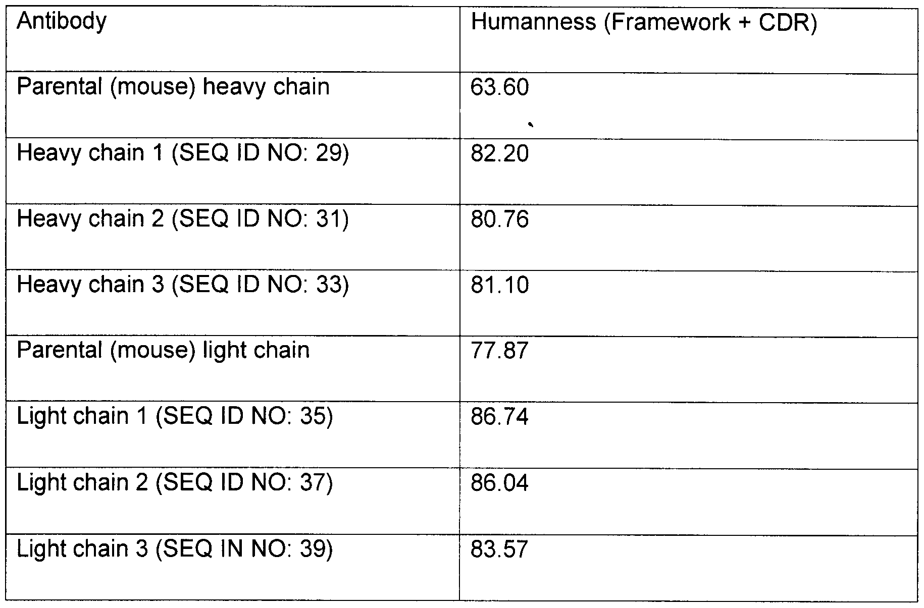

Claims
Priority Applications (7)
| Application Number | Priority Date | Filing Date | Title |
|---|---|---|---|
| US16/092,743 US11958900B2 (en) | 2017-04-14 | Anti-age antibodies for treating neurodegenerative disorders | |
| KR1020187031932A KR20180133452A (en) | 2016-04-15 | 2017-04-14 | Anti-aging antibodies to treat neurodegenerative diseases |
| AU2017250301A AU2017250301A1 (en) | 2016-04-15 | 2017-04-14 | Anti-age antibodies for treating neurodegenerative disorders |
| CA3057829A CA3057829A1 (en) | 2016-04-15 | 2017-04-14 | Anti-age antibodies for treating neurodegenerative disorders |
| CN201780037571.4A CN109311975A (en) | 2016-04-15 | 2017-04-14 | For treating the anti-AGE antibody of neurodegenerative disorder |
| EP17737078.0A EP3443007A1 (en) | 2016-04-15 | 2017-04-14 | Anti-age antibodies for treating neurodegenerative disorders |
| IL262222A IL262222A (en) | 2016-04-15 | 2018-10-09 | Anti-age antibodies for treating neurodegenerative disorders |
Applications Claiming Priority (2)
| Application Number | Priority Date | Filing Date | Title |
|---|---|---|---|
| US201662323471P | 2016-04-15 | 2016-04-15 | |
| US62/323,471 | 2016-04-15 |
Publications (1)
| Publication Number | Publication Date |
|---|---|
| WO2017181116A1 true WO2017181116A1 (en) | 2017-10-19 |
Family
ID=59297340
Family Applications (1)
| Application Number | Title | Priority Date | Filing Date |
|---|---|---|---|
| PCT/US2017/027773 WO2017181116A1 (en) | 2016-04-15 | 2017-04-14 | Anti-age antibodies for treating neurodegenerative disorders |
Country Status (7)
| Country | Link |
|---|---|
| EP (1) | EP3443007A1 (en) |
| KR (1) | KR20180133452A (en) |
| CN (1) | CN109311975A (en) |
| AU (1) | AU2017250301A1 (en) |
| CA (1) | CA3057829A1 (en) |
| IL (1) | IL262222A (en) |
| WO (1) | WO2017181116A1 (en) |
Cited By (17)
| Publication number | Priority date | Publication date | Assignee | Title |
|---|---|---|---|---|
| US9993535B2 (en) | 2014-12-18 | 2018-06-12 | Siwa Corporation | Method and composition for treating sarcopenia |
| WO2018191718A1 (en) * | 2017-04-13 | 2018-10-18 | Siwa Corporation | Humanized monoclonal advanced glycation end-product antibody |
| US10226531B2 (en) | 2010-09-27 | 2019-03-12 | Siwa Corporation | Selective removal of age-modified cells for treatment of atherosclerosis |
| US10358502B2 (en) | 2014-12-18 | 2019-07-23 | Siwa Corporation | Product and method for treating sarcopenia |
| WO2020041625A1 (en) | 2018-08-23 | 2020-02-27 | Siwa Corporation | Anti carboxymethyl lysine antibodies and ultrasound for removing age-modified cells |
| US10584180B2 (en) | 2014-09-19 | 2020-03-10 | Siwa Corporation | Anti-AGE antibodies for treating inflammation and auto-immune disorders |
| US10858449B1 (en) | 2017-01-06 | 2020-12-08 | Siwa Corporation | Methods and compositions for treating osteoarthritis |
| US10925937B1 (en) | 2017-01-06 | 2021-02-23 | Siwa Corporation | Vaccines for use in treating juvenile disorders associated with inflammation |
| US10960234B2 (en) | 2010-11-22 | 2021-03-30 | Siwa Corporation | Selective removal of cells having accumulated agents |
| US10961321B1 (en) | 2017-01-06 | 2021-03-30 | Siwa Corporation | Methods and compositions for treating pain associated with inflammation |
| US10995151B1 (en) | 2017-01-06 | 2021-05-04 | Siwa Corporation | Methods and compositions for treating disease-related cachexia |
| WO2021222758A1 (en) * | 2020-05-01 | 2021-11-04 | Siwa Corporation | Methods of treating infections |
| US11213585B2 (en) | 2016-06-23 | 2022-01-04 | Siwa Corporation | Vaccines for use in treating various diseases and disorders |
| US11261241B2 (en) | 2008-05-23 | 2022-03-01 | Siwa Corporation | Methods, compositions and apparatuses for facilitating regeneration |
| US11518801B1 (en) | 2017-12-22 | 2022-12-06 | Siwa Corporation | Methods and compositions for treating diabetes and diabetic complications |
| US11833202B2 (en) | 2016-02-19 | 2023-12-05 | Siwa Corporation | Method and composition for treating cancer, killing metastatic cancer cells and preventing cancer metastasis using antibody to advanced glycation end products (AGE) |
| US11958900B2 (en) | 2017-04-14 | 2024-04-16 | Siwa Corporation | Anti-age antibodies for treating neurodegenerative disorders |
Families Citing this family (1)
| Publication number | Priority date | Publication date | Assignee | Title |
|---|---|---|---|---|
| CN110283246A (en) * | 2019-06-20 | 2019-09-27 | 长春西诺生物科技有限公司 | A kind of cat source genetic engineering antibody of anti-feline panleucopenia virus |
Citations (10)
| Publication number | Priority date | Publication date | Assignee | Title |
|---|---|---|---|---|
| US4217344A (en) | 1976-06-23 | 1980-08-12 | L'oreal | Compositions containing aqueous dispersions of lipid spheres |
| US4911928A (en) | 1987-03-13 | 1990-03-27 | Micro-Pak, Inc. | Paucilamellar lipid vesicles |
| US4917951A (en) | 1987-07-28 | 1990-04-17 | Micro-Pak, Inc. | Lipid vesicles formed of surfactants and steroids |
| US5702704A (en) | 1991-12-20 | 1997-12-30 | The Rockefeller University | Antibodies to in vivo advanced glycosylation endproducts |
| US6380165B1 (en) | 1997-09-19 | 2002-04-30 | The Picower Institute For Medical Research | Immunological advanced glycation endproduct crosslink |
| US6387373B1 (en) | 1993-01-15 | 2002-05-14 | Novavax, Inc. | Vaccines containing paucilsmellar lipid vesicles as immunological adjuvants |
| WO2009143411A2 (en) | 2008-05-23 | 2009-11-26 | Siwa Corporation | Methods, compositions and apparatus for facilitating regeneration |
| US20100226932A1 (en) | 2006-02-22 | 2010-09-09 | Novavax, Inc. | Adjuvant and Vaccine Compositions |
| US20130131006A1 (en) * | 2010-05-11 | 2013-05-23 | Gachon University Of Industry-Academic Cooperation Foundation | Method for inhibiting the induction of cell death by inhibiting the synthesis or secretion of age-albumin in cells of the mononuclear phagocyte system |
| US20140234343A1 (en) * | 2011-11-10 | 2014-08-21 | Gachon University Of Medicine Science Industry-Academic Cooperation Foundation | Composition for prevention or treatment of ischemic cardiac disease, comprising inhibitor against age-albumin synthesis or release of mononuclear phagocyte system cells as active ingredient |
Family Cites Families (2)
| Publication number | Priority date | Publication date | Assignee | Title |
|---|---|---|---|---|
| US6410598B1 (en) * | 1994-02-03 | 2002-06-25 | Michael P. Vitek | Compositions and methods for advanced glycosylation endproduct-mediated modulation of amyloidosis |
| TWI557112B (en) * | 2012-03-05 | 2016-11-11 | 百靈佳殷格翰國際股份有限公司 | Inhibitors of beta-secretase |
-
2017
- 2017-04-14 AU AU2017250301A patent/AU2017250301A1/en active Pending
- 2017-04-14 CN CN201780037571.4A patent/CN109311975A/en active Pending
- 2017-04-14 WO PCT/US2017/027773 patent/WO2017181116A1/en active Application Filing
- 2017-04-14 EP EP17737078.0A patent/EP3443007A1/en active Pending
- 2017-04-14 CA CA3057829A patent/CA3057829A1/en active Pending
- 2017-04-14 KR KR1020187031932A patent/KR20180133452A/en not_active Application Discontinuation
-
2018
- 2018-10-09 IL IL262222A patent/IL262222A/en unknown
Patent Citations (10)
| Publication number | Priority date | Publication date | Assignee | Title |
|---|---|---|---|---|
| US4217344A (en) | 1976-06-23 | 1980-08-12 | L'oreal | Compositions containing aqueous dispersions of lipid spheres |
| US4911928A (en) | 1987-03-13 | 1990-03-27 | Micro-Pak, Inc. | Paucilamellar lipid vesicles |
| US4917951A (en) | 1987-07-28 | 1990-04-17 | Micro-Pak, Inc. | Lipid vesicles formed of surfactants and steroids |
| US5702704A (en) | 1991-12-20 | 1997-12-30 | The Rockefeller University | Antibodies to in vivo advanced glycosylation endproducts |
| US6387373B1 (en) | 1993-01-15 | 2002-05-14 | Novavax, Inc. | Vaccines containing paucilsmellar lipid vesicles as immunological adjuvants |
| US6380165B1 (en) | 1997-09-19 | 2002-04-30 | The Picower Institute For Medical Research | Immunological advanced glycation endproduct crosslink |
| US20100226932A1 (en) | 2006-02-22 | 2010-09-09 | Novavax, Inc. | Adjuvant and Vaccine Compositions |
| WO2009143411A2 (en) | 2008-05-23 | 2009-11-26 | Siwa Corporation | Methods, compositions and apparatus for facilitating regeneration |
| US20130131006A1 (en) * | 2010-05-11 | 2013-05-23 | Gachon University Of Industry-Academic Cooperation Foundation | Method for inhibiting the induction of cell death by inhibiting the synthesis or secretion of age-albumin in cells of the mononuclear phagocyte system |
| US20140234343A1 (en) * | 2011-11-10 | 2014-08-21 | Gachon University Of Medicine Science Industry-Academic Cooperation Foundation | Composition for prevention or treatment of ischemic cardiac disease, comprising inhibitor against age-albumin synthesis or release of mononuclear phagocyte system cells as active ingredient |
Non-Patent Citations (81)
| Title |
|---|
| A BIERHAUS ET AL: "AGEs and their interaction with AGE-receptors in vascular disease and diabetes mellitus. I. The AGE concept", CARDIOVASCULAR RESEARCH, vol. 37, no. 3, 1 March 1998 (1998-03-01), GB, pages 586 - 600, XP055399820, ISSN: 0008-6363, DOI: 10.1016/S0008-6363(97)00233-2 * |
| AHMED, E. K. ET AL.: "Protein Modification and Replicative Senescence of WI-38 Human Embryonic Fibroblasts", AGING CELLS, vol. 9, 2010, pages 252,260 |
| AHMED, E.K. ET AL.: "Protein Modification and Replicative Senescence of WI-38 Human Embryonic Fibroblasts", AGING CELLS, vol. 9, no. 252, 2010, pages 260 |
| AMELI, S. ET AL.: "Effect of Immunization With Homologous LDL and Oxidized LDL on Early Atherosclerosis in Hypercholesterolemic Rabbits", ARTERIOSCLEROSIS, THROMBOSIS, AND VASCULAR BIOLOGY, vol. 16, 1996, pages 1074 |
| ANDO K ET AL.: "Membrane Proteins of Human Erythrocytes Are Modified by Advanced Glycation End Products During Aging in the Circulation", BIOCHEMICAL AND BIOPHYSICAL RESEARCH COMMUNICATIONS, vol. 258, 1999, pages 123 - 27, XP002729025, DOI: doi:10.1006/bbrc.1999.0606 |
| ANDO, K. ET AL.: "Membrane Proteins of Human Erythrocytes Are Modified by Advanced Glycation End Products during Aging in the Circulation", BIOCHEM BIOPHYS RES COMMUN., vol. 258, no. 123, 1999, pages 125 |
| ANDO, K. ET AL.: "Membrane Proteins of Human Erythrocytes Are Modified by Advanced Glycation End Products during Aging in the Circulation", BIOCHEM BIOPHYS RES COMMUN.,, vol. 258, no. 123, 1999, pages 125 |
| AROIAN R. ET AL.: "Pore-Forming Toxins and Cellular Non-Immune Defenses (CNIDs", CURRENT OPINION IN MICROBIOLOGY, vol. 10, 2007, pages 57 - 61, XP005884635 |
| BAKER, D. J. ET AL.: "Clearance of p16lnk4a-positive senescent cells delays ageing-associated disorders", NATURE, vol. 479, 2011, pages 232 - 236, XP055074325, DOI: doi:10.1038/nature10600 |
| BAKER, D.J. ET AL.: "Clearance of p16lnk4a-positive senescent cells delays ageing-associated disorders", NATURE, vol. 479, 2011, pages 232 - 236, XP055074325, DOI: doi:10.1038/nature10600 |
| BHAT, R. ET AL.: "Astrocyte Senescence as a Component of Alzheimer's Disease", PLOS ONE, vol. 7, no. 9, September 2012 (2012-09-01), pages 1 - 10 |
| BIERHAUS A: "AGEs and their interaction with AGE-receptors in vascular disease and diabetes mellitus. I. The AGE concept", CARDIOVASC RES, vol. 37, no. 3, 1998, pages 586 - 600, XP055399820, DOI: doi:10.1016/S0008-6363(97)00233-2 |
| BIRAN, A. ET AL.: "Senescent cells communicate via intercellular protein transfer", GENES & DEVELOPMENT, vol. 29, 2015, pages 791 - 802 |
| BLAHD, W., SARCOPENIA WITH AGING, 3 August 2014 (2014-08-03) |
| BLASKO, C. ET AL.: "Encyclopedia of Neuroscience", 2009, ACADEMIC PRESS, article "Glial cells: astrocytes and oligodendrocytes during normal brain aging", pages: 743 - 747 |
| CHEN, K. S. ET AL.: "Monoclonal antibody therapy for malignant glioma", GLIOMA: IMMUNOTHERAPEUTIC APPROACHES, 2012, pages 121 - 141 |
| CHINTA, S. J. ET AL.: "Environmental stress, ageing and glial cell senescence: a novel mechanistic link to Parkinson's disease?", J INTERN MED, vol. 273, 2013, pages 429 - 436 |
| CML (N-EPSILON-(CARBOXYMETHYL)LYSINE) ASSAYS AND REAGENTS, 15 December 2014 (2014-12-15), Retrieved from the Internet <URL:www.cellbiolabs.com/cml-assays> |
| CML-BSA PRODUCT DATA SHEET, 2010 |
| CRUZ-JENTOFT, A. J. ET AL.: "Sarcopenia: European consensus on definition and diagnosis", AGE AND AGEING, vol. 39, 13 April 2010 (2010-04-13), pages 412 - 423, XP009167538, DOI: doi:10.1093/ageing/afq034 |
| DANYSZ, W. ET AL.: "Alzheimer's disease, (3-amyloid, glutamate, NMDA receptors and memantine - searching for the connections", BRITISH JOURNAL OF PHARMACOLOGY, vol. 167, 2012, pages 324 - 352 |
| DAS, M. M.; SVENDSEN, C. N.: "Astrocytes show reduced support of motor neurons with aging that is accelerated in a rodent model of ALS", NEUROBIOLOGY OF AGING, vol. 36, 2015, pages 1130 - 1139 |
| DE GENST, E. ET AL.: "Antibody repertoire development in camelids", DEVELOPMENTAL & COMPARATIVE IMMUNOLOGY, vol. 30, 11 July 2005 (2005-07-11), pages 187 - 198, XP025088320, DOI: doi:10.1016/j.dci.2005.06.010 |
| DI, G-H. ET AL.: "IL-6 Secreted from Senescent Mesenchymal Stem Cells Promotes Proliferation and migration of Breast Cancer Cells", PLOS ONE, vol. 9, no. 11, 2014, pages e113572 |
| DOBSON, D.M.: "The structural basis of protein folding and its links with human disease", PHIL. TRANS. R. SOC. LOND. B, vol. 356, 2001, pages 133 - 145 |
| FEIGE, M. J. ET AL.: "The structural analysis of shark IgNAR antibodies reveals evolutionary principles of immunoglobulins", PROCEEDINGS OF THE NATIONAL ACADEMY OF SCIENCES OF THE UNITED STATES OF AMERICA, vol. 111, no. 22, 3 June 2014 (2014-06-03), pages 8155 - 8160, XP055123270, DOI: doi:10.1073/pnas.1321502111 |
| FIELDING, R. A. ET AL.: "Sarcopenia: an undiagnosed condition in older adults. Current consensus definition: prevalence, etiology, and consequences", JOURNAL OF THE AMERICAN MEDICAL DIRECTORS ASSOCIATION, vol. 12, no. 4, May 2011 (2011-05-01), pages 249 - 256 |
| FLANARY, B. E. ET AL.: "Evidence That Aging And Amyloid Promote Microglial Cell Senescence", REJUVENATION RESEARCH,, vol. 10, no. 1, March 2007 (2007-03-01), pages 61 - 74 |
| FU, M.-X. ET AL.: "The Advanced Glycation End Product, NE-(Carboxymehtyl)lysine, Is a Product of both Lipid Peroxidation and Glycoxidation Reactions", J. BIOL. CHEM., vol. 271, 1996, pages 9982 - 9986, XP055171871, DOI: doi:10.1074/jbc.271.17.9982 |
| FURTHERMORE, AHMED, E. K. ET AL., ONE OF THE MAJOR CAUSES OF SPONTANEOUS DAMAGE TO CELLULAR AND EXTRACELLULAR PROTEINS |
| GAO, S. H. ET AL.: "Monoclonal antibody humanness score and its applications", BMC BIOTECHNOLOGY, vol. 13, 5 July 2013 (2013-07-05), pages 55, XP021156542, DOI: doi:10.1186/1472-6750-13-55 |
| GARCIA-MATAS, S. ET AL.: "Dysfunction of astrocytes in senescence-accelerated mice SAMP8 reduces their neuroprotective capacity", AGING CELL, vol. 7, 2008, pages 630 - 640 |
| GOLDE, T.E. ET AL.: "Proteinopathy-induced neuronal senescence: a hypothesis for brain failure in Alzheimer's and other neurodegenerative diseases", ALZHEIMER'S RESEARCH & THERAPY, vol. 1, no. 5, 13 October 2009 (2009-10-13) |
| GRIFFIN, L.M. ET AL.: "Analysis of heavy and light chain sequences of conventional camelid antibodies from Camelus dromedarius and Camelus bactrianus species", JOURNAL OF IMMUNOLOGICAL METHODS, vol. 405, 18 January 2014 (2014-01-18), pages 35 - 46 |
| GROOT, K. ET AL.: "Vascular Endothelial Damage and Repair in Antineutrophil Cytoplasmic Antibody-Associated Vasculitis", ARTHRITIS AND RHEUMATISM, vol. 56, no. 11, 2007, pages 3847,3847 |
| HAMERS-CASTERMAN, C. ET AL.: "Naturally occurring antibodies devoid of light chains", NATURE, vol. 363, 3 June 1993 (1993-06-03), pages 446 - 448, XP002535892, DOI: doi:10.1038/363446a0 |
| JANA HADRABOVA ET AL.: "Chicken immunoglobulins for prophylaxis: Effect of inhaled antibodies on inflammatory parameters in rat airways", JOURNAL OF APPLIED BIOMEDICINE, 5 May 2014 (2014-05-05) |
| KAJSTURA, J. ET AL.: "Myocite Turnover in the Aging Human Heart", CIRC. RES., vol. 107, no. 11, 2010, pages 1374 - 86 |
| KATCHER, H. L.: "Studies that shed new light on aging", BIOCHEMISTRY (MOSCOW), vol. 78, no. 9, 2013, pages 1061 - 1070, XP035358815, DOI: doi:10.1134/S0006297913090137 |
| KATO, S. ET AL.: "Advanced glycation endproduct-modified superoxide dismutase-1 (SODI)-positive inclusions are common to familial amyotrophic lateral sclerosis patients with SOD1 gene mutations and transgenic mice expressing human SOD1 with a G85R mutation", ACTA NEUROPATHOL, vol. 100, 2000, pages 490 - 505 |
| KEYHOLE LIMPET HEMOCYANIN, 18 April 2014 (2014-04-18) |
| KING, O.D. ET AL.: "The tip of the iceberg: RNA-binding proteins with prion-like domains in neurodegenerative disease", BRAIN RES., vol. 1462, 2012, pages 61 - 80, XP028521741, DOI: doi:10.1016/j.brainres.2012.01.016 |
| KIRSTEIN, M. ET AL.: "Receptor-specific Induction of Insulin-like Growth Factor I in Human Monocytes by Advanced Glycosylation End Product-modified Proteins", J. CLIN. INVEST., vol. 90, no. 439, 1992, pages 439 - 440 |
| LEONARD, B.W. ET AL.: "Subventricular zone neural progenitors from rapid brain autopsies of elderly subjects with and without neurodegenerative disease", THE JOURNAL OF COMPARATIVE NEUROLOGY, vol. 515, 2009, pages 269 - 294 |
| LINDSEY JB ET AL.: "Receptor For Advanced Glycation End-Products (RAGE) and soluble RAGE (sRAGE): Cardiovascular Implications", DIABETES VASCULAR DISEASE RESEARCH, vol. 6, no. 1, 2009, pages 7 - 14 |
| LIU, D. ET AL.: "Senescent Human Fibroblasts Increase the Early Growth of Xenograft Tumors via Matrix Metalloproteinase Secretion", CANCER RES, vol. 67, 2007, pages 3117 - 3126 |
| LUESSI, F. ET AL.: "Neurodegeneration in multiple sclerosis: novel treatment strategies", EXPERT REV. NEUROTHER., vol. 9, 2012, pages 1061 - 1077, XP055337068, DOI: doi:10.1586/ern.12.59 |
| MAASS, D. R. ET AL.: "Alpaca (Lama pacos) as a convenient source of recombinant camelid heavy chain antibodies (VHHs", JOURNAL OF IMMUNOLOGICAL METHODS, vol. 324, no. 1-2, 31 July 2007 (2007-07-31), pages 13 - 25, XP022138109, DOI: doi:10.1016/j.jim.2007.04.008 |
| MANESSO, E. ET AL.: "Dynamics of β-Cell Turnover: Evidence for β-Ce Turnover and Regeneration from Sources of β-Cells other than β-ce Replication in the HIP Rat", AM. J. PHYSIOL. ENDOCRINOL. METAB., vol. 297, 2009, pages E323,E324 |
| MATUS ET AL.: "Invasive Cell Fate Requires G1 Cell-Cycle arrest and Histone Deacetylase-Mediated Changes in Gene Expression", DEVELOPMENTAL CELL, vol. 35, 2015, pages 162 - 174, XP029308527, DOI: doi:10.1016/j.devcel.2015.10.002 |
| MERA, K. ET AL.: "An autoantibody against NE-(carboxyethyl)lysine (CEL): Possible involvement in the removal of CEL-modified proteins by macrophages", BIOCHEMICAL AND BIOPHYSICAL RESEARCH COMMUNICATIONS, vol. 407, 12 March 2011 (2011-03-12), pages 420 - 425 |
| MEUTER A. ET AL.: "Markers of cellular senescence are elevated in murine blastocysts cultured in vitro: molecular consequences of culture in atmospheric oxygen", J ASSIST REPROD GENET., 10 August 2014 (2014-08-10) |
| MURPHY, J. F.: "Trends in cancer immunotherapy", CLINICAL MEDICAL INSIGHTS: ONCOLOGY, vol. 14, no. 4, 2010, pages 67 - 80 |
| MUYLDERMANS, S. ET AL.: "Sequence and structure of V domain from naturally occurring camel heavy chain immunoglobulins lacking light chains", PROTEIN ENGINEERING, vol. 7, no. 9, 1994, pages 1129 - 1135 |
| NAYLOR, R. M. ET AL.: "Senescent cells: a novel therapeutic target for aging and age-related diseases", CLINICAL PHARMACOLOGY & THERAPEUTICS, vol. 93, no. 1, 5 December 2012 (2012-12-05), pages 105 - 116, XP002710105, DOI: doi:10.1038/clpt.2012.193 |
| NELSON, G., A: "senescent cell bystander effect: senescence-induced senescence", AGING CELL, vol. 11, 2012, pages 345 - 349 |
| NGUYEN, V.K. ET AL.: "Camel heavy-chain antibodies: diverse germline V H and specific mechanisms enlarge the antigen-binding repertoire", THE EUROPEAN MOLECULAR BIOLOGY ORGANIZATION JOURNAL, vol. 19, no. 5, 2000, pages 921 - 930, XP002194418, DOI: doi:10.1093/emboj/19.5.921 |
| NIE, H. ET AL.: "Impaired glial glutamate uptake induces extrasynaptic glutamate spillover in the spinal sensory synapses of neuropathic rats", JOURNAL OF NEUROPHYSIOLOGY, vol. 103, 2010, pages 2570 - 2580 |
| PATEL, M. ET AL.: "Sequence of the dog immunoglobulin alpha and epsilon constant region genes", IMMUNOGENETICS, vol. 41, 1995, pages 282 - 286, XP002037155, DOI: doi:10.1007/BF00172152 |
| PRADAT, P.-F. ET AL.: "Abnormalities of satellite cells function in amyotrophic lateral sclerosis", AMYOTROPHIC LATERAL SCLEROSIS, vol. 12, 2011, pages 264 - 271 |
| R. YAMANAKA: "Glioma: Immunotherapeutic Approaches", 2012, article CHEN, K.S. ET AL.: "MONOCLONAL ANTIBODY THERAPY FOR MALIGNANT GLIOMA (chapter 10)", pages: 132 - 141 |
| RAYESS, H. ET AL.: "Cellular senescence and tumor suppressor gene p16", INTJ CANCER, vol. 130, 2012, pages 1715 - 1725, XP055282399, DOI: doi:10.1002/ijc.27316 |
| REDDY, S. ET AL.: "NE-(carboxymethyl)lysine is a dominant advanced glycation end product (AGE) antigen in tissue proteins", BIOCHEMISTRY, vol. 34, 1 August 1995 (1995-08-01), pages 10872 - 10878, XP002088366, DOI: doi:10.1021/bi00034a021 |
| ROLL, P. ET AL.: "Anti-CD20 Therapy in Patients with Rheumatoid Arthritis", ARTHRITIS & RHEUMATISM, vol. 58, no. 6, 2008, pages 1566 - 1575, XP055030411, DOI: doi:10.1002/art.23473 |
| ROLLAND, Y. ET AL.: "Sarcopenia: its assessment, etiology, pathogenesis, consequences and future perspectives", J. NUTR. HEALTH AGING,, vol. 12, no. 7, 2008, pages 433 - 450 |
| ROMAGOSA, C. ET AL.: "p16lnk4a overexpression in cancer: a tumor suppressor gene associated with senescence and high-grade tumors", ONCOGENE, vol. 30, 2011, pages 2087 - 2097 |
| ROUGER ET AL.: "Systemic Delivery of Allogenic Muscle Stem Cells Induces Long-Term Muscle Repair and Clinical Efficacy in Duchenne Muscular Dystrophy Dogs", THE AMERICAN JOURNAL OF PATHOLOGY, vol. 179, no. 5, November 2011 (2011-11-01), pages 2501 - 2518, XP009171616, DOI: doi:10.1016/j.ajpath.2011.07.022 |
| SARCOPENIA, 14 November 2014 (2014-11-14) |
| STRIETZEL, C.J. ET AL.: "In vitro functional characterization of feline IgGs", VETERINARY IMMUNOLOGY AND IMMUNOPATHOLOGY, vol. 158, 2014, pages 214 - 223 |
| TAKAMI, A. ET AL.: "Treatment of primary central nervous system lymphoma with induction of complement-dependent cytotoxicity by intraventricular administration of autologous-serum-supplemented rituximab", CANCER SCI., vol. 97, January 2006 (2006-01-01), pages 80 - 83 |
| TAKEDA, A. ET AL.: "Advanced glycation end products co-localize with astrocyes and microglial cells in Alzheimer's disease brain", ACTA NEUROPATHOLOGICA,, vol. 95, 1998, pages 555 - 558 |
| VIRELLA, G. ET AL.: "Autoimmune Response to Advanced Glycosylation End-Products of Human LDL", JOURNAL OF LIPID RESEARCH, vol. 44, 2003, pages 487 - 493 |
| VLASSARA ET AL.: "High-affinity-receptor-mediated Uptake and Degradation of Glucose-modified Proteins: A Potential Mechanism for the Removal of Senescent Macromolecules", PROC. NATL. ACAD. SCI. USAI, vol. 82, 1985, pages 5588,5591 |
| VLASSARA, H. ET AL.: "Advanced Glycosylation Endproducts on Erythrocyte Cell Surface Induce Receptor-Mediated Phagocytosis by Macrophages", J. EXP. MED., vol. 166, 1987, pages 539,545 |
| VLASSARA, H. ET AL.: "Advanced Glycosylation Endproducts on Erythrocyte Cell Surface Induce Receptor-Mediated Phagocytosis by Macrophages", J. EXP. MED., vol. 166, no. 539, 1987, pages 545 |
| VLASSARA, H. ET AL.: "High-affinity-receptor-mediated Uptake and Degradation of Glucose-modified Proteins: A Potential Mechanism for the Removal of Senescent Macromolecules", PROC. NATL. ACAD. SCI. USA, vol. 82, 1985, pages 5588,5591 |
| WAGNER, B. ET AL.: "The complete map of the Ig heavy chain constant gene region reveals evidence for seven IgG isotypes and for IgD in the horse", THE JOURNAL OF IMMUNOLOGY, vol. 173, 2004, pages 3230 - 3242 |
| WESOLOWSKI ET AL.: "Single domain antibodies: promising experimental and therapeutic tools in infection and immunity", MEDICAL MICROBIOLOGY AND IMMUNOLOGY, vol. 198, 16 June 2009 (2009-06-16), pages 157 - 174 |
| WHAT IS SARCOPENIA?, 2014 |
| WRIGHT, W. E.: "Myoblast Senescence in Muscular Dystrophy", EXP CELL RES, vol. 157, 1985, pages 343 - 354, XP024858006, DOI: doi:10.1016/0014-4827(85)90119-3 |
| YAN, S.F. ET AL.: "Soluble RAGE: therapy & biomarker in unraveling the RAGE axis in chronic disease and aging", BIOCHEMICAL PHARMACOLOGY, vol. 79, no. 10, 15 May 2010 (2010-05-15), pages 1379 - 1386, XP026939420, DOI: doi:10.1016/j.bcp.2010.01.013 |
Cited By (21)
| Publication number | Priority date | Publication date | Assignee | Title |
|---|---|---|---|---|
| US11261241B2 (en) | 2008-05-23 | 2022-03-01 | Siwa Corporation | Methods, compositions and apparatuses for facilitating regeneration |
| US10226531B2 (en) | 2010-09-27 | 2019-03-12 | Siwa Corporation | Selective removal of age-modified cells for treatment of atherosclerosis |
| US10960234B2 (en) | 2010-11-22 | 2021-03-30 | Siwa Corporation | Selective removal of cells having accumulated agents |
| US10584180B2 (en) | 2014-09-19 | 2020-03-10 | Siwa Corporation | Anti-AGE antibodies for treating inflammation and auto-immune disorders |
| US9993535B2 (en) | 2014-12-18 | 2018-06-12 | Siwa Corporation | Method and composition for treating sarcopenia |
| US11872269B2 (en) | 2014-12-18 | 2024-01-16 | Siwa Corporation | Method and composition for treating sarcopenia |
| US10358502B2 (en) | 2014-12-18 | 2019-07-23 | Siwa Corporation | Product and method for treating sarcopenia |
| US11873345B2 (en) | 2014-12-18 | 2024-01-16 | Siwa Corporation | Product and method for treating sarcopenia |
| US11833202B2 (en) | 2016-02-19 | 2023-12-05 | Siwa Corporation | Method and composition for treating cancer, killing metastatic cancer cells and preventing cancer metastasis using antibody to advanced glycation end products (AGE) |
| US11213585B2 (en) | 2016-06-23 | 2022-01-04 | Siwa Corporation | Vaccines for use in treating various diseases and disorders |
| US10961321B1 (en) | 2017-01-06 | 2021-03-30 | Siwa Corporation | Methods and compositions for treating pain associated with inflammation |
| US10995151B1 (en) | 2017-01-06 | 2021-05-04 | Siwa Corporation | Methods and compositions for treating disease-related cachexia |
| US10925937B1 (en) | 2017-01-06 | 2021-02-23 | Siwa Corporation | Vaccines for use in treating juvenile disorders associated with inflammation |
| US10858449B1 (en) | 2017-01-06 | 2020-12-08 | Siwa Corporation | Methods and compositions for treating osteoarthritis |
| US10919957B2 (en) | 2017-04-13 | 2021-02-16 | Siwa Corporation | Humanized monoclonal advanced glycation end-product antibody |
| US11542324B2 (en) | 2017-04-13 | 2023-01-03 | Siwa Corporation | Humanized monoclonal advanced glycation end-product antibody |
| WO2018191718A1 (en) * | 2017-04-13 | 2018-10-18 | Siwa Corporation | Humanized monoclonal advanced glycation end-product antibody |
| US11958900B2 (en) | 2017-04-14 | 2024-04-16 | Siwa Corporation | Anti-age antibodies for treating neurodegenerative disorders |
| US11518801B1 (en) | 2017-12-22 | 2022-12-06 | Siwa Corporation | Methods and compositions for treating diabetes and diabetic complications |
| WO2020041625A1 (en) | 2018-08-23 | 2020-02-27 | Siwa Corporation | Anti carboxymethyl lysine antibodies and ultrasound for removing age-modified cells |
| WO2021222758A1 (en) * | 2020-05-01 | 2021-11-04 | Siwa Corporation | Methods of treating infections |
Also Published As
| Publication number | Publication date |
|---|---|
| EP3443007A1 (en) | 2019-02-20 |
| IL262222A (en) | 2018-12-31 |
| CA3057829A1 (en) | 2017-10-19 |
| US20190119371A1 (en) | 2019-04-25 |
| CN109311975A (en) | 2019-02-05 |
| AU2017250301A1 (en) | 2018-11-15 |
| KR20180133452A (en) | 2018-12-14 |
Similar Documents
| Publication | Publication Date | Title |
|---|---|---|
| CA3000815C (en) | Anti-age antibodies and methods of use thereof | |
| US11873345B2 (en) | Product and method for treating sarcopenia | |
| WO2017181116A1 (en) | Anti-age antibodies for treating neurodegenerative disorders | |
| US10889634B2 (en) | Anti-age antibodies and methods of use thereof | |
| AU2017219749B2 (en) | Method and composition for treating cancer, killing metastatic cancer cells and preventing cancer metastasis using antibody to advanced glycation end products (AGE) | |
| US11542324B2 (en) | Humanized monoclonal advanced glycation end-product antibody | |
| US20210253737A1 (en) | Methods and compositions for treating disease-related cachexia | |
| WO2023177390A1 (en) | Humanized monoclonal advanced glycation end-product antibody for treating pancreatic cancer | |
| AU2018251183B2 (en) | Humanized monoclonal advanced glycation end-product antibody | |
| US11958900B2 (en) | Anti-age antibodies for treating neurodegenerative disorders | |
| GRUBER | Patent 3000815 Summary |
Legal Events
| Date | Code | Title | Description |
|---|---|---|---|
| NENP | Non-entry into the national phase |
Ref country code: DE |
|
| ENP | Entry into the national phase |
Ref document number: 20187031932 Country of ref document: KR Kind code of ref document: A |
|
| ENP | Entry into the national phase |
Ref document number: 2017250301 Country of ref document: AU Date of ref document: 20170414 Kind code of ref document: A |
|
| WWE | Wipo information: entry into national phase |
Ref document number: 2017737078 Country of ref document: EP |
|
| ENP | Entry into the national phase |
Ref document number: 2017737078 Country of ref document: EP Effective date: 20181115 |
|
| 121 | Ep: the epo has been informed by wipo that ep was designated in this application |
Ref document number: 17737078 Country of ref document: EP Kind code of ref document: A1 |
|
| ENP | Entry into the national phase |
Ref document number: 3057829 Country of ref document: CA |